-
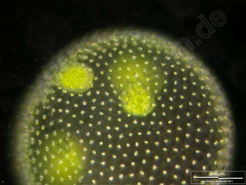
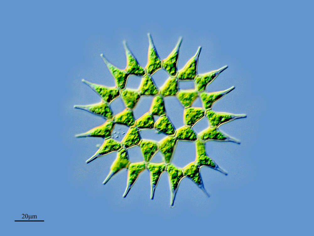
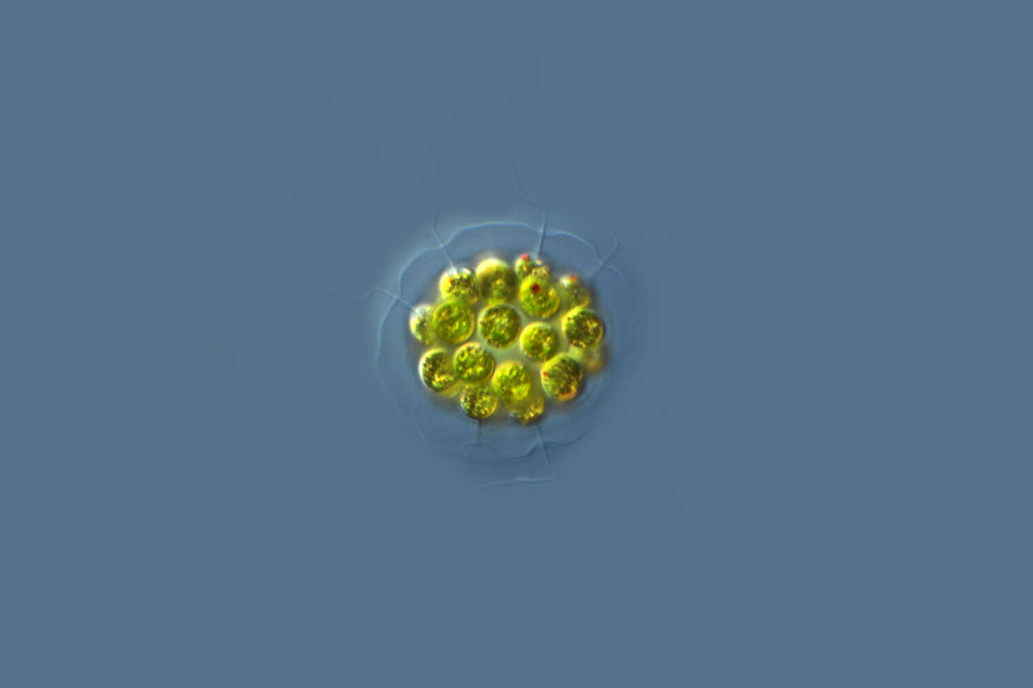
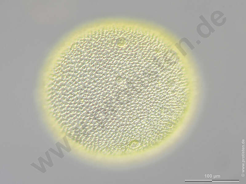
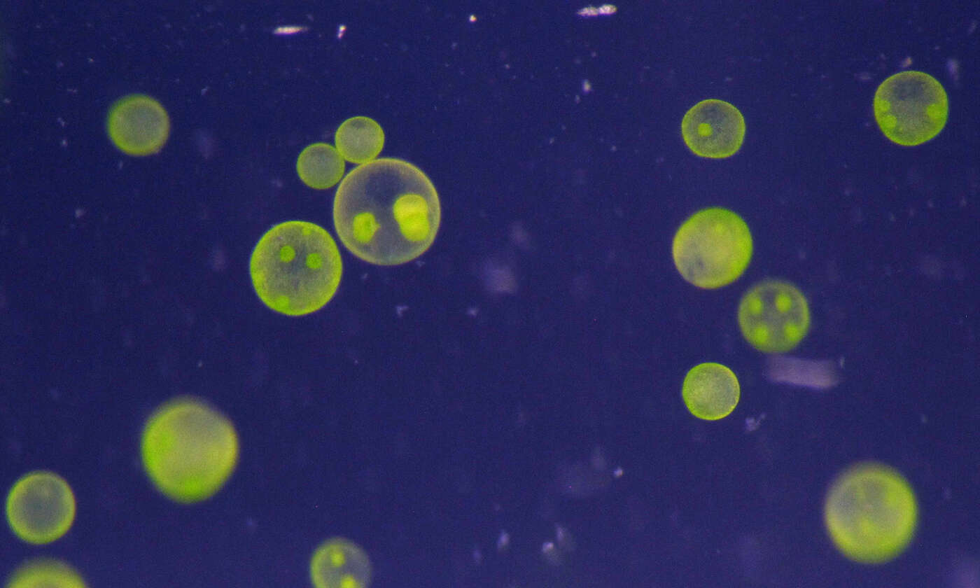
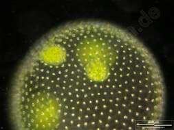
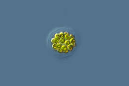
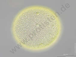
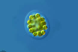
Cell colony with daugther colonies. Scale bar indicates 100 m. Sample from ponds situated in the vicinity of Lake Constance. The image was built up using several photomicrographic frames with manual stacking technique. Images were taken using Zeiss Universal with Olympus C7070 CCD camera.For permission to use of (high-resolution) images please contact postmaster@protisten.de.
-
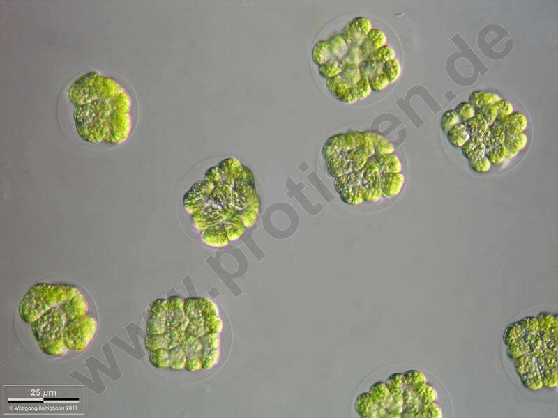
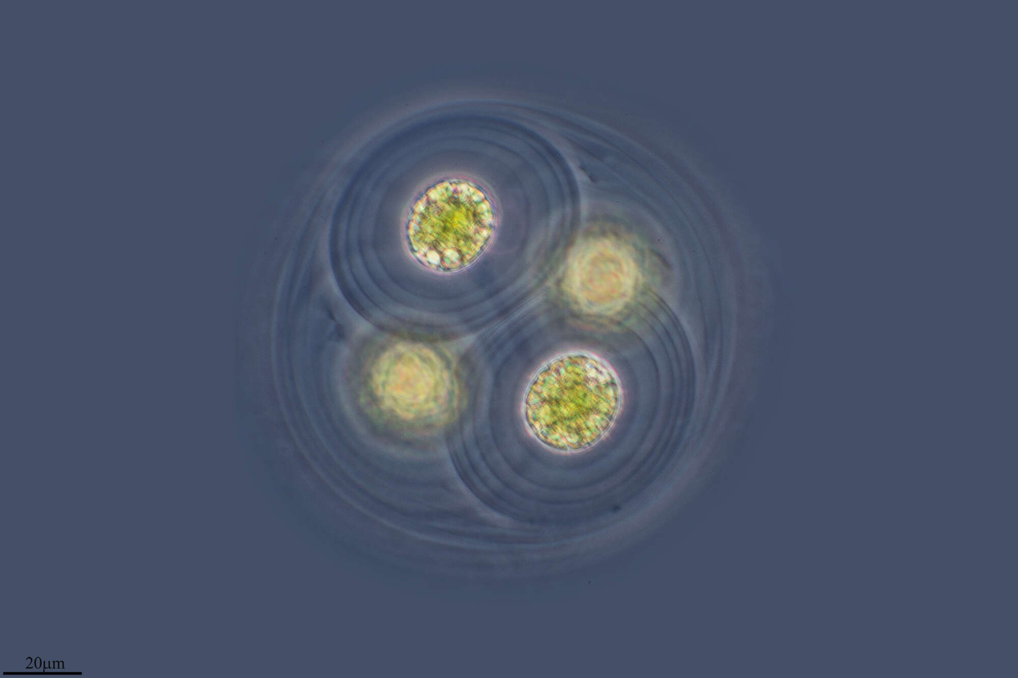
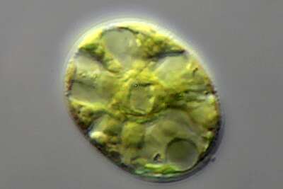
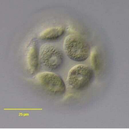
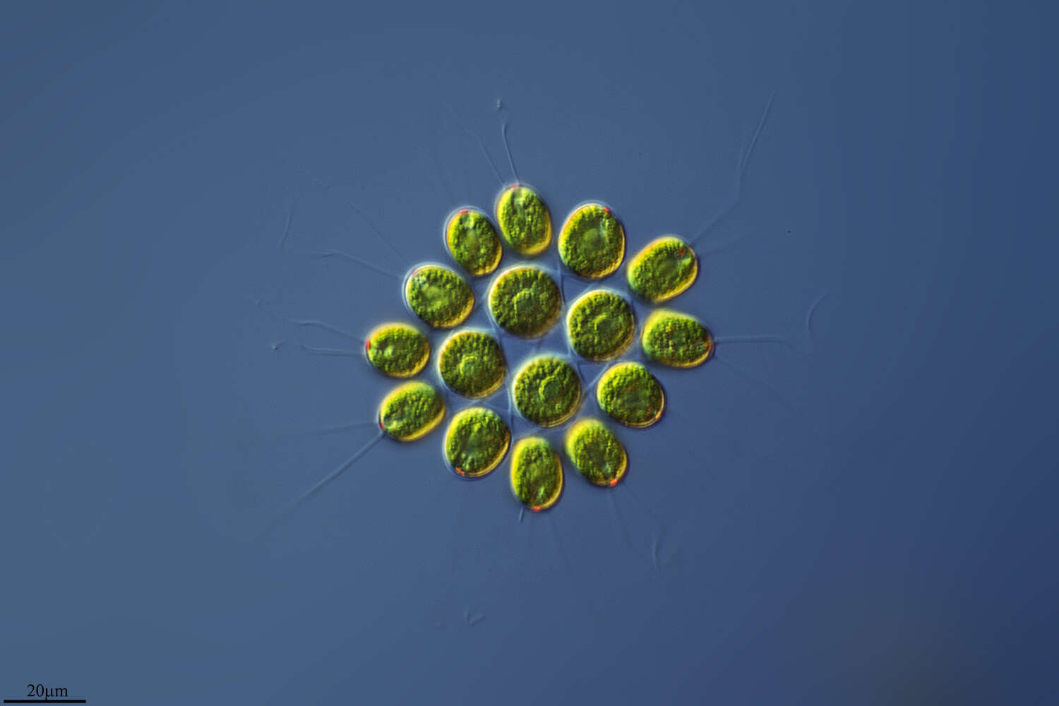
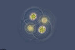
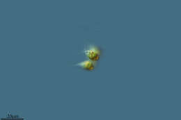
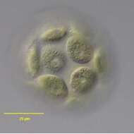
Young colonies, cell divisions are almost over, the flagellae are not yet fully formed. Scale bar indicates 25 m.Sample from the Lake Constance (vicinity of Bodman). The image was built up using several photomicrographic frames with manual stacking technique. Images were taken using Zeiss Universal with Olympus C7070 CCD camera.For permission to use of (high-resolution) images please contact postmaster@protisten.de.
-
Camargo, Cantabria, Spain
-
Lumbreras, La Rioja, Espaa
-
Madrid, Madrid, Spain
-
-
Logrono, La Rioja, Spain
-
Ribadelago de Franco, Castille and Leon, Spain
-
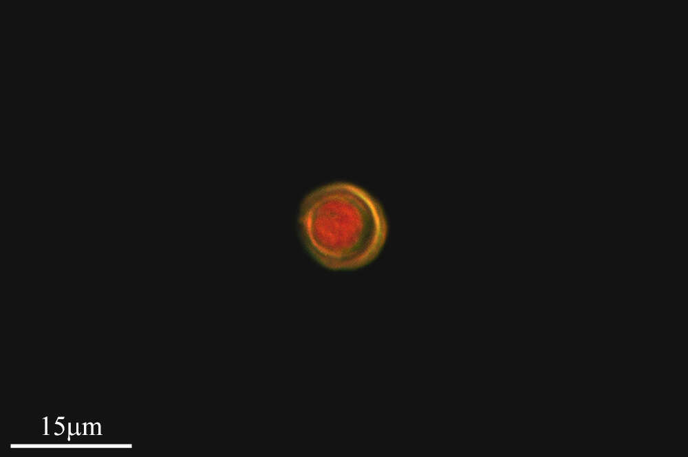
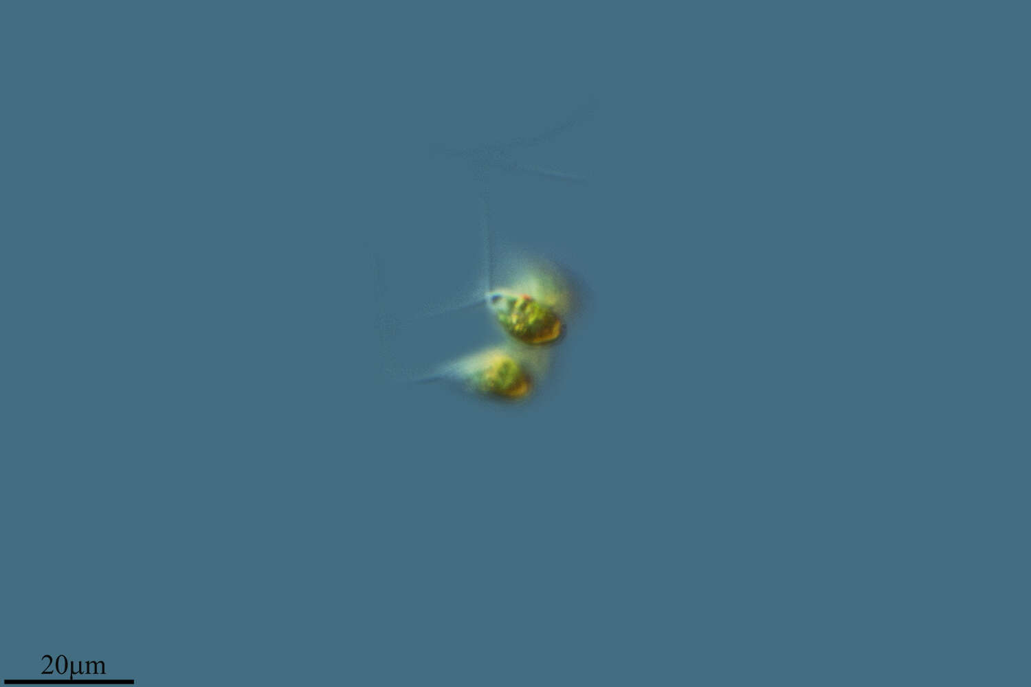
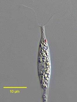
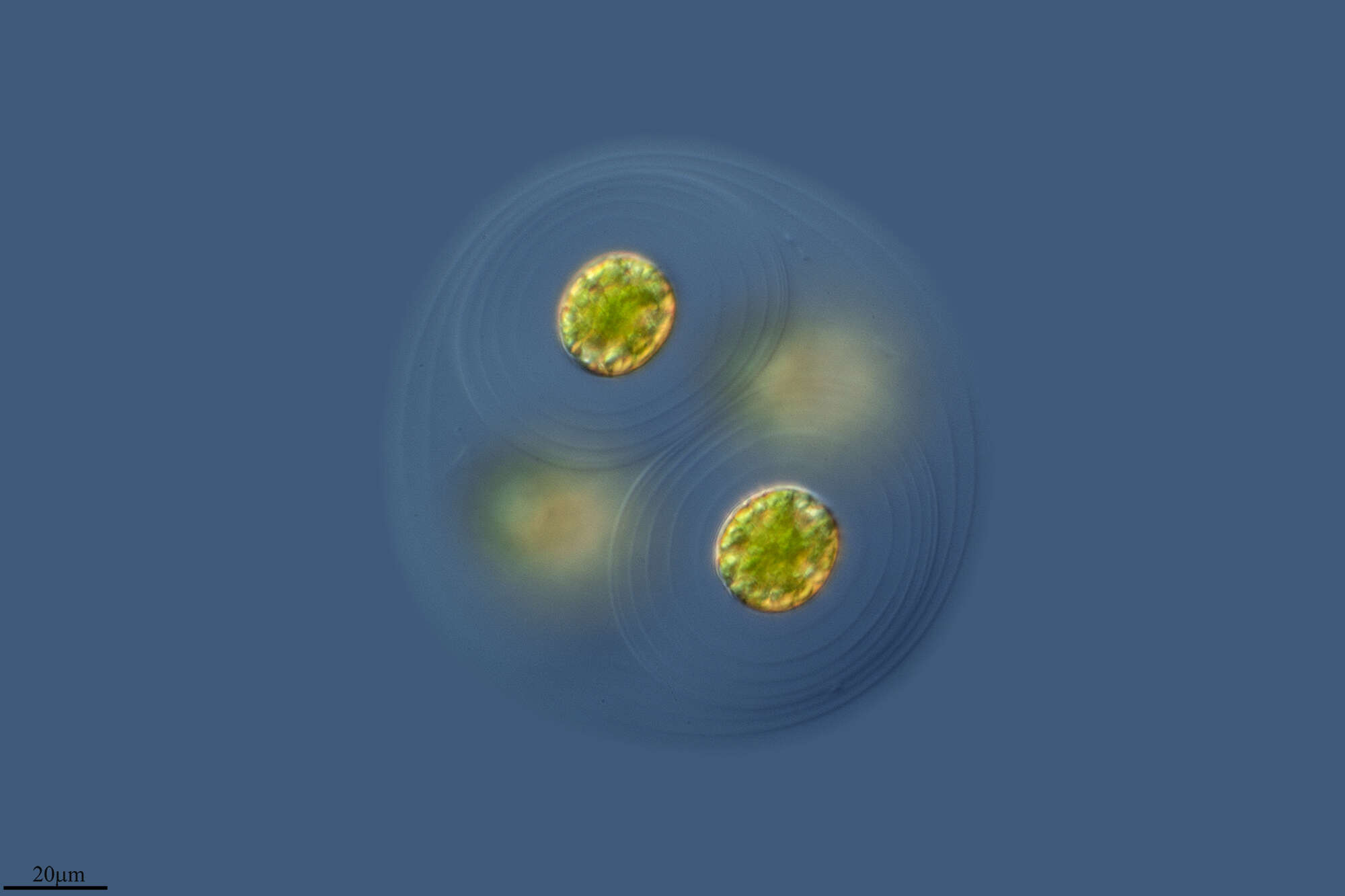
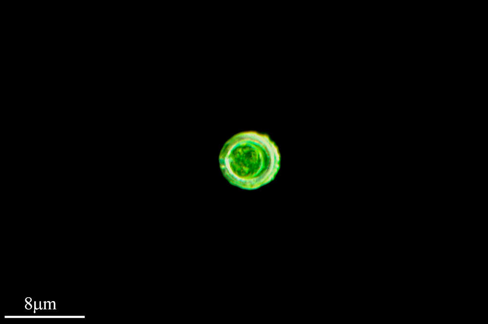
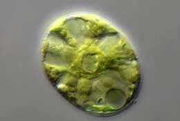
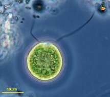
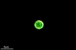
Close-up view. In the center of the cell we see a pyrenoid amidst the stellate chloroplast. In addition, the right cell shows the nucleus and the contractile vacuole. The scale bar indicates 10 m. Sample from sphagnum pond Dosenmoor near Neumuenster (Schleswig-Holstein, Germany). Images were taken using Zeiss Axioplan with Canon 600D CCD camera.For permission to use of (high-resolution) images please contact postmaster@protisten.de.
-
Ribadelago de Franco, Castille and Leon, Spain
-
All Biocode files are based on field identifications to the best of the researcher’s ability at the time.
-
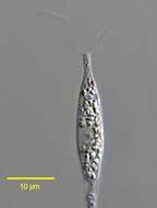
Hyalogonium klebsii (Klebs) Pascher,1927, a colorless volvocid flagellate with two equal anterior flagella. The body is elongate and fusiform. A red stigma is present on the left. Many small starch grains are seen in the cytoplasm.The nucleus is central. This genus may be confused with the euglenid Cyclidiopsis but Hyalogonium is smaller, biflagellate with thinner flagella, and lacks large paramylon bodies and the anterior canal opening. From standing rainwater pool near Boise, Idaho December 2005. DIC.
-
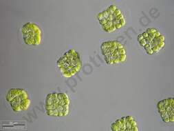
Chlamydomonas (clam-ee-doe-moan-ass), a solitary volvocid (flagellated green algal cell). Cell surrounded by a cellulosic wall, with two similar flagella emerging from near the apex. The photosynthetic pigments are located within a cup-shaped chloroplast which has a large pyrenoid with associated polysaccharide materials located posteriorly. The nucleus is located within the cup. Animations by Rosemary Arbur of flagellar beat patterns are available
here.Differential interference contrast.
-
Chlamydomonas (clam-ee-dough-moan-ass) iconic volvocid motile green alga, with two similar flagella inserting into the anterior end of the cell. Photosynthetic pigments include chlorophyll B which gives the cells their bright green colour. Phase contrast micrograph.
-
In vivo surface view of the volvocid flagellate, Volvulina steinii Playfair,1915. Collected from a temporary rainwater puddle on a grass lawn in Boise, Idaho 43° 36' 49.03" N 116° 13' 23.77" W elev. 2674 ft. March 2006. DIC.
-
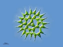
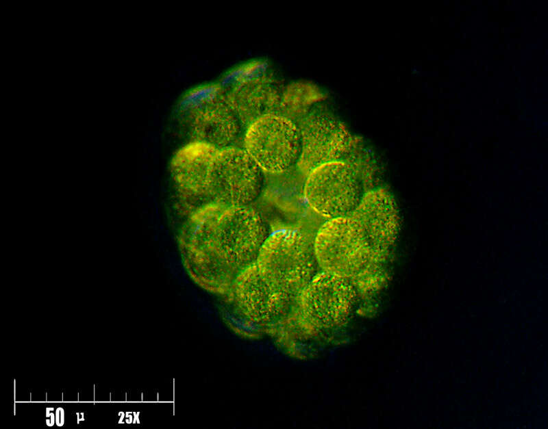
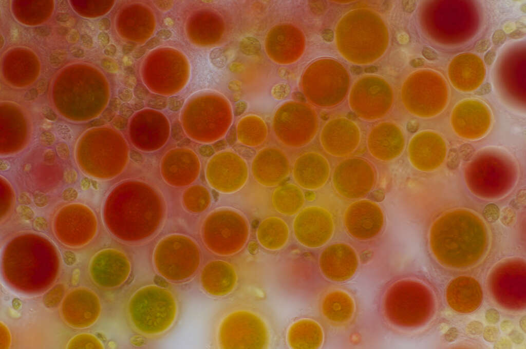
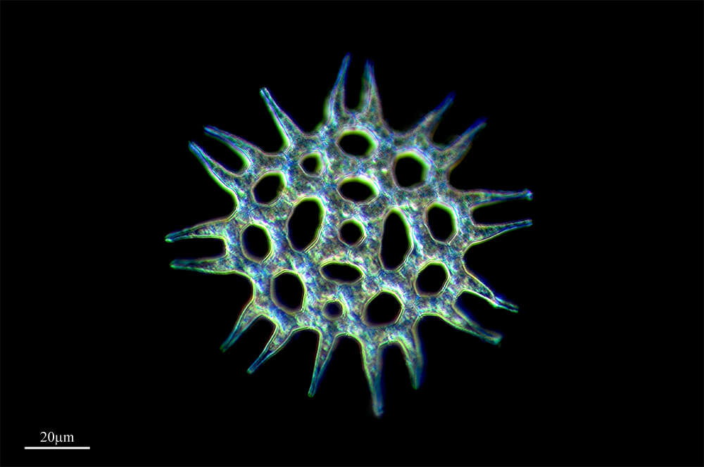
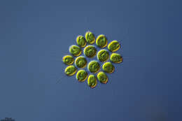
Scale bar indicates 100 m. Sample from the Domnental pond of Kronshagen near Kiel. The image was built up using several photomicrographic frames with manual stacking technique. Images were taken using Zeiss Axioplan with Olympus OM-D-E-M5 MKII.For permission to use of (high-resolution) images please contact postmaster@protisten.de.
-
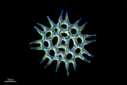
Akinets in brightfield
-
Boone, North Carolina, United States
-
Casas de Fadoncino, Castille and Leon, Spain
-
Camargo, Cantabria, Spain
-
Lumbreras, La Rioja, Espaa
-
Santa Coloma, La Rioja, Spain
-
Logroo, La Rioja, Espaa
-
San Martin De Castaneda, Castille and Leon, Spain