-
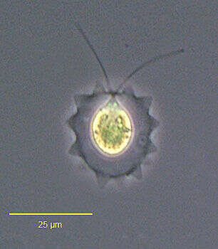
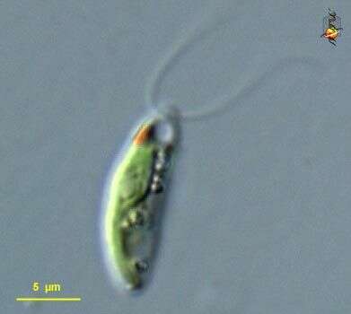
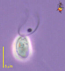
Portrait of Lobomonas stellata (Chodat), a volvocid flagellate. The ellipsoid to pear-shaped protoplast is separated from the cell wall by a space containing gelatinous material. The cell wall has irregularly spaced conical protrusions. There is one large cup-shaped chloroplast. A pyrenoid is located posteriorly. A peripheral stigma is located in the anterior 1/3 of the cell. Two equal flagella are about the length of the cell body. From freshwater pond near Boise, Idaho. Phase contrast.
-
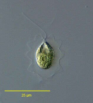
Portrait of Lobomonas stellata (Chodat), a volvocid flagellate. The ellipsoid to pear-shaped protoplast is separated from the cell wall by a space containing gelatinous material. The cell wall has irregularly spaced conical protrusions. There is one large cup-shaped chloroplast. A pyrenoid is located posteriorly. A peripheral stigma is located in the anterior 1/3 of the cell. Two equal flagella are about the length of the cell body. From freshwater pond near Boise, Idaho.DIC.
-
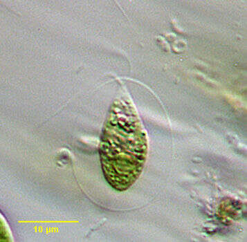
Pseudocarteria, a volvocid flagellate distinguished from the similar genus Carteria by absence of an anterior papilla. Four approximately equal-length flagella and single large chloroplast. Prominent stigma. From freshwater pond near Boise, Idaho. Oblique illumination.
-
-
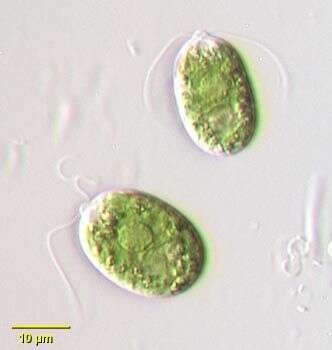
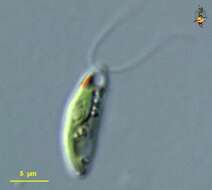
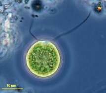
Chlamydomonas (clam-ee-doe-moan-ass) a common volvocid (green alga) flagellate. Cells vary in shape from elongate to rounded, this being one of the more elongate cells. With a cell wall, a cup-shaped chloroplasts with chlorophyll B, a red eyespot located external to the plastid, and two equal flagella emerging from the anterior pole of the cell. Differential interference contrast. Animations by Rosemary Arbur of flagellar beat patterns are available
here. Material from Nymph Creek and Nymph Lake, thermal sites within Yellowstone Park, photograph by Kathy Sheehan and David Patterson.
-
Chlamydomonas (clam-ee-dough-moan-ass) a common volvocid (green alga) flagellate. Cells vary in shape from elongate to rounded, this being one of the more elongate cells. With a cell wall, a cup-shaped chloroplasts with chlorophyll B, a red eyespot located external to the plastid, and two equal flagella emerging from the anterior pole of the cell. These cells undergo a form of sexual reproduction referred to as conjugation in which two similar to near similar cells fuse and exchange genetic information. Animations by Rosemary Arbur of flagellar beat patterns are available
here. Differential interference contrast. Material from Nymph Creek and Nymph Lake, thermal sites within Yellowstone National Park, photograph by Kathy Sheehan and David Patterson.
-
Portrait of Vitreochlamys fluviatilis, formerly Sphaerellopsis fluviatilis. The genus name, Sphaerellopsis (Korchikoff, 1925) was preoccupied by an Ascomycete fungus. This fungus Sphaerellopsis filum (Cooke, 1883) is a hyperparasite of another fungus, willow rust (Melampsora). Batko renamed this volvocid flagellate genus Vitreochlamys. This genus is similar to Chlamydomonas (some consider it synonymous) but differs from it by having a protoplast and surrounding gelatinous sheath that are fusiform. There are two equal length flagella. The nucleus is central. There is a large cup-shaped chloroplast and a posterior pyrenoid. Two anterior contractile vacuoles are located near the flagellar bases. There is a small anterior stigma. From a freshwater pond near Boise, Idaho. Oblique illumination.
-
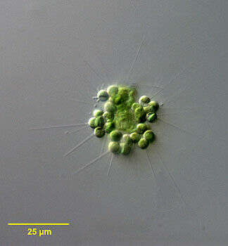
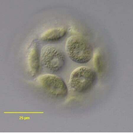
Portrait of the planktonic green alga Errerella bornhemiensis Conrad, 1913. Clusters of spherical cells are arranged in pyramidal clusters. Each cell bears one stout spine on its free face. Single cup-shaped chloroplast. The similar Micractinium has multiple finer spines on each cell. Collected from freshwater pond near Boise, Idaho July 2004. DIC.
-
In vivo portrait of the volvocid flagellate, Volvulina steinii Playfair,1915. Collected from a temporary rainwater puddle on a grass lawn in Boise, Idaho 43°36'49.03" N 116° 13' 23.77" W elev. 2674 ft.March 2006.Brightfield.
-
Portartit of the volvocid flagellate, Volvulina steinii Playfair,1915. The cells are hemispherical with the flattened face of each toward the exterior in contact with the thin investing gelatinous envelope.The inner limit of the envelope is visible here. the thickness of the investing layer of the gelatinous envelope is indicated here by debris adhering to its exterior surface.Each of the 16 cells in the colony bears two equal-length flagella (seen here in the cell at 12 o'clock). Only cells at the "anterior" end of the colony have eyespots. each cell has two contractile vacuoles (seen here in the cell at 12 o'clock.Collected from a temporary rainwater puddle on a grass lawn in Boise, Idaho In vivo portrait of the volvocid flagellate, 43°36'49.03" N 116° 13' 23.77" W elev. 2674 ft. elev. 2674 ft. March 2006. DIC.
-
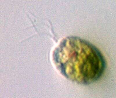
Portrait of Lobomonas stellata (Chodat), a volvocid flagellate. The ellipsoid to pear-shaped protoplast is separated from the cell wall by a space containing gelatinous material. The cell wall has irregularly spaced conical protrusions. There is one large cup-shaped chloroplast. A pyrenoid is located posteriorly. A peripheral stigma is located in the anterior 1/3 of the cell. Two equal flagella are about the length of the cell body. From freshwater pond near Boise, Idaho.DIC.
-
-
-
In vivo portartit of slightly compressed volvocid flagellate, Volvulina steinii Playfair,1915 showing the two contractile vacuoles. The single nucleus of several cells is visible. Only the "anterior" cells of the colony have red eyespots. Collected from a temporary rainwater puddle on a grass lawn in Boise, Idaho. 43°36'49.03" N 116° 13' 23.77" W elev. 2674 ft. March 2006. DIC.
-
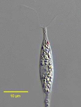
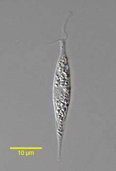
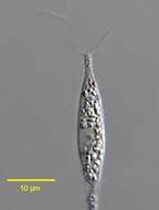
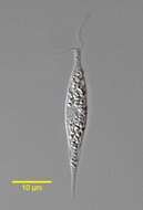
Hyalogonium klebsii (Klebs) Pascher,1927, a colorless volvocid flagellate with two equal anterior flagella. The body is elongate and fusiform. A red stigma is present on the left. Many small starch grains are seen in the cytoplasm.The nucleus is central. This genus may be confused with the euglenid Cyclidiopsis but Hyalogonium is smaller, biflagellate with thinner flagella, and lacks large paramylon bodies and the anterior canal opening. From standing rainwater pool near Boise, Idaho December 2005. DIC.
-
Chlamydomonas (clam-ee-doe-moan-ass), a solitary volvocid (flagellated green algal cell). Cell surrounded by a cellulosic wall, with two similar flagella emerging from near the apex. The photosynthetic pigments are located within a cup-shaped chloroplast which has a large pyrenoid with associated polysaccharide materials located posteriorly. The nucleus is located within the cup. Animations by Rosemary Arbur of flagellar beat patterns are available
here.Differential interference contrast.
-
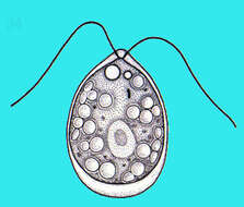
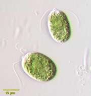
Chlamydomonas (clam-ee-dough-moan-ass) iconic volvocid motile green alga, with two similar flagella inserting into the anterior end of the cell. Photosynthetic pigments include chlorophyll B which gives the cells their bright green colour. Phase contrast micrograph.
-
In vivo surface view of the volvocid flagellate, Volvulina steinii Playfair,1915. Collected from a temporary rainwater puddle on a grass lawn in Boise, Idaho 43° 36' 49.03" N 116° 13' 23.77" W elev. 2674 ft. March 2006. DIC.
-
Hyalogonium klebsii (Klebs) Pascher,1927, a colorless volvocid flagellate with two equal anterior flagella. The body is elongate and fusiform. A red stigma is present on the left. Many small starch grains are seen in the cytoplasm.The nucleus is central. There are two anterior contractile vacuoles.This genus may be confused with the euglenid Cyclidiopsis but Hyalogonium is smaller, biflagellate with thinner flagella, and lacks large paramylon bodies and the anterior canal opening. From standing rainwater pool near Boise, Idaho December 2005. DIC.
-
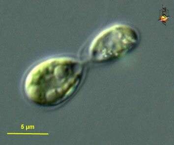
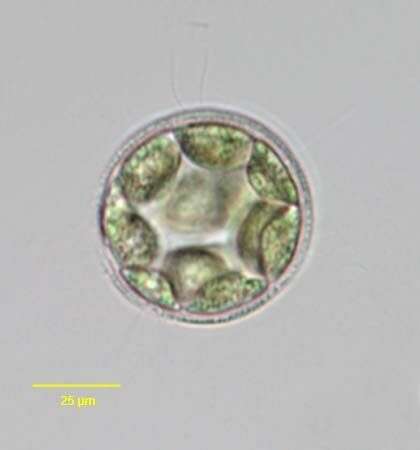
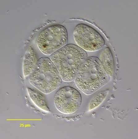
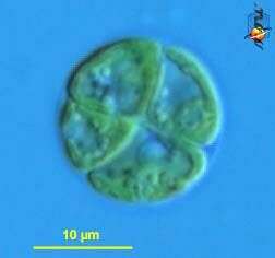
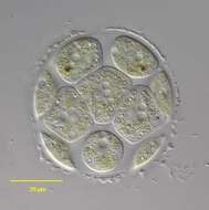
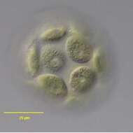
Chlamydomonas (clam-ee-doe-moan-ass), a solitary volvocid (flagellated green algal cell). Cell surrounded by a cellulosic wall, this is a division form in which four daughter cells are being produced at the same time. Animations by Rosemary Arbur of flagellar beat patterns are available
here. Differential interference contrast.
-
Phase contrast microscopy.
-
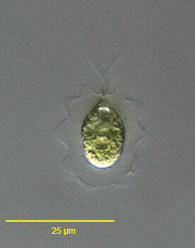
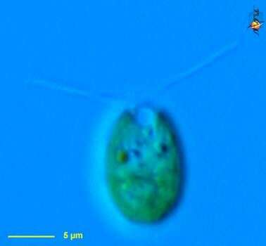
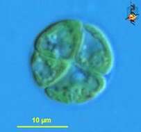
Chlamydomonas (clam-ee-doe-moan-ass), a solitary volvocid (flagellated green algal cell). Cell surrounded by a cellulosic wall, with two similar flagella emerging from near the apex. The photosynthetic pigments are located within a cup-shaped chloroplast which has a large pyrenoid with associated polysaccharide materials located posteriorly. The nucleus is located within the cup. This image shows the small red eyespot (orange colour here) and one anterior contractile vacuole. Differential interference contrast.
-
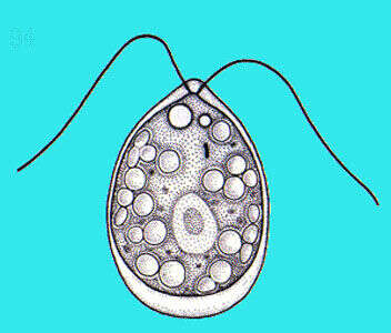
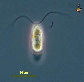
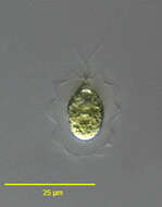
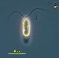
Chlamydomonas, a volvocid flagellate. The genus is very large probably with many synonymous species. Two equal length flagella emerge from a prominent anterior papilla in this species. A small contractile vacuole can be seen just posterior to the flagellar insertion site. A well-demarcated central nucleus can be seen in these images. A very small stigma is present. There is a single large cup shaped chloroplast in this species. A large pyrenoid is present in the posterior half of the cell. Some species have a gelatinous sheath although the cell wall is closely applied to the protoplast in this species. From freshwater pond near Boise, Idaho. Oblique illumination.
-
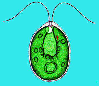
Chlamydomonas (clam-ee-doe-moan-ass), a solitary volvocid (flagellated green algal cell). Cell surrounded by a cellulosic wall, with two similar flagella emerging from near the apex. The photosynthetic pigments are located within a cup-shpaed chloroplast which has a large pyrenoid with associated polysaccharide materials. Many taxa described (there are books on this genus). Eyespot located within plastid. Flagella beat with a breast-stroke pattern. Phase contrast.