-
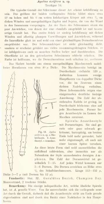
Original description and illustration.
-
From Hooper, 2008.
-
Original description and illustrations.
-
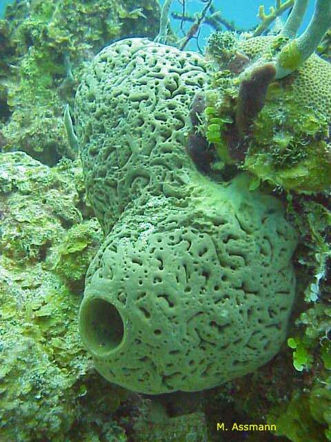
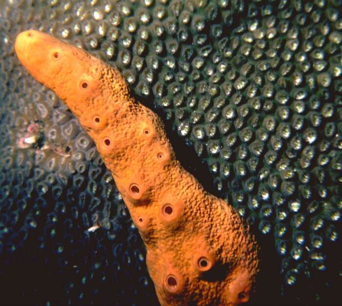
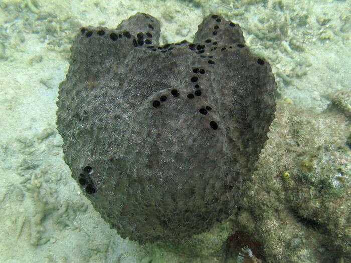
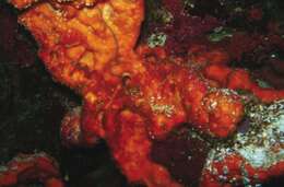
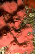
Agelas cerebrum Assmann et al. 2001, ZMA Por. 15603, in situ on Bahamian reefs, photo M. Assmann.
-
Bahamas, photo F. Parra Velandia.
-
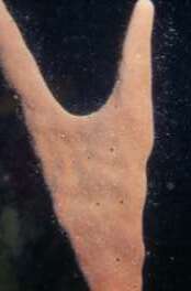
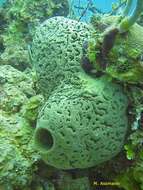
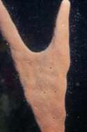
Agelas conifera specimen from the Curaçao reefs
-
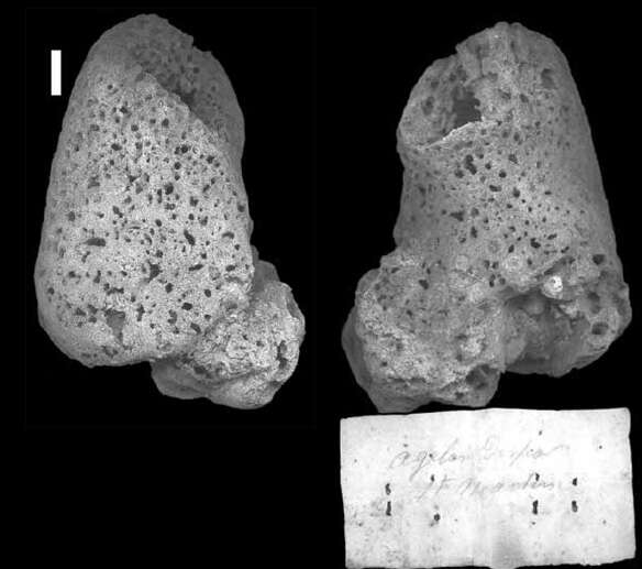
Holotype ZMA 00607 seen from opposite sides. Photo R.W.M. van Soest.
-
-
Preserved specimen, ZMA Por. 03611, from reef at Blauwbaai, Curaçao, coll. S. Weinberg & E. Westinga, 15-20 m, November 1975. Photo L.A. van der Laan.
-
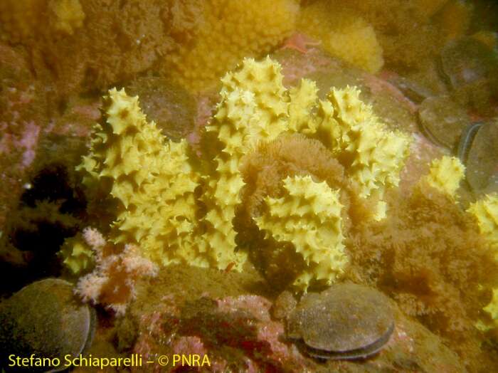
In situ photo of ZMA Por. 11036 (holotype), from 5-15 m, off Ciudad Velha, Sao Tiago, Cape Verde Islands, coll. & photo R.W.M. van Soest, field nr. CANCAP 7 stat. D01A/14, 21 August 1986.
-
Specimen MNRJ 15665, collected on mangrove root.
-
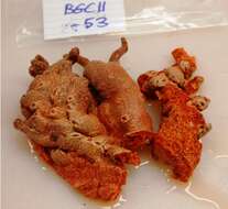
On deck photo of Por. ZMA 22459, from Dry Tortugas, Florida, 55-60 m, coll. & photo Jason Cruce, field nr,. BGC11-53, 13 May 2011.
-
Van Soest, 1978: pl. VII fig. 2, from Barbados, off Holetown, depth 100 m, reg. nr. ZMA Por. 04309, photo L.A. van der Laan
-
-
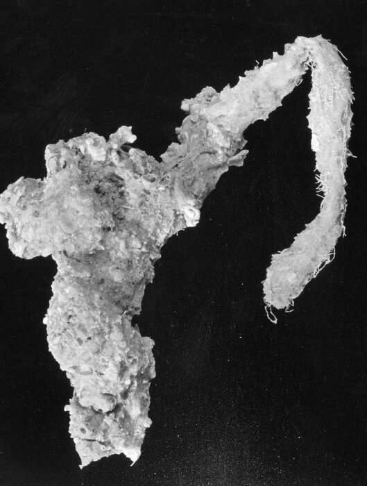
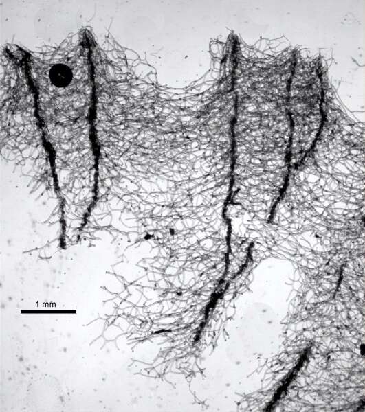
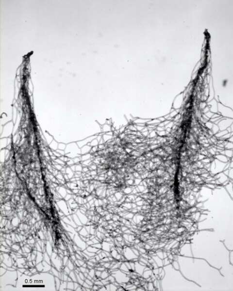
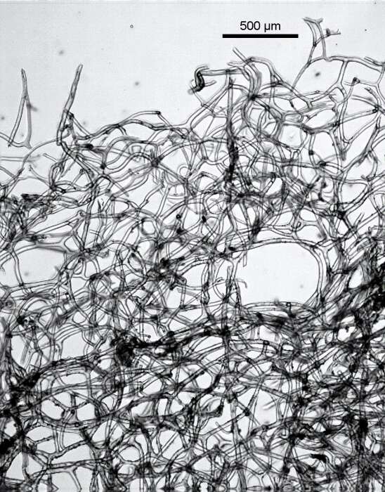
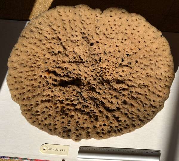
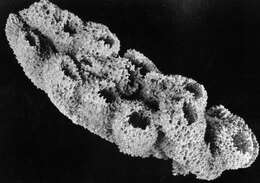
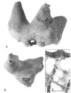
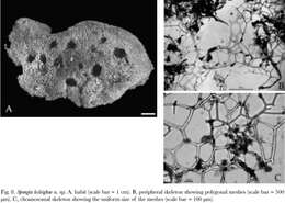
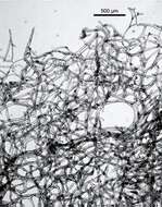
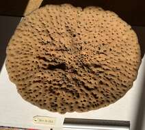
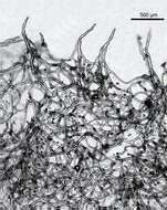
A, Holotype, NMNZ Por. 433 (= SDCC/NZ048), side view. Note damaged and healed middle turret, with abnormal ocscule on the side of the turret. B, Holotype, top view. C, Detail of the fibre skeleton from paratype (SDCC/ NZ059), showing vertically orientated, cored primary fibres, uncored or partially cored secondaries and the fine canals throughout the sponge. The armoured surface is visible at the at top of the photomicrograph (scale bar = 500 µm)
-
From Helmy et al. 2004
-
-
Primary fibers and conules cored by foreign particles
-
Detail of conules and primary fibers
-
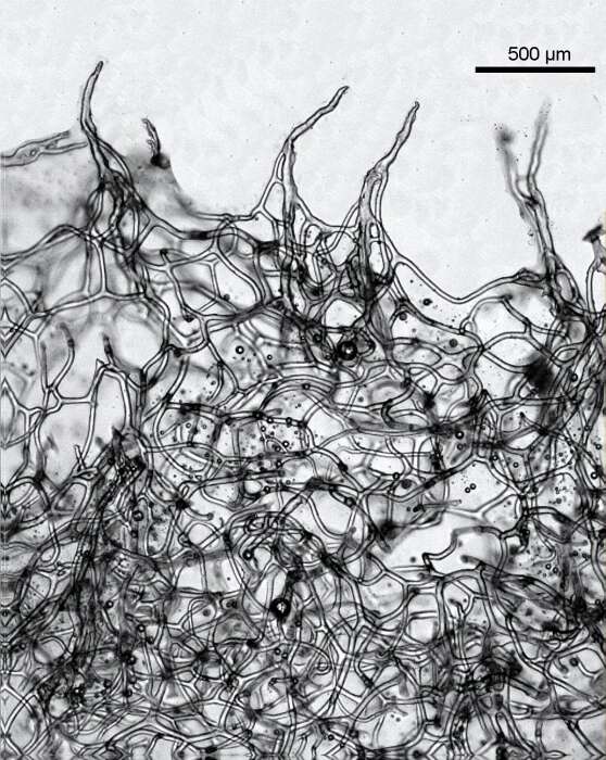
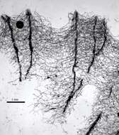
Skeleton, the primary fibers are evoid of foreign bodies
-
-
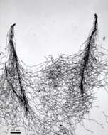
Fibers are devoid of foreign bodies
-
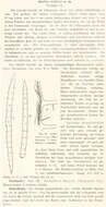
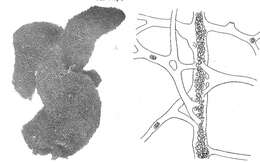
Original illustratiosn of type material.
-
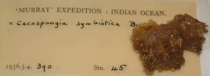
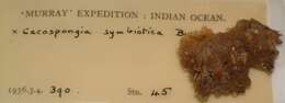
Photo of one of the paratypes from stat. 45.