-
-
-
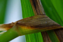
Portrait of the colorless sulfur bacterium Beggiatoa alba (VAUCHER, 1803) TREVISAN, 1845.
-
-
-
-
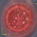
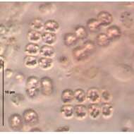
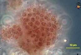
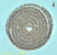
Thiocystis (thigh-owe-cyst-is) is a red sulphur bacterium (or purple sulphur bacterium). It is found in sediments above the reduced zone. It oxidizes hydrogen sulphide producing elemental sulphur which is deposited within the bacterial cell as granules of sulphur. Forms aggregates and individual cells are often hard to distinguish. The cellular nature of these aggregates is hard to determine at first glance. This detail shows that the mass is made of individual cells and the cells have granular inclusions. Phase contrast.
-
-
Thiocystis (thigh-owe-cyst-is) is a red sulphur bacterium (or purple sulphur bacterium). It is found in sediments above the reduced zone. It oxidizes hydrogen sulphide producing elemental sulphur which is deposited within the bacterial cell as granules of sulphur. Forms aggregates and individual cells are often hard to distinguish. Phase contrast.
-
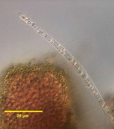
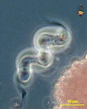
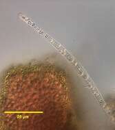
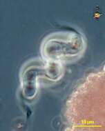
Thiospirillum (thigh-ow-spire-ill-um) is one of the sulphur bacterium. Rather like Spirillum, cells take the form of short-cork-screw and have flagella at both ends of the cells. The flagella occur in very substantial tufts so can be seen easily seen with the light microscope. This cell was rather large. Phase contrast.
-
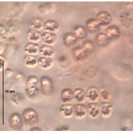
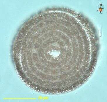
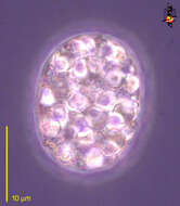
Achromatium (a-chrome-ace-ee-um) one of the larger bacteria, relatively common, and usually distinguished by the presence of large sulphur deposits inside the cell. Phase contrast.
-
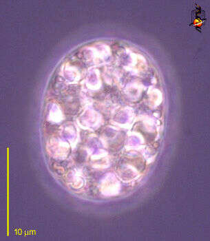
Achromatium (a-chrome-ace-ee-um) is one of the non-photosynthetic sulphur bacteria. It is a heterotrophic bacterium which relies on a supply of organic matter to assist the degradation of reduced sulphur to elemental sulphur - which is then deposited as slightly pink granules inside the cell. Large (for a bacterium). Differential interference contrast.
-
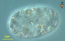
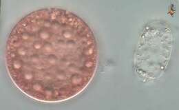
This picture compares Achromatium (a-chrome-ace-ee-um) (right) - a heterotrophic sulphur bacterium, from the red or purple sulphur bacterium, Chromatium. Differential interference contrast.
-
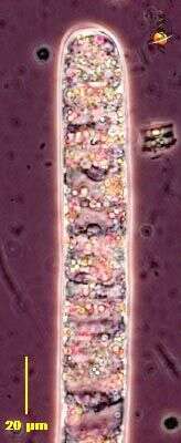
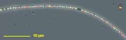
Beggiatoa (beg-ee-a-toe-a) is a colourless sulphur bacterium which occurs as filaments of various widths. Beggiatoa is found in sediments above the reduced zone. It oxidizes hydrogen sulphide, producing elemental sulphur which is deposited within the bacterial cell as sulphur granules and give this filament its opaque appearance. Very long. These cells can glide, a good trait for organisms which live in a habitat the characteristics of which are changing depending on whether there is or is not overlying water or if or if not there is sunlight. Phase contrast.
-
Beggiatoa (beg-ee-a-toe-a), commonly seen on the surface of very rich sediments - such as those of salt marshes. The bacteria metabolise reduced hydrogen sulphide and produce granules of sulphur as a by product. They can be seen as refractile elements within the cells which make up this filament. Phase contrast.
-
Beggiatoa (beg-ee-a-toe-a), commonly seen on the surface of very rich sediments - such as those of salt marshes. The bacteria metabolise reduced hydrogen sulphide and produce granules of sulphur as a by product. They can be seen within the cells which make up this filament. Differential interference contrast.
-
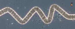
Beggiatoa (beg-ee-a-toe-a) is a colourless sulphur bacterium which occurs as filaments of various widths. Beggiatoa is found in sediments above the reduced zone. It oxidizes hydrogen sulphide, producing elemental sulphur which is deposited within the bacterial cell as sulphur granules and give this filament its opaque appearance. Layers of Beggiatoa are often seen on the surface of muds or other places where there is a lot of decaying organic matter (around dead dogs is a good place) and looks like white tissue paper lying over the sediment. These cells can glide, a good trait for organisms which live in a habitat the characteristics of which are changing depending on whether there is or is not overlying water or if or if not there is sunlight. Phase contrast.
-
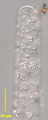
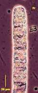
Beggiatoa (beg-ee-a-toe-a) is a colourless sulphur bacterium which occurs as filaments of various widths. Beggiatoa is found in sediments above the reduced zone. It oxidizes hydrogen sulphide, producing elemental sulphur which is deposited within the bacterial cell as sulphur granules. In this delicate filament, the individual sulphur grains can be seen as pink refractile inclusions. Phase contrast.
-
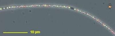
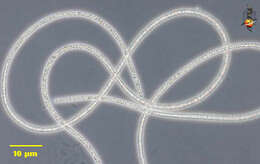
Beggiatoa (beg-ee-a-toe-a) is a colourless sulphur bacterium which occurs as filaments of various widths. Beggiatoa is found in sediments above the reduced zone. It oxidizes hydrogen sulphide, producing elemental sulphur which is deposited within the bacterial cell as sulphur granules visible inside this filament . May be very long. These cells can glide, a good trait for organisms which live in a habitat the characteristics of which are changing depending on whether there is or is not overlying water or if or if not there is sunlight. They don t usually make these cute spirals. Differential interference contrast.
-
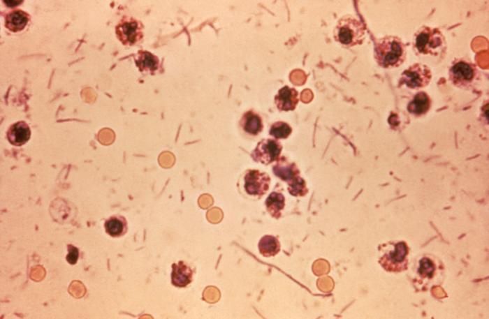
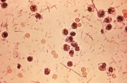
This photomicrograph revealed stool exudates in a patient with shigellosis, which is also known as Shigella dysentery, or Bacterial dysentery.Created: 1980
-
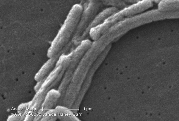
Under a very high magnification of 25000X, this scanning electron micrograph (SEM) depicted a grouping of Gram-negative Legionella pneumophila bacteria. Please note that youre able to see a number of the flagella emanating from these organisms. Please see PHIL 11092 through 11152 for additional SEMs of these organisms, specifically PHIL 11106 for a colorized version of this image, which better features the flagella.Youll note that a number of these bacteria seem to display an elongated-rod morphology. L. pneumophila are known to most frequently exhibit this configuration when grown in broth, however, they can also elongate when plate-grown cells age, as it was in this case, especially when theyve been refrigerated. The usual L. pneumophila morphology consists of stout, fat bacilli, which is the case for the vast majority of the organisms depicted here.Created: 2009
-
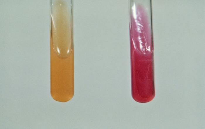
This urease test, based on the process involving the hydrolysis of urea, was performed to help identify the Gram-negative enteric bacterium Yersinia enterocolitica.Created: 1976
-
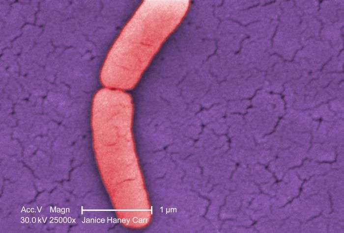
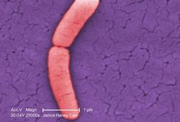
Under a very high magnification of 25000X, this colorized scanning electron micrograph (SEM) revealed the presence of a single Gram-negative Salmonella typhimurium bacterium, which was imaged right at the point where it was undergoing the process of cell division, resulting in the formation of two separate organisms. This dividing bacterium had been isolated from a pure culture. See PHIL 10994 for a black and white version of this image.Created: 2009
-
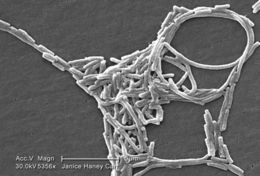
Under a moderately-high magnification of 5356X, this scanning electron micrograph (SEM) depicted a grouping of Gram-negative Legionella pneumophila bacteria. Note that in some of these views youre able to see a number of the flagella emanating from these organisms. Please see PHIL 11092 through 11152 for additional SEMs of these organisms, specifically PHIL 11104 for a colorized version of this image.Youll note that a number of these bacteria seem to display an elongated-rod morphology. L. pneumophila are known to most frequently exhibit this configuration when grown in broth, however, they can also elongate when plate-grown cells age, as it was in this case, especially when theyve been refrigerated. The usual L. pneumophila morphology consists of stout, fat bacilli, which is the case for the vast majority of the organisms depicted here.Created: 2009