-
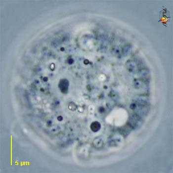
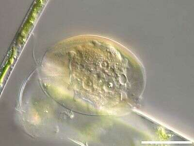
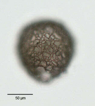
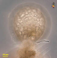
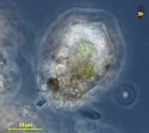
Microchlamys (mike-row-clam-iss) is a testate amoeba, but one with a flexible shell or lorica. Optical section through the cell. Two nuclei with dark nucleoli and the light coloured contractile vacuole are evident. Phase contrast.
-
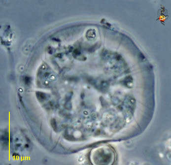
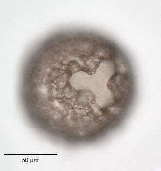
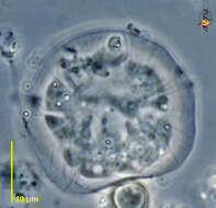
Microchlamys (mike-row-clam-iss) is a testate amoeba, but one with a flexible shell or lorica. As can be seen in this image, the cell is deformed by pushing against an object in the real world, and the shell itself is folded. The amoeba inside produces lobose pseudopodia. Phase contrast.
-
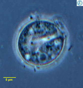
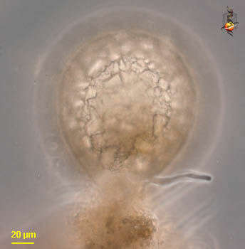
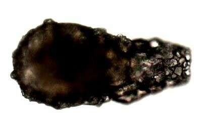
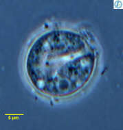
Microchlamys, a shelled amoeba in which the lorica is thin, organic and flexible. In this image the lorica is folded. The lorica seems to be transparent when first formed, with age it becomes brown. From Lake Donghu, China. Phase contrast micrograph.
-
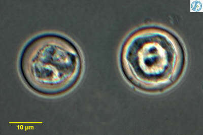
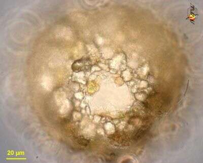
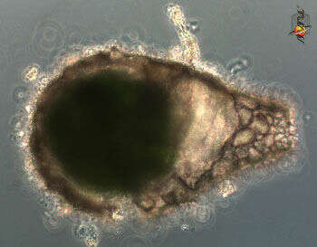
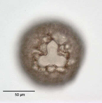
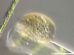
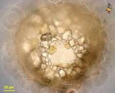
Microchlamys, a shelled amoeba in which the lorica is thin, organic and flexible. The lorica seems to be transparent when first formed, with age it becomes brown. The lorica has a central ventral aperture through which pseudopodia extend. This image shows the cell from the ventral side. From Lake Donghu, China. Phase contrast micrograph.
-
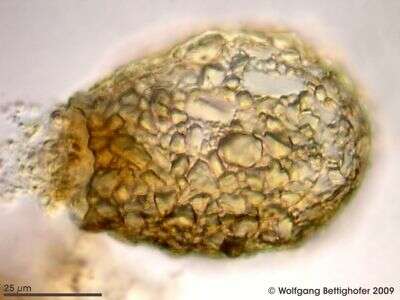
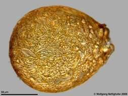
Scale bar indicates 25 µm. Sample from a wetland at the Pillersee (Tyrol, Austria). The image was built up using several photomicrographic frames with manual stacking technique. Images were taken using Zeiss Universal with Olympus C7070 CCD camera.Image under Creative Commons License V 3.0 (CC BY-NC-SA).
-
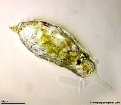
Test of a freshwater Difflugina. Multi layer image using 28 frames generating depth of focus, stacked manually using Corel Photopaint. Collected from a creek sediment. This image was taken using Zeiss Universal with Olympus C7070 CCD camera.
-
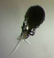
Difflugia (dif-flew-gee-a) is a testate amoeba, in which the amoeba is enclosed within a shell that is comprised of an organic cement and variously sized inorganic granules. This individual has produced one pseudopodium from the opening or aperture which is at the bottom of the test and is surrounded by accumulated debris. Phase contrast.
-
Difflugia (dif-flew-gee-a) is a testate amoeba, in which the amoeba is enclosed within a shell that is comprised of an organic cement and variously sized inorganic granules. This image shows the aperture through which the pseudopodia emerge. Phase contrast.
-
-
Difflugia (diff-flew-gee-a) a testate amoeba. The test is comprised of cemented inorganic granules. There is an aperture at one end of the cell, from which lobose pseudopodia emerge. Phase contrast.
-
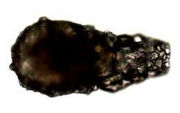
Difflugia is an amoeba that lives within a shell or 'test' comprised of small particles of sand and grit. Pseudopodia, used for movement or to capture food, if present would extend from a hole or aperture at the right end of the test.
-
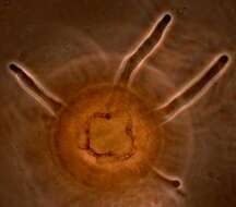
The aperture of the shell of Difflugia from which four pseudopodia are emerging.
-
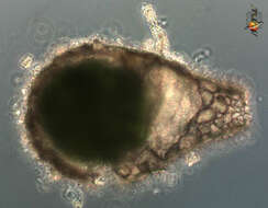
This shelled amoeba has a test that includes small sand granules that have been glued together. The cytoplasm of the enclosed amoeba is darh green because the cytoplasm includes large numbers of symbiotic algae. The aperture is to the right in this image. No pseudopodia are visible. Phase contrast microscopy.
-
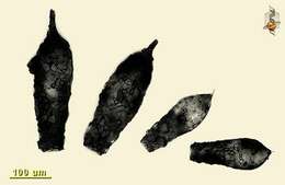
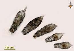
Small collection of Difflugia from one site illustrating the variation in shape and size within a population. Image two of two, this one assembled using Mr Zeiss' nice software - extended focus option.
-
Small collection of Difflugia from one site illustrating the variation in shape and size within a population. Image one of two, this one assembled using one of those software programs that cost an arm and a leg and you are only allowed restricted use of it. Ho hum.
-
Members of the Difflugia group build agglutinated shells selecting quartz grains. This depth of focus picture shows shell together with pseudopod and food vacuole. Based on 6 pictures depth of focus was generated using MicroPicS by Bernhard Wiedemann and Photoshop. More pictures in ZIP archive. Collected from bottom sediments of a rain storage reservoir in Kiel (Schleswig-Holstein, Germany). Images were taken using Zeiss Universal with Olympus C7070 CCD camera.
-
Difflugia spec. living in the sediment of a sphangnum pond. Sample from sphagnum pond near Bergenhusen (Schleswig-Holstein, Germany). This image was taken using Zeiss Universal with Olympus C7070 CCD camera.
-
The xenosoms derived frome pennate diatoms on the test of Difflugia acuminata are clearly visible. The multi layer image was built up using 13 brightfield frames with a manual stacking technique using Corel Photopaint. The specimen was gathered in a tiny freshwater pond at the island of Hiddensee (Baltic Sea, Germany) which shows a fascinating biodiversity of naked and testate amoebae. Images were taken using Zeiss Standard with Olympus C7070 CCD camera.
-
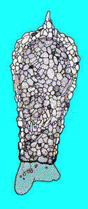
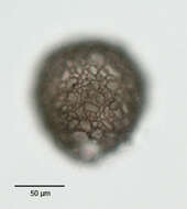
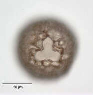
Anterolateral view of the test of Difflugia gramen (Penard, 1902). Collected from a freshwater pond near Boise, Idaho. June 2005. Brightfield.
-
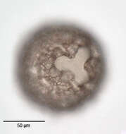
Slightly oblique view of the trilobed aperture of the test of Difflugia gramen (Penard, 1902). Collected from a freshwater pond near Boise, Idaho. June 2005. Brightfield.
-
View of the trilobed aperture of the test of Difflugia gramen (Penard, 1902). Collected from a freshwater pond near Boise, Idaho. June 2005. Brightfield.
-
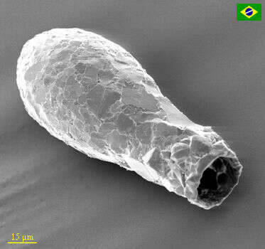
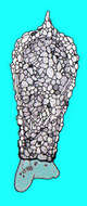
Scanning electron micrograph of a D. lanceolata. The clean contour is diagnostic for the species.
-
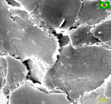
Scanning electron micrograph of test composition of a D. lanceolata. Note the circular ring-like units of organic cement matrix that holds the quartz particles together.
-
This video shows a Difflugia pulling itself and test by protruding a pseudopod, retracting it, protruding another, so on and so on. A good example of creeping organism. This video provided by Dan Lahr.