-
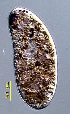
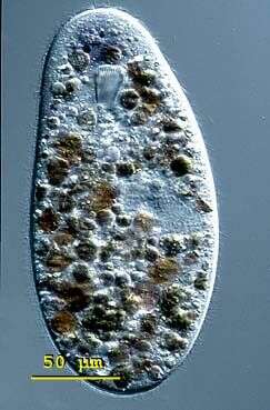
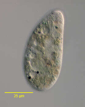
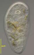
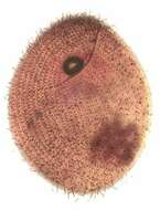
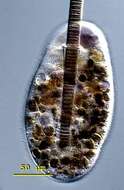
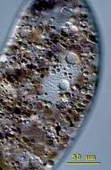
Nassulopsis (nass-you-lop-sis). Nassulopsis elegans is a conspicuous pink- or blue coloured nassulid found in pond and lake plankton. This cell was collected from floating colonies of Oscillatoria in a freshwater pond near Konstanz, Germany. The body is cylindrical in shape and measures 150 - 300 X 50 - 100 microns. Nassulopsis can be distinguished from Nassula and Obertrumia by 5-7 contractile vacuoles arranged in a ventral row. The macronucleus is cylindrical with rounded ends. The cytopharyngeal basket is located in the anterior third of the cell. The cells fill up with food vacuoles of different colour (green, orange), depending on stage of digestion. Free-swimming cell, 250 microns long. Differential interference contrast.
-
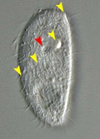
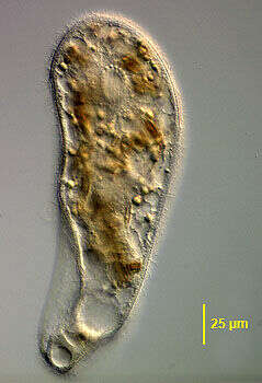
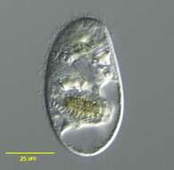
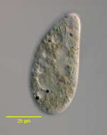
Portrait of the nassophorean ciliate, Chilodontopsis depressa (Perty, 1852). The cell is dorsoventrally flattened. The right side is convex meeting the straight left side at a rostrum. The somatic ciliature is denser on the ventral side with an indistinct feathery hypostomial frange of longer cilia slanting posteriorly to the cytostome from the rostrum anteriorly to the right margin of the body (yellow arrowheads). The right ventral kineties curve around the anterior end to meet the straight left ventral kineties to the left of the cytostome (red arrowhead) at the line of the hypostomial frange. The cytopharyngeal basket or cyrtos is seen anteriorly (red arrowhead). A distinctive large contractile vacuole fills the posterior end of the cell. The central macronucleus and micronucleus are spherical. C. depressa feeds on bacteria, diatoms and green algae. Collected from freshwater pond near Boise, Idaho September 2003. DIC optics
-
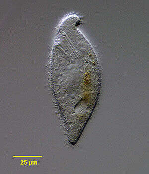
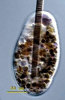
Zosterodasys transversa (Kahl,1928) Foissner,1994. (previously known as Chilodontopsis vorax), a large nassulid ciliate. Z. transversa is elongate and dorsoventrally flattened. Both dorsal and ventral surfaces bear uniform longitudinal rows of somatic cilia. A distinctive synhymenion or hypostomial frange runs obliquely posterior to the cytostome around half the cell circumference. Unlike the similar Synhymenia there is no preoral suture. The cytoplasm is highly vacuolated. There are multiple contractile vacuoles. The macronucleus is ovoid. There is a very prominent anterior cytopharyngeal basket composed of stout nematodesmata. From freshwater pond near Boise, Idaho. DIC.
-
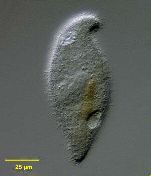
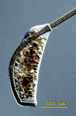
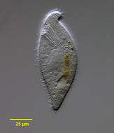
Portrait of the nassophorean marine ciliate, Orthodonella hamatus (Bhatia, 1936) . The elongate cell is dorsoventrally flattened with the anterior end bent to the left in a blunt hook shape. The posterior is bluntly tapered. The oral aperture is right anterior. There is a cytopharyngeal apparatus composed of stout nematodesmata. The longitudinal somatic kineties are uniform. There is a sigmoid post-oral frange extending obliquely from the anterior end just posterior to the cytostome almost to the right edge (not seen in this image). The ovoid macronucleus and adjacent micronucleus are located in the mid-cell. A posterior peripheral contractile vacuole is seen in this image. O. hamatus is also found in freshwater habitats. Orthodonella is a monotypic genus. Collected from a commercial saltwater aquarium in Boise, Idaho February 2004. DIC optics.
-
Nassulopsis (nass-you-lop-sis). Nassulopsis elegans is a conspicuous pink- or blue coloured nassulid found in pond and lake plankton. This cell was collected from floating colonies of Oscillatoria in a freshwater pond near Konstanz, Germany. The body is cylindrical in shape and measures 150 - 300 X 50 - 100 microns. Nassulopsis can be distinguished from Nassula and Obertrumia by 5-7 contractile vacuoles arranged in a ventral row. The macronucleus is cylindrical with rounded ends. The cytopharyngeal basket is located in the anterior third of the cell. The cells fill up with food vacuoles of different colour (green, orange), depending on stage of digestion. Slightly flattened cell, 260 microns long. Differential interference contrast.
-
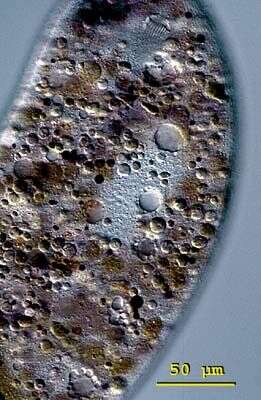
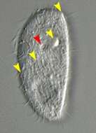
Portrait of the nassophorean ciliate, Chilodontopsis depressa (Perty, 1852). This image shows a coronal optical section through the cell center. The cell is dorsoventrally flattened. The right side is convex meeting the straight left side at a rostrum. The somatic ciliature (not seen here) is denser on the ventral side with an indistinct feathery hypostomial frange of cilia slanting posteriorly to the cytostome from the rostrum anteriorly to the right margin of the body. The right ventral kineties curve around the anterior end to meet the straight left ventral kineties to the left of the cytostome at the line of the hypostomial frange. The cytopharyngeal basket or cyrtos is seen anteriorly. A distinctive large contractile vacuole fills the posterior end of the cell. The central macronucleus and micronucleus are spherical. C. depressa feeds on bacteria, diatoms (seen in this cell) and green algae. Collected from freshwater pond near Boise, Idaho September 2003. DIC optics
-
Zosterodasys transversa (Kahl,1928) Foissner,1994. (previously known as Chilodontopsis vorax), a large nassulid ciliate. Z. transversa is elongate and dorsoventrally flattened. Both dorsal and ventral surfaces bear uniform longitudinal rows of somatic cilia. A distinctive synhymenion or hypostomial frange runs obliquely posterior to the cytostome around half the cell circumference (seen well in accompanying images). Unlike the similar Synhymenia there is no preoral suture. The cytoplasm is highly vacuolated. There are multiple contractile vacuoles. The macronucleus is ovoid. There is a very prominent anterior cytopharyngeal basket composed of stout nematodesmata. From freshwater pond near Boise, Idaho. Differential interference contrast.
-
Portrait of the nassophorean marine ciliate, Orthodonella hamatus (Bhatia, 1936). The elongate cell is dorsoventrally flattened with the anterior end bent to the left in a blunt hook shape. The posterior is bluntly tapered. The oral aperture is right anterior. There is a cytopharyngeal apparatus composed of stout nematodesmata (seen clearly in this image). The longitudinal somatic kineties are uniform. There is a sigmoid post-oral frange extending obliquely from the anterior end just posterior to the cytostome almost to the right edge (not seen in this image). The ovoid macronucleus and adjacent micronucleus are located in the mid-cell. A posterior peripheral contractile vacuole is seen in this image. O. hamatus is also found in freshwater habitats. Orthodonella is a monotypic genus. Collected from a commercial saltwater aquarium in Boise, Idaho February 2004. DIC optics.
-
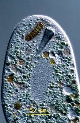
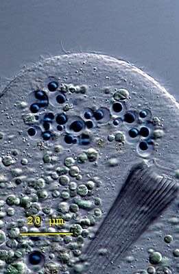
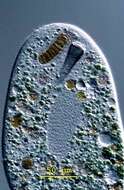
Nassulopsis (nass-you-lop-sis). Nassulopsis elegans is a conspicuous pink- or blue coloured nassulid found in pond and lake plankton. This cell was collected from floating colonies of Oscillatoria in a freshwater pond near Konstanz, Germany. The body is cylindrical in shape and measures 150 - 300 X 50 - 100 microns. Nassulopsis can be distinguished from Nassula and Obertrumia by 5-7 contractile vacuoles arranged in a ventral row. The macronucleus is cylindrical with rounded ends. The cytopharyngeal basket is located in the anterior third of the cell. The cells fill up with food vacuoles of different colour (green, orange), depending on stage of digestion. The cytopharyngeal basket is anterior. To the right of the basket is an ingested cyanobacteria. Below the basket an ovoid-shaped macronucleus is visible. Differential interference contrast.
-
Ventral infraciliature of the nassophorean ciliate, Chilodontopsis depressa (Perty, 1852). The cell is dorsoventrally flattened. The right side is convex meeting the straight left side at a rostrum. The somatic ciliature is denser on the ventral side with an indistinct feathery hypostomial frange of cilia slanting posteriorly to the cytostome from the rostrum anteriorly to the right margin of the body (seen well here). The right ventral kineties curve around the anterior end to meet the straight left ventral kineties to the left of the cytostome at the line of the hypostomial frange. The cytopharyngeal basket or cyrtos is seen anteriorly. A distinctive large contractile vacuole fills the posterior end of the cell (not seen here). The central macronucleus and micronucleus are spherical (darkly stained in this image). C. depressa feeds on bacteria, diatoms and green algae. Collected from freshwater pond near Boise, Idaho January 2005. Stained by a silver carbonate technic (see Foissner, W.Europ. J. Protistol.27,313-330;1991). Brightfield.
-
Ventrolateral surface view of the nassophorean marine ciliate, Orthodonella hamatus (Bhatia, 1936). The elongate cell is dorsoventrally flattened with the anterior end bent to the left in a blunt hook shape. The posterior is bluntly tapered. The oral aperture is right anterior. There is a cytopharyngeal apparatus composed of stout nematodesmata (not seen in this image). The longitudinal somatic kineties are uniform. There is a sigmoid post-oral frange extending obliquely from the anterior end just posterior to the cytostome almost to the right edge (seen well in this image). The ovoid macronucleus and adjacent micronucleus are located in the mid-cell. A posterior peripheral contractile vacuole is seen in this image. O. hamatus is also found in freshwater habitats. Orthodonella is a monotypic genus. Collected from a commercial saltwater aquarium in Boise, Idaho February 2004. DIC optics.
-
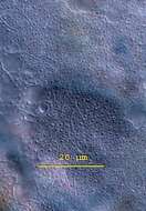
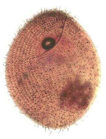
Nassulopsis (nass-you-lop-sis). Nassulopsis elegans is a conspicuous pink- or blue coloured nassulid found in pond and lake plankton. This cell was collected from floating colonies of Oscillatoria in a freshwater pond near Konstanz, Germany. The body is cylindrical in shape and measures 150 - 300 X 50 - 100 microns. Nassulopsis can be distinguished from Nassula and Obertrumia by 5-7 contractile vacuoles arranged in a ventral row. The macronucleus is cylindrical with rounded ends. The cytopharyngeal basket is located in the anterior third of the cell. The cells fill up with food vacuoles of different colour (green, orange), depending on stage of digestion. This picture shows the pellicle of Nassulopsis elegans with its net like ornamentation. A mucocyst lies at the centre of each square. Differential interference contrast.
-
Portrait of Scaphidiodon. Slightly oblique view of large hypostome ciliate with prominent cytopharyngeal basket. Dorsal surface unciliated. Lobular posterior protrusion on dorsal surface. Multiple small peripheral contractile vacuoles. From freshwater pond near Boise, Idaho. Brightfield.
-
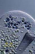
Nassulopsis (nass-you-lop-sis). Nassulopsis elegans is a conspicuous pink- or blue coloured nassulid found in pond and lake plankton. This cell was collected from floating colonies of Oscillatoria in a freshwater pond near Konstanz, Germany. The body is cylindrical in shape and measures 150 - 300 X 50 - 100 microns. Nassulopsis can be distinguished from Nassula and Obertrumia by 5-7 contractile vacuoles arranged in a ventral row. The macronucleus is cylindrical with rounded ends. The cytopharyngeal basket is located in the anterior third of the cell. The cells fill up with food vacuoles of different colour (green, orange), depending on stage of digestion. This picture shows the mouth and an anterior pigment spot consisting of 20 - 50 vacuoles filled with strongly coloured blue or violet oil. Differential interference contrast.
-
Nassulopsis (nass-you-lop-sis). Nassulopsis elegans is a conspicuous pink- or blue coloured nassulid found in pond and lake plankton. This cell was collected from floating colonies of Oscillatoria in a freshwater pond near Konstanz, Germany. The body is cylindrical in shape and measures 150 - 300 X 50 - 100 microns. Nassulopsis can be distinguished from Nassula and Obertrumia by 5-7 contractile vacuoles arranged in a ventral row. The macronucleus is cylindrical with rounded ends. The cytopharyngeal basket is located in the anterior third of the cell. The cells fill up with food vacuoles of different colour (green, orange), depending on stage of digestion. This image shows a specimen of Nassulopsis elegans beginning to ingest Oscillatoria. The cyanobacteria measures 18 (m in diameter. Differential interference contrast.
-
Nassulopsis (nass-you-lop-sis). Nassulopsis elegans is a conspicuous pink- or blue coloured nassulid found in pond and lake plankton. This cell was collected from floating colonies of Oscillatoria in a freshwater pond near Konstanz, Germany. The body is cylindrical in shape and measures 150 - 300 X 50 - 100 microns. Nassulopsis can be distinguished from Nassula and Obertrumia by 5-7 contractile vacuoles arranged in a ventral row. The macronucleus is cylindrical with rounded ends. The cytopharyngeal basket is located in the anterior third of the cell. The cells fill up with food vacuoles of different colour (green, orange), depending on stage of digestion. This image shows a specimen of Nassulopsis elegans elongated and deformed by ingesting a cyanobacteria (Oscillatoria).. Differential interference contrast
-
Nassulopsis (nass-you-lop-sis). Nassulopsis elegans is a conspicuous pink- or blue coloured nassulid found in pond and lake plankton. This cell was collected from floating colonies of Oscillatoria in a freshwater pond near Konstanz, Germany. The body is cylindrical in shape and measures 150 - 300 X 50 - 100 microns. Nassulopsis can be distinguished from Nassula and Obertrumia by 5-7 contractile vacuoles arranged in a ventral row. The macronucleus is cylindrical with rounded ends. The cytopharyngeal basket is located in the anterior third of the cell. The cells fill up with food vacuoles of different colour (green, orange), depending on stage of digestion. This image shows a compressed specimen of Nassulopsis elegans with a view of a row of contractile vacuoles. Differential interference contrast.
-
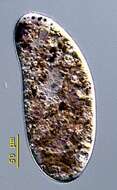
Ventral view of the nassulid ciliate, Nassulopsis elegans (Ehrenberg,1833)Foissner,1994.Collected from a freshwater pond near Boise, Idaho.DIC.
-
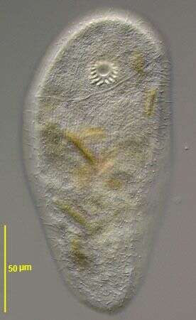
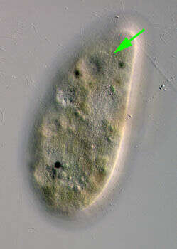
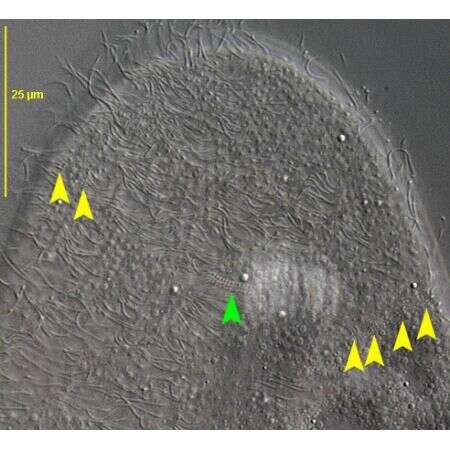
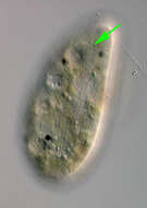
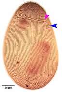
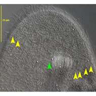
Surface view of the nassulid ciliate,Nassulopsis elegans(Ehrenberg,1833)Foissner,1994. There is a transverse hypostomial frange (synhymenium) of polykinetids which nearly encircles the anterior end of the cell (green arrow).Each element of the frange consists of 6-9 basal bodies.Collected from a freshwater pond near Boise, Idaho.DIC.
-
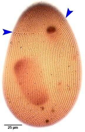
Ventral infraciliature of Nassulopsis elegans (EHRENBERG,1833) FOISSNER,1994. The synhymenium (blue arrowheads) is composed of 60-75 small nassulid organelles. It spans the ventral surface and wraps around on the right and left side of the dorsal surface leaving only a small gap between the right and left ends. Collected from the slow-moving runoff of a freshwater pond near Boise, Idaho.Stained by the silver carbonate technique (Foissner,W. Europ. J. Protistol.27:313-330;1991).Brightfield.
-
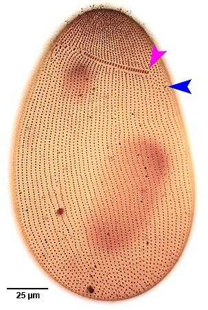
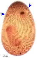
Dorsal infraciliature of Nassulopsis elegans (EHRENBERG,1833) FOISSNER,1994. The synhymenium is composed of 60-75 small nassulid organelles. It spans the ventral surface and wraps around on the right and left side of the dorsal surface leaving only a small gap between the right (pink arrowhead) and left ends (blue arrowhead). The dorsal component of the synhymenium is composed of more closely spaced nassulid organelles.Collected from the slow-moving runoff of a freshwater pond near Boise, Idaho.Stained by the silver carbonate technique (Foissner,W. Europ. J. Protistol.27:313-330;1991).Brightfield.
-
Dorsal view of the nassulid ciliate, Nassulopsis elegans (Ehrenberg,1833)Foissner,1994.The yellow arrowheads indicate nassulid organelles composed of two rows with three basal bodies each.The green arrowhead indicates the dorsal terminus of the left part of the synhymenion consisting of about seven more closely spaced nassulid organelles.Collected from a freshwater irrigation canal near Boise,Idaho.DIC.
-
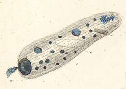
Originally described by Ehrenberg under the name Nassula elegans.
-
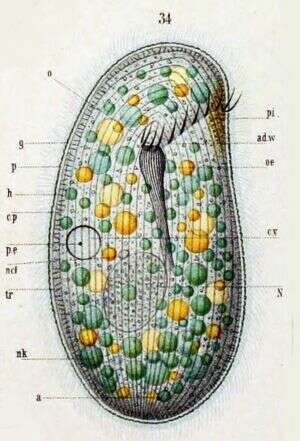
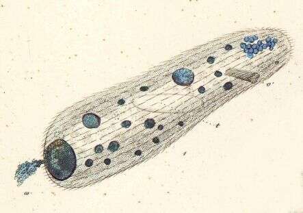
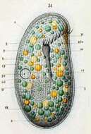
Originally described by Schewiakoff under the name Nassula elegans. Although the species Nassula elegans Ehrenberg has been transferred to the genus Nassulopsis Faure-Fremiet, 1959 (Foissner, 1994), the organism described and illustrated by Schewiakoff under the original name does not meet all the morphological criteria for the new taxonomic placement (multiple contractile vacuoles, hypostomial frange that spans the ventral face). Schewiakoff is very firm on the presence of only one contractile vacuole in all his specimens, and his illustration of the frange is unambiguous. It seems likely, then, that his Nassula elegans was, in fact, a malnourished variety of Nassula ornata, as described by Edna McNally (Biol. Bull. October 1, 1926 vol. 51 no. 4 237-244 ). Key to Schewiakoff's abbreviations: a -- Anus ad. w -- Adoral ciliated zone cp -- Cortical plasma cv -- Contractile vacuole g -- Gelationous layer h -- Ectoplasm of a homogenous appearance oe -- Throat N -- Macronucleus ncl -- Micronucleus nk -- Food particle o -- Mouth p -- Pellicle pe -- Excretory pore of the contractile vacuole pi -- Pigmented spot tr -- Trichocysts