-
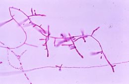
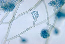
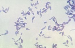
Magnified under a low magnification of 40X, this photomicrograph depicts the microconidia of the fungus Trichophyton mariatii.Created: 1979
-
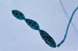
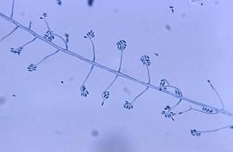
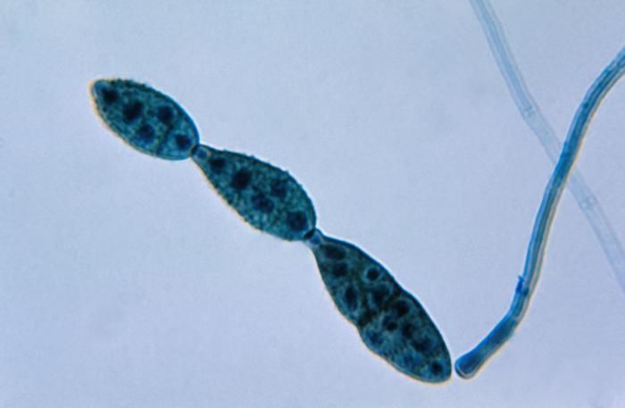
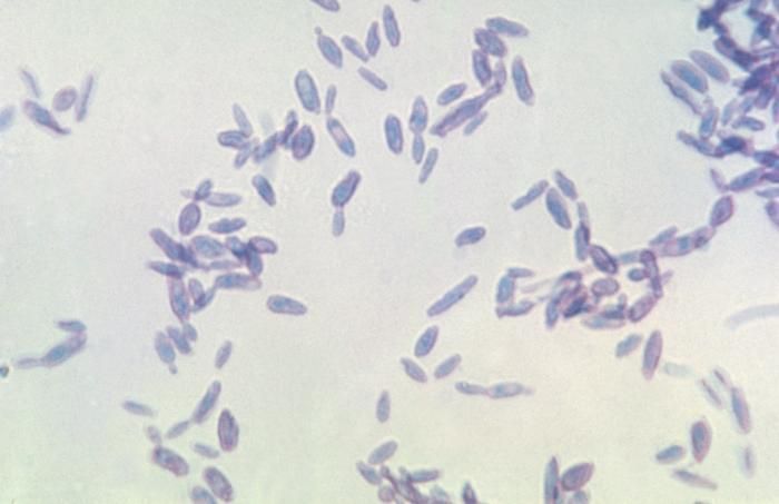
This photomicrograph shows a chain of conidia of a Alternaria sp. fungus, which can be a cause of phaeohyphomycosis.Created: 1955
-
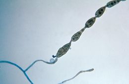
This photomicrograph shows a chain of conidia of a Alternaria sp. fungus, which can be a cause of phaeohyphomycosis.Created: 1955
-
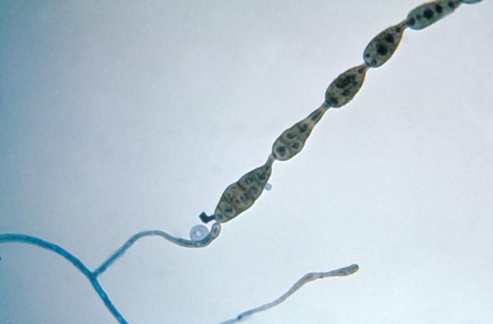
This photomicrograph shows a chain of conidia of a Alternaria sp. fungus, which can be a cause of phaeohyphomycosis.Created: 1955
-
This photomicrograph shows a chain of conidia of a Alternaria sp. fungus, which can be a cause of phaeohyphomycosis.Created: 1955
-
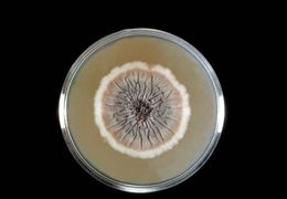
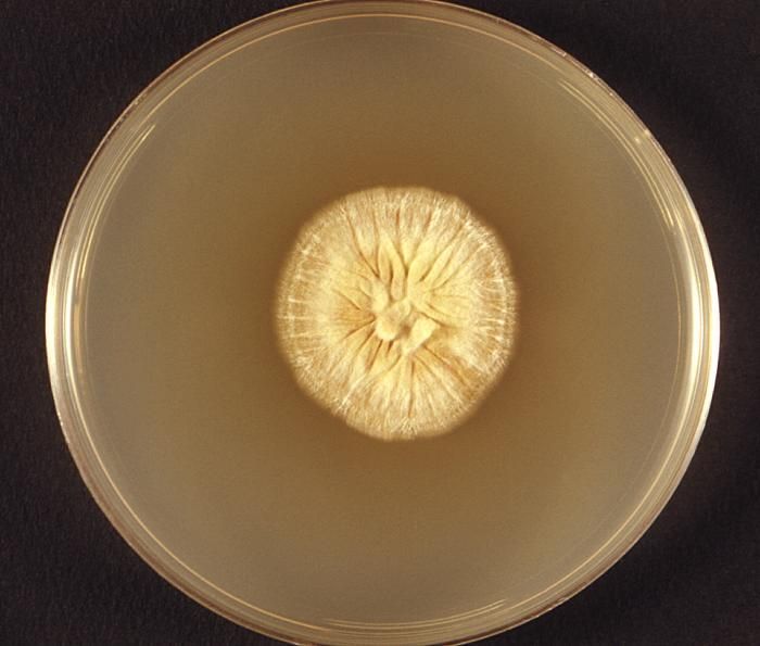
This photograph featured a Petri dish, which had been used to culture a colony of dermatophytic fungus, Microsporum ferrugineum.Dermatophytes are types of fungi that cause common skin, hair and nail infections. Infections caused by these fungi are also known by the names tinea and ringworm. It is important to emphasize that ringworm is not caused by a worm, but rather by a type of fungus called a dermatophyte. One example of a very common dermatophyte infection is athletes foot, which is also called tinea pedis. Another common dermatophyte infection affecting the groin area is jock itch, also known as tinea cruris.Created: 1974
-
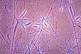
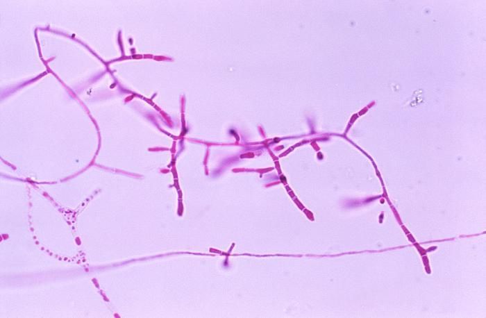
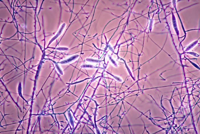
This photomicrograph depicts the mycelia, conidiophores, and conidia of the fungus Microsporum gallinae.Created: 1978
-
This micrograph depicts the conidia-laden conidiophores of a fungal organism of the genus Exophiala.Created: 1971
-
This micrograph depicts the conidia-laden conidiophores of a fungal organism of the genus Exophiala.Created: 1971
-
Magnified 500X, this photomicrograph revealed the presence of Sporothrix sp. fungal organisms that were isolated from a peat moss specimen.Created: 1971
-
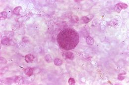
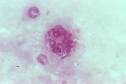
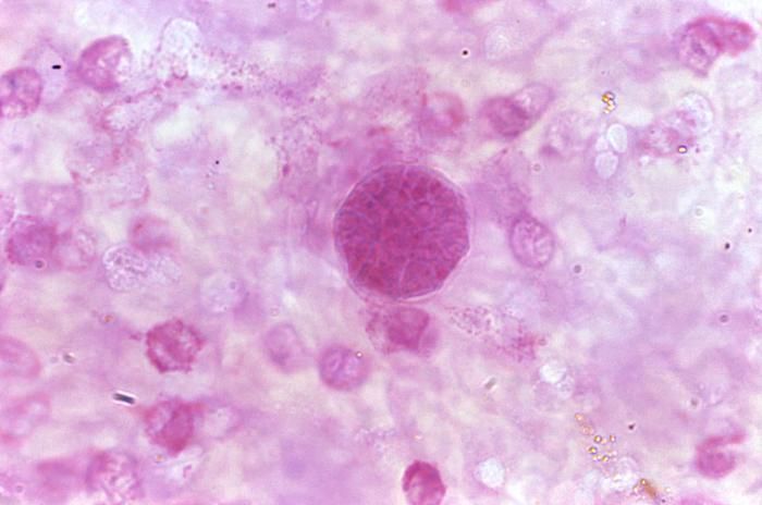
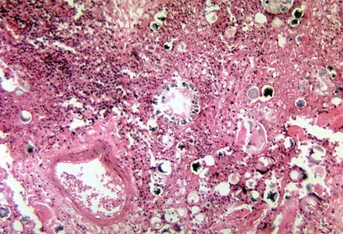
This photomicrograph revealed some of the histopathologic characteristics found within a pus specimen, which was prepared using periodic acid-Schiff (PAS), and which had been harvested from a skin lesion in a case of cutaneous coccidioidomycosis. In this particular specimen youll note the chlamydospore, or immature spherule of a Coccidioides immitis fungal organism. As the reproductive structure of this, as well as other types of fungi, this spherule is also known as a chlamydoconidium, and contains the organisms endospores.Created: 1975
-
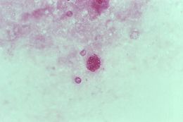
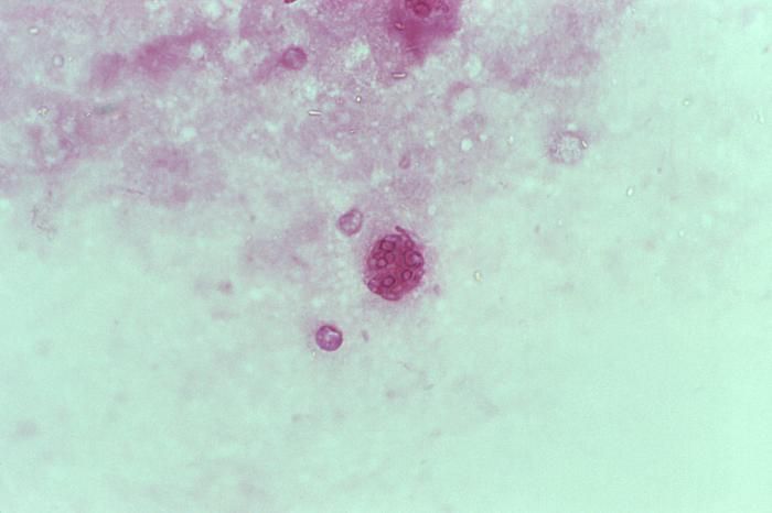
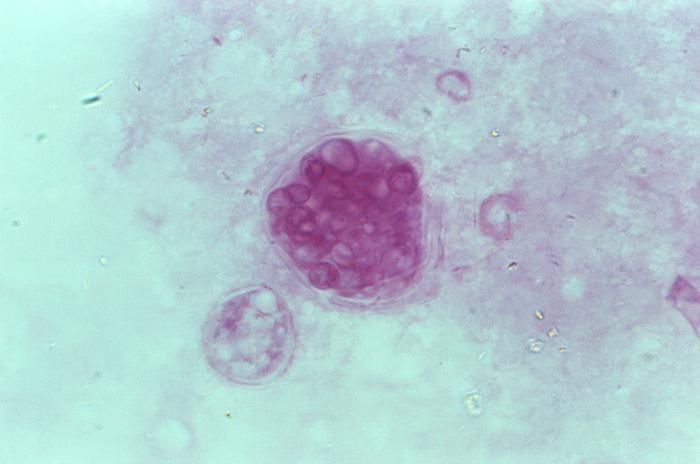
This photomicrograph revealed some of the histopathologic characteristics found within a pus specimen, which was prepared using periodic acid-Schiff (PAS), and which had been harvested from a skin lesion in a case of cutaneous coccidioidomycosis. In this particular specimen youll note the chlamydospore, or mature spherule of a Coccidioides immitis fungal organism. As the reproductive structure of this, as well as other types of fungi, this spherule is also known as a chlamydoconidium, and contains the organisms endospores.Created: 1975
-
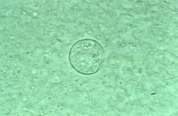
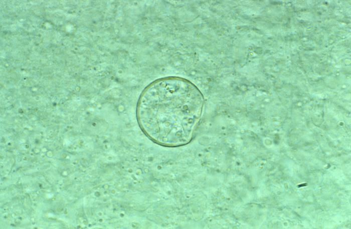
This photomicrograph revealed some of the histopathologic characteristics found within a pus specimen, which was prepared using potassium hudroxide (KOH), and which had been harvested from a skin lesion in a case of cutaneous coccidioidomycosis. In this particular specimen youll note the chlamydospore, or spherule of a Coccidioides immitis fungal organism. As the reproductive structure of this, as well as other types of fungi, this spherule is also known as a chlamydoconidium, and contains the organisms endospores.Created: 1975
-
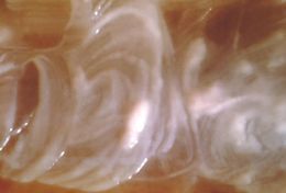
This was a Sabourauds dextrose agar culture of Coccidioides immitis with chloramphenicol and cycloheximide after 2 wks.Created: 1979
-
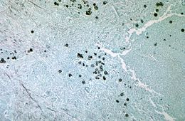
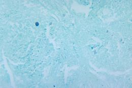
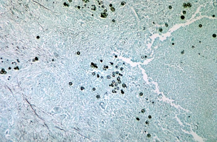
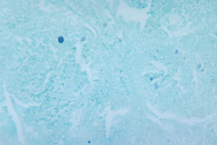
Using methenamine silver stain, this photomicrograph reveals spherules of Coccidioides immitis in brain tissue.Created: 1964
-
This methenamine silver stained photomicrograph reveals spherules of Coccidioides immitis fungus.Created: 1964
-
Using methenamine silver stain, this photomicrograph reveals spherules of the fungus Coccidioides immitis.Created: 1964
-
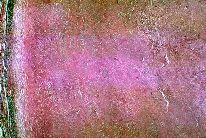
Note the histopathologic changes in a case of coccidioidomycosis of the lung showing a large fibrocaseous nodule.Created: 1964
-
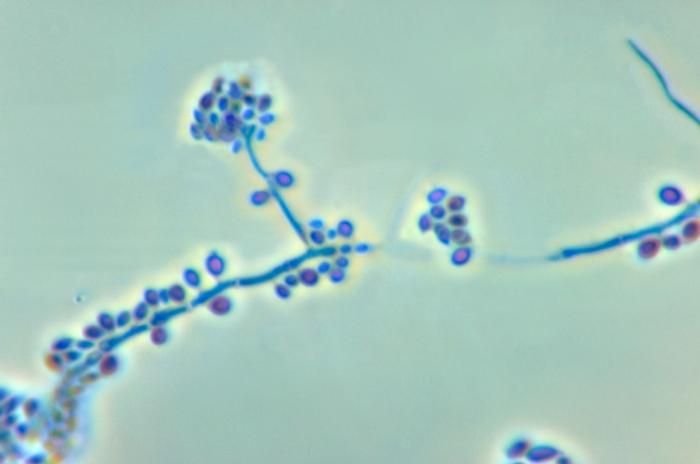
This photomicrograph reveals the conidiophores and conidia of the fungus Sporothrix schenckii.Created: 1972
-
Shown here is a photomicrograph of the fungus Sporothrix schenckii during yeast phase.Created: 1964
-
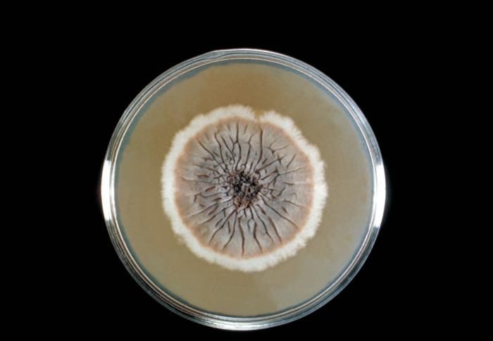
This Sabouraud's dextrose agar plate culture is growing the fungus Sporothrix schenckii.Created: 1964
-
Shown here is a close-up of a Sporothrix schenckii culture during yeast phase.Created: 1964