-
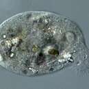
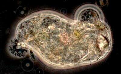
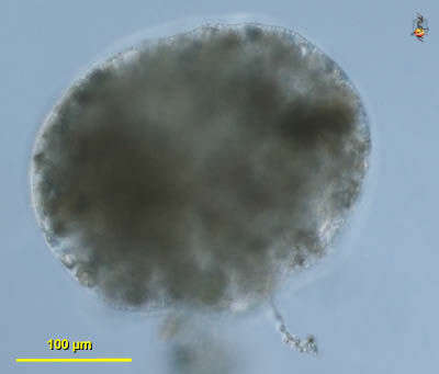
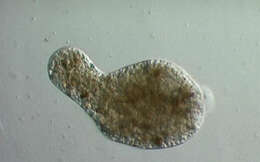
Pelomyxa is an amoeboid organism that inhabits mud in freshwater sites where there is little or no oxygen. The bright bits inside are pieces of sand.
-
-
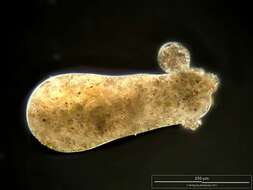
Pelomyxa (peal-o-mix-a), a large pelobiont which developed some reputation as possibly the most primitive eukaryote. This argument was based on the fact that it does not have mitochondria, conventional dictyosomes if any, flagella are aberrant, and nuclear division was also thought to be aberrant. The arguments for a primitive status now seem to be unsound. Cytoplasm with small particles of sand. eats algae and detritus. Moves with fountain-flow motion (cytoplasm moving forward up the centre of the cell and then spilling out near the front. Posterior end crumpled, to form a uroid. Phase contrast micrograph.
-
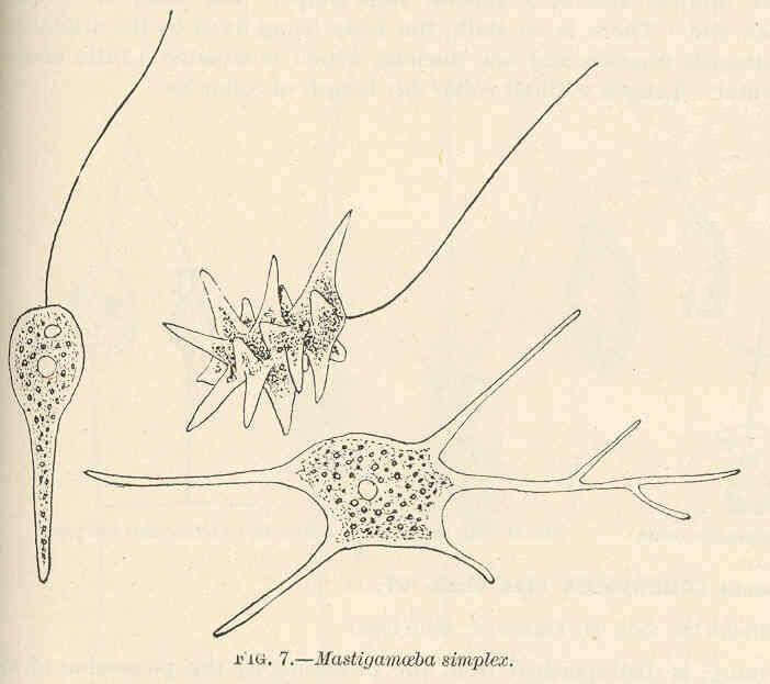
Mastigamoeba simplex.
-
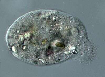
Pelomyxa, an unusual amoeba from areas with little oxygen. The front of the cell is to the upper right, and the crumpled structure, a uroid, is at the back. This organism eats algae and the cytoplasm also contains sand grains.
-
Synonym - Phreatamoeba balamuthi. Isolated from Gambia. ATCC 30984.
-
Pelomyxa (peal-o-mix-a), a large pelobiont which developed some reputation as possibly the most primitive eukaryote. This argument was based on the fact that it does not have mitochondria, conventional dictyosomes if any, flagella are aberrant, and nuclear division was also thought to be aberrant. The arguments for a primitive status now seem to be unsound. Cytoplasm with small particles of sand. eats algae and detritus. Moves with fountain-flow motion (cytoplasm moving forward up the centre of the cell and then spilling out near the front. With short inactive flagella over the surface of the cell. Posterior end crumpled, to form a uroid. Phase contrast micrograph.
-
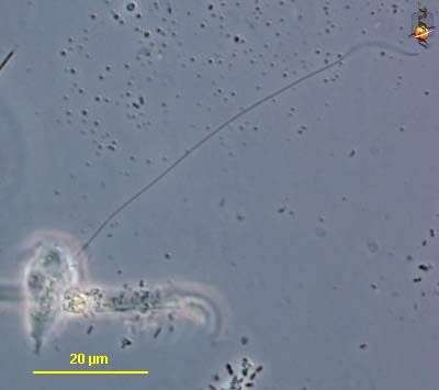
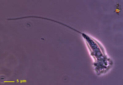
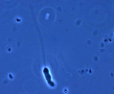
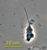
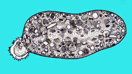
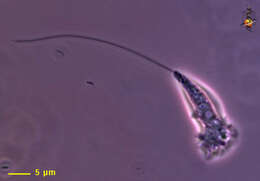
Mastigamoeba (massed-ig-a-me-ba) One of about 5 genera of pelobionts. These organisms are unusual in lacking mitochondria, dictyosomes and were for some time regarded as the most primitive of all eukaryotes. Most species which have been studied can adopt a variety of morphologies - including flagellates, amoebae, and cysts. The flagellates typically have one very long flagellum which beats in a fairly ineffectual fashion - and mostly progress by gliding. Body of cell most usually adopts an amoeboid form - such as that illustrated here. Typically found in habitats with little or no oxygen. Phase contrast.
-
ATCC 30984.
-
Pelomyxa (peal-o-mix-a), a large pelobiont which developed some reputation as possibly the most primitive eukaryote. This argument was based on the fact that it does not have mitochondria, conventional dictyosomes if any, flagella are aberrant, and nuclear division was also thought to be aberrant. The arguments for a primitive status now seem to be unsound. Cytoplasm with small particles of sand. eats algae and detritus. Moves with fountain-flow motion (cytoplasm moving forward up the centre of the cell and then spilling out near the front. Posterior end crumpled, to form a uroid. Differential interference contrast.
-
Mastigamoeba (massed-ig-a-me-ba) One of about 5 genera of pelobionts. These organisms are unusual in lacking mitochondria, dictyosomes and were for some time regarded as the most primitive of all eukaryotes. Most species which have been studied can adopt a variety of morphologies - including flagellates, amoebae, and cysts. The flagellates typically have one very long flagellum which beats in a fairly ineffectual fashion - and mostly progress by gliding. Body of cell most usually adopts an amoeboid form - such as that illustrated here. Typically found in habitats with little or no oxygen. Phase contrast.
-
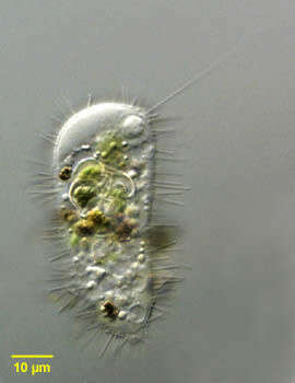
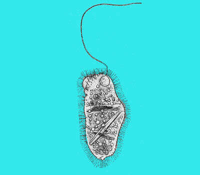
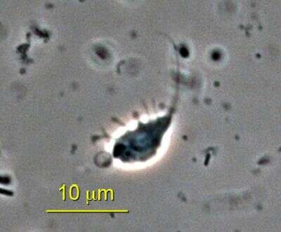
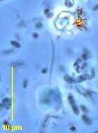
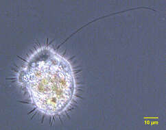
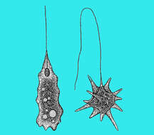
Portrait of Mastigina setosa a flagellated pelobiont. The ameboid monopodial cells have radiating "setae" which are 6-10 micron long filaments each of which has a basal granule (seen in this image). There is a single, long, lazily beating flagellum, which is closely associated with the nucleus (seen at the 12 o clock position adjacent to the cell membrane in this image). Electron microscopy shows the base of the flagellum connected to the nucleus by a cone of microtubules. The flagellum has a variable arrangement of microtubules. The nucleus contains a large endosome. Ingested algae are seen in food vacuoles. There is vigorous cytoplasmic streaming resulting in ameboid locomotion. The flagellum appears to contribute little to motility. Mastigina and the other pelobionts lack mitochondria and dictyosomes. Obvious ameboid locomotion and cytoplasmic streaming may help differentiate Mastigina from Mastigamoeba, a similar pelobiont. From slow-moving organically enriched freshwater runoff stream near Boise, Idaho. Differential interference contrast. Differential interference contrast optics.
-
Pelomyxa (peal-o-mix-a), a large pelobiont which developed some reputation as possibly the most primitive eukaryote. This argument was based on the fact that it does not have mitochondria, conventional dictyosomes if any, flagella are aberrant, and nuclear division was also thought to be aberrant. The arguments for a primitive status now seem to be unsound. Cytoplasm with small particles of sand. eats algae and detritus. Moves with fountain-flow motion (cytoplasm moving forward up the centre of the cell and then spilling out near the front. Posterior end crumpled, to form a uroid. Phase contrast micrograph.
-
Mastigamoeba (massed-ig-a-me-ba) One of about 5 genera of pelobionts. These organisms are unusual in lacking mitochondria, dictyosomes and were for some time regarded as the most primitive of all eukaryotes. Most species which have been studied can adopt a variety of morphologies - including flagellates, amoebae, and cysts. The flagellates typically have one very long flagellum which beats in a fairly ineffectual fashion - and mostly progress by gliding. Body of cell most usually adopts an amoeboid form. Typically found in habitats with little or no oxygen. Phase contrast.
-
Portrait of Mastigina setosa a flagellated pelobiont. The ameboid monopodial cells have radiating "setae" which are 6-10 micron long filaments. There is a single, long, lazily beating flagellum, which is closely associated with the nucleus (seen at the 1 o clock position adjacent to the cell membrane in this image). Electron microscopy shows the base of the flagellum connected to the nucleus by a cone of microtubules. The flagellum has a variable arrangement of microtubules. The nucleus contains a large nucleolus. Ingested algae are seen in food vacuoles. There is vigorous cytoplasmic streaming resulting in ameboid locomotion. The flagellum appears to contribute little to motility. Mastigina and the other pelobionts lack mitochondria and dictyosomes. Obvious ameboid locomotion and cytoplasmic streaming may help differentiate Mastigina from Mastigamoeba, a similar pelobiont. From slow-moving organically enriched freshwater runoff stream near Boise, Idaho. Phase contrast.
-
-
-
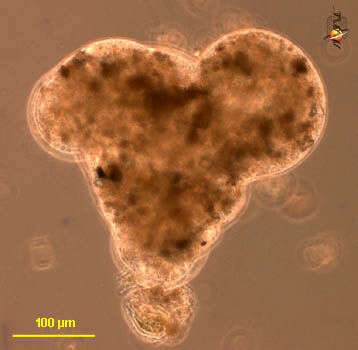
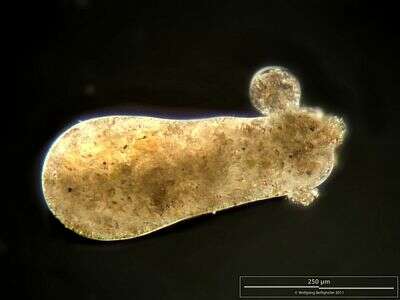
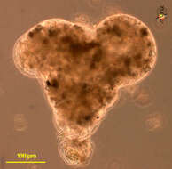
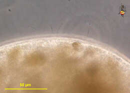
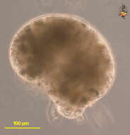
Specimen of medium cell size with typical monopodial locomotion. Scale bar indicates 250 µm. Sample from a freshwater pond on the island of Hiddensee (Baltic Sea, Germany). This image was taken using Zeiss Universal with Olympus C7070 CCD camera.Image under Creative Commons License V 3.0 (CC BY-NC-SA).
-
-
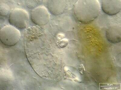
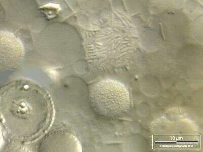
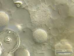
Cellular detail at high magnification. Among many vacuoles, a shell of an engulfed testacea out of the group of euglyphida is visible. Scale bar indicates 10 µm. Sample from a freshwater pond on the island of Hiddensee (Baltic Sea, Germany). This image was taken using Zeiss Universal with Olympus C7070 CCD camera.Image under Creative Commons License V 3.0 (CC BY-NC-SA).
-
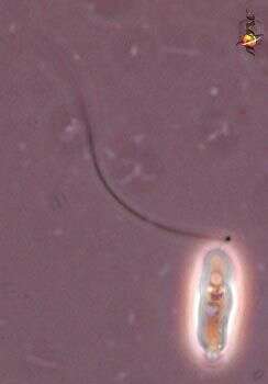
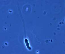
This cell is typical of many smaller mastigamoebae. There is a single long anterior flagellum, a tapering body that is capable of producing pseudopodia. identities of taxa in this area are not clear as many large species can produce smaller forms at certain stages in the life cycle. Phase contrast microscopy.
-
Cellular detail at high magnification. In the lower left a cross-section through the nucleus is visible. The nucleolus is a parietal layer beneath the cell membrane. The nuclei are enveloped with a layer of symbiotic bacteria. This is seen in the upper center of the image. Pelomyxa don´t own mitochondria, and these bacteria act like mitochondria. Scale bar indicates 10 µm. Sample from a freshwater pond on the island of Hiddensee (Baltic Sea, Germany). This image was taken using Zeiss Universal with Olympus C7070 CCD camera.Image under Creative Commons License V 3.0 (CC BY-NC-SA).
-
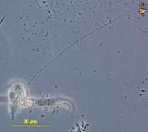
Small cell, one very long flagellum extending from a more homogeneous anterior region of the cell (this is where the nucleus is). The rest of the cell has inclusions. the body is flexible or amoeboid. Phase contrast microscopy.
-
Video showing how this amoebae collected at Cedar Swamps in Woods Hole moves around with a single anterior protruding pseudopode. This video made by Dan Lahr under a Zeiss Discovery V12, believe it or not, a dissecting scope.