-
Rafael F. Almeida, Isabel R. Guesdon, Marcelo R. Pace, Renata M.S. Meira
Phytokeys
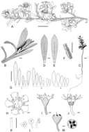
Figure 3.
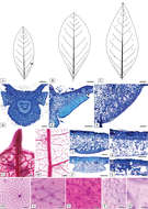
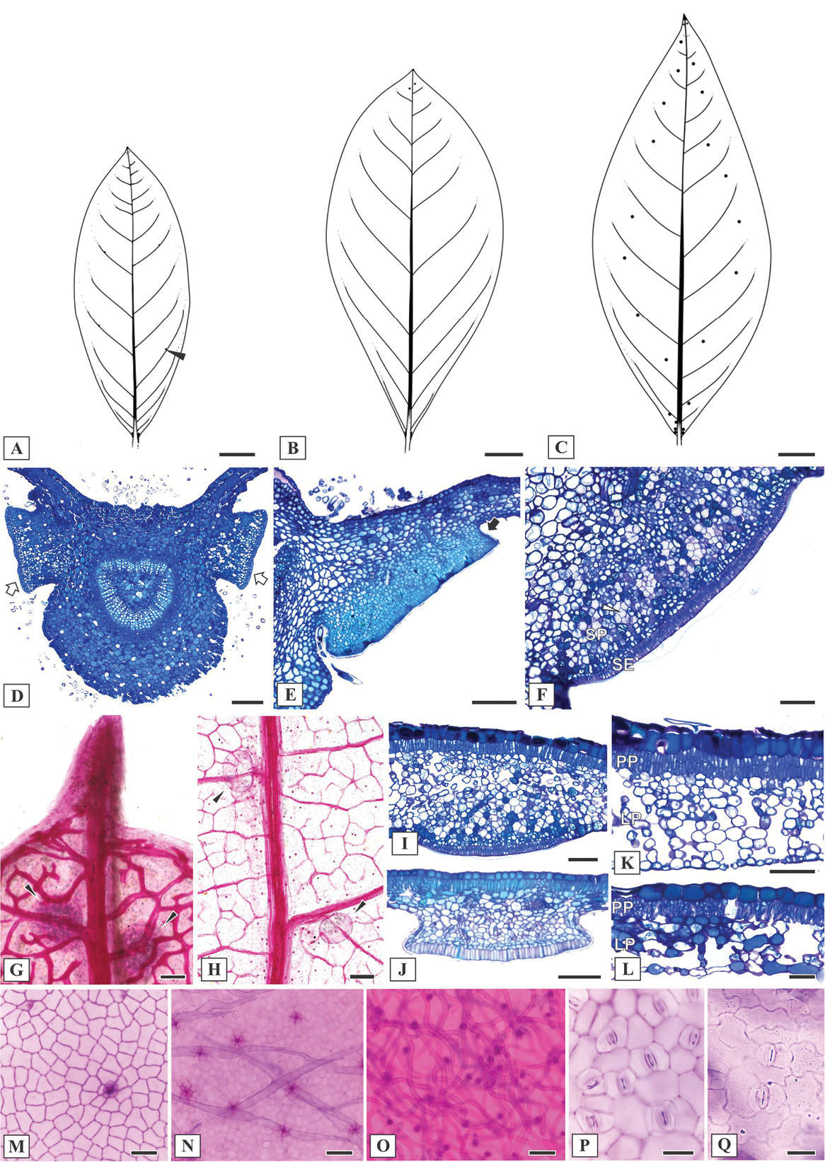
Leaf morphoanatomy of Mcvaughia species. A patterns of leaf glands distribution on the abaxial leaf surface of M.bahianaB patterns of leaf glands distribution on the abaxial leaf surface of M.piauhiensisC patterns of leaf glands distribution on the abaxial leaf surface of M.sergipanaD transverse section of leaf base showing the basilaminar pair of stalked glands (white arrows) E basilaminar leaf gland with a stalk (black arrow) in M.piauhiensisF basilaminar gland in M.sergipana showing a sessile position (SE= anatomical arrangement with secretory epidermis, SP= vascularized secretory parenchyma) G–H laminar glands on the apex of cleared leaves of M.sergipana and M.bahiana respectively, note the apical tooth (G) I sessile laminar glands in M.sergipanaJ stalked laminar gland in M.piauhiensisK–L transverse sections of the leaf blade; mesophyll with uniserial palisade-like parenchyma and spongy parenchyma composed by several or few layers in M.sergipana and M.bahiana, respectively; note the idioblast with druse crystals at the mesophyll (white arrow) and the stomata distribution at the abaxial leaf surface (black arrow) M–N adaxial epidermis surface of M.piauhiensis and M.sergipana, showing scars of malpighiaceous trichomes O abaxial epidermis surface of trichomes abundance in M.bahianaP–Q outline of the anticlinal epidermal cell walls: straight in M.sergipana (P) and sinuous in M.bahiana (Q). Laminar scale bars: 1 cm (A–C), 100 μm (D, F–K, N–O), 150 μm (E), 50 μm (L–M, P–Q).
-
Rafael F. Almeida, Isabel R. Guesdon, Marcelo R. Pace, Renata M.S. Meira
Phytokeys
Figure 4.
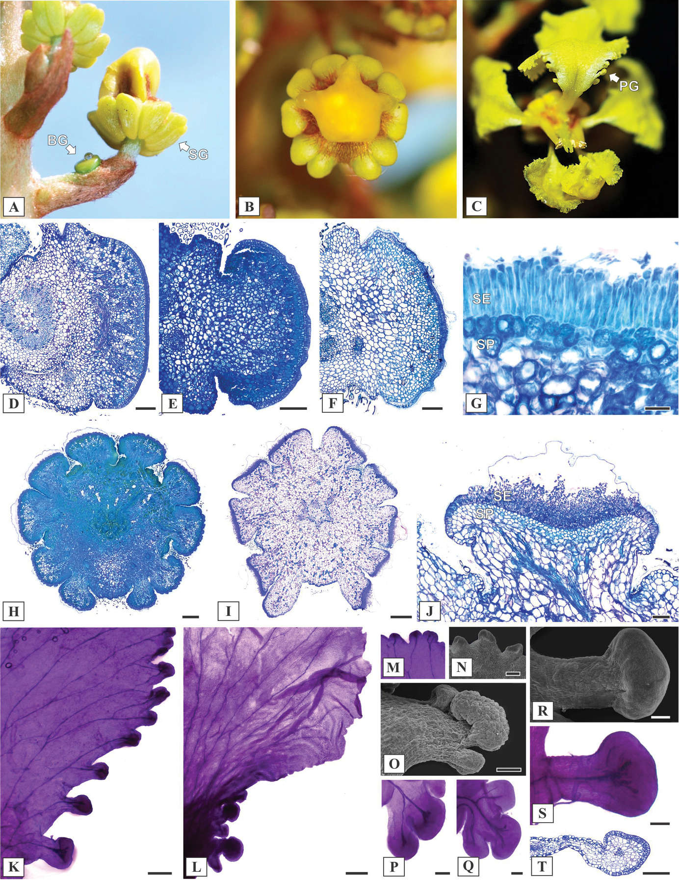
Reproductive morphoanatomy of Mcvaughia species. A inflorescence during development, showing a bracteole gland (BG) and Sepal glands (SG) B ten sepal glands encircling the calyx C Petal glands (PG) along the margin of posterior petal D–F transverse section of bracteole glands in M.sergipana, M.bahiana and M.piauhiensis, respectively G anatomical arrangement of bracteole gland, with a palisade-like secretory epidermis (SE) and secretory parenchyma (SP) H–I transverse section of floral bud and anthesis flower in Mcvaughiabahiana and M.sergipana; calyx gland pair displaced at the anterior sepal J calyx gland structure, showing a secretory epidermis (SE) and vascularized secretory parenchyma (SP) K–L petal glands on the margin of petals in M.sergipana and M.bahiana respectively M–N detail of the petal glands at the apex of the petal limb in M.sergipana, cleared and in SEM image O–Q petal glands positioned at the base, M.bahiana on SEM image, M.bahiana and M.piauhiensis cleared R–T conspicuous and stalked petal glands at the base of M.sergipana, in SEM image, cleared and longitudinal section. Scale bars: 200 μm (D), 150 μm (E–F), 50 μm (G), 500 μm (H–I), 100 μm (J, P–S), 300 μm (L–M), 200 μm (N, T).
-
Yanqin Xu, Linjian Liu, Shaoxiong Liu, Yiming He, Renqing Li, Fei Ge
Phytokeys
Figure 1.
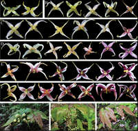
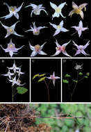
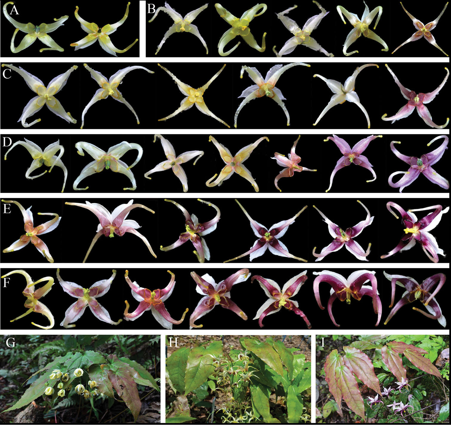
The flower colour variations of different populations of E.acuminatum. A SCMP, mainly yellow B SCSL, mainly yellow, occasionally purple-yellow at the base of petals C CQNC, mainly yellow, occasionally rose-purple D SCSS, from yellow to purple E SCEM, mainly purple, occasionally purple-yellow F SCYJ, mainly purple, occasionally purple-yellow G Individual with yellow flowers H Individual with purple-yellow flowers I Individual with purple flowers.
-
Yanqin Xu, Linjian Liu, Shaoxiong Liu, Yiming He, Renqing Li, Fei Ge
Phytokeys
Figure 2.
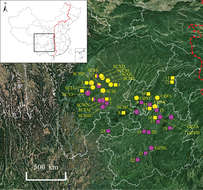
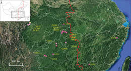
Geographical variation in flower colour patterns within E.acuminatum. The circles and boxes represent wild populations and herbarium specimens, respectively. The colour of the circles and boxes represent flower colour. The 19 population codes are shown in Table 1. The left area of the red dotted line is western China. The information of 23 representative herbarium specimens is as follow: 1. B. Y. Peng 47070; 2. B. L. Guo 0540; 3. B. L. Guo 0608; 4. B. L. Guo 0607; 5. Sichuan Econ. Pl. Exped. 0013; 6. F. T. Wang 23329; 7. G. H. Yang 54343; 8. K. Y. Lang 3002; 9. Sichuan Econ. Pl. Exped. 0030; 10. T. C. Pan & G. F. Wu 105; 11. Sanxia Exped. 0729; 12. Sanxia Exped. 0821; 13. K. J. Guan et al. 0273; 14. X. B. Zhang 19; 15. Jinfoshan Exped. 0202; 16. Q. H. Chen et al. 9411; 17. J. M. Yuan 003; 18. Z. S. Zhang et al. 401131; 19. Y. Tsiang 4994; 20. S. Z. He 96410; 21. P. Zhao 807; 22. S. W. Teng 0008; 23. Z. Y. Wu 60.
-
Yanqin Xu, Linjian Liu, Shaoxiong Liu, Yiming He, Renqing Li, Fei Ge
Phytokeys
Figure 3.
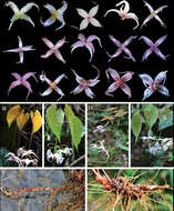
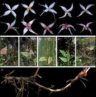
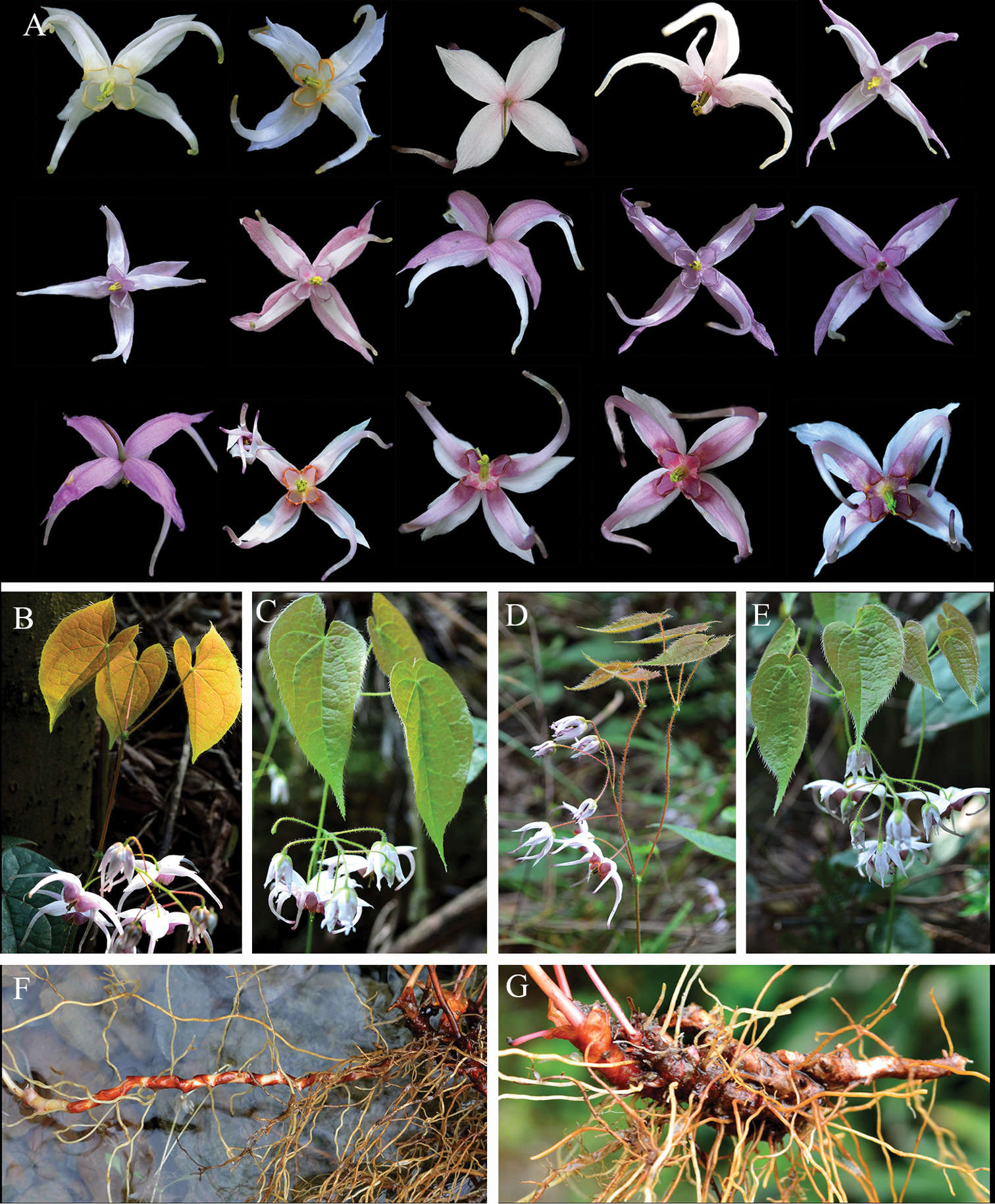
The variations and morphological characters of E.leptorrhizum. A the colour variations of inner sepals and petals B–E variations of the number and arrangement of stem-leaves F long creeping rhizome G stout and compact rhizome.
-
Yanqin Xu, Linjian Liu, Shaoxiong Liu, Yiming He, Renqing Li, Fei Ge
Phytokeys
Figure 4.
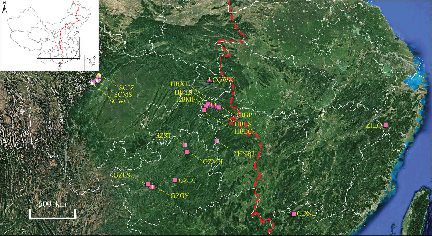
Geographical variation in flower colour patterns within E.leptorrhizum (boxes), E.pauciflorum (circles), E.mikinorii (diamonds) and E.glandulosopilosum (triangle). The colour of the boxes, circles, diamonds and triangle represent flower colour. The population codes are shown in Table 1. The left area of the red dotted line is western China.
-
Yanqin Xu, Linjian Liu, Shaoxiong Liu, Yiming He, Renqing Li, Fei Ge
Phytokeys
Figure 5.
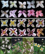
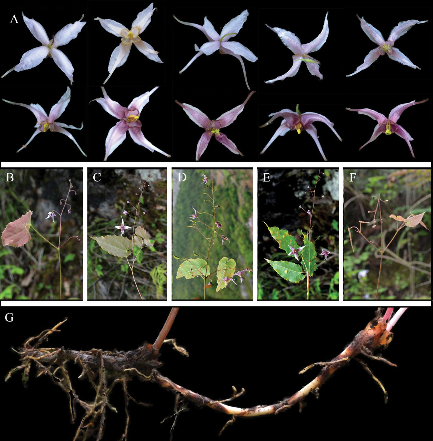
The variations and morphological characters of E.pauciflorum. A colour variations of inner sepals and petals B–D variations of the number and arrangement of stem-leaves E long creeping and thread-like rhizome.
-
Yanqin Xu, Linjian Liu, Shaoxiong Liu, Yiming He, Renqing Li, Fei Ge
Phytokeys
Figure 4.
Geographical variation in flower colour patterns within E.leptorrhizum (boxes), E.pauciflorum (circles), E.mikinorii (diamonds) and E.glandulosopilosum (triangle). The colour of the boxes, circles, diamonds and triangle represent flower colour. The population codes are shown in Table 1. The left area of the red dotted line is western China.
-
Yanqin Xu, Linjian Liu, Shaoxiong Liu, Yiming He, Renqing Li, Fei Ge
Phytokeys
Figure 6.
The variations and morphological characters of E.mikinorii. A colour variations of inner sepals B colour variations of petals C individuals that with different flower colour.
-
Yanqin Xu, Linjian Liu, Shaoxiong Liu, Yiming He, Renqing Li, Fei Ge
Phytokeys
Figure 4.
Geographical variation in flower colour patterns within E.leptorrhizum (boxes), E.pauciflorum (circles), E.mikinorii (diamonds) and E.glandulosopilosum (triangle). The colour of the boxes, circles, diamonds and triangle represent flower colour. The population codes are shown in Table 1. The left area of the red dotted line is western China.
-
Yanqin Xu, Linjian Liu, Shaoxiong Liu, Yiming He, Renqing Li, Fei Ge
Phytokeys
Figure 7.
The variations and morphological characters of E.glandulosopilosum. A variations of flower colour B–F variations of the number and arrangement of stem-leaves G creeping and slender rhizome.
-
Yanqin Xu, Linjian Liu, Shaoxiong Liu, Yiming He, Renqing Li, Fei Ge
Phytokeys
Figure 4.
Geographical variation in flower colour patterns within E.leptorrhizum (boxes), E.pauciflorum (circles), E.mikinorii (diamonds) and E.glandulosopilosum (triangle). The colour of the boxes, circles, diamonds and triangle represent flower colour. The population codes are shown in Table 1. The left area of the red dotted line is western China.
-
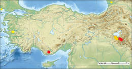
Figure 3.
Distribution of the taxa. (square)Crataegusazarolusvar.senobaaensis; (star) Crataegusmonogynavar.odemisii var. nov. (circle) Crataegusyaltirikii (Near East topographic map-blank.svg).
-
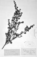
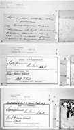
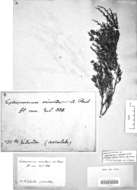
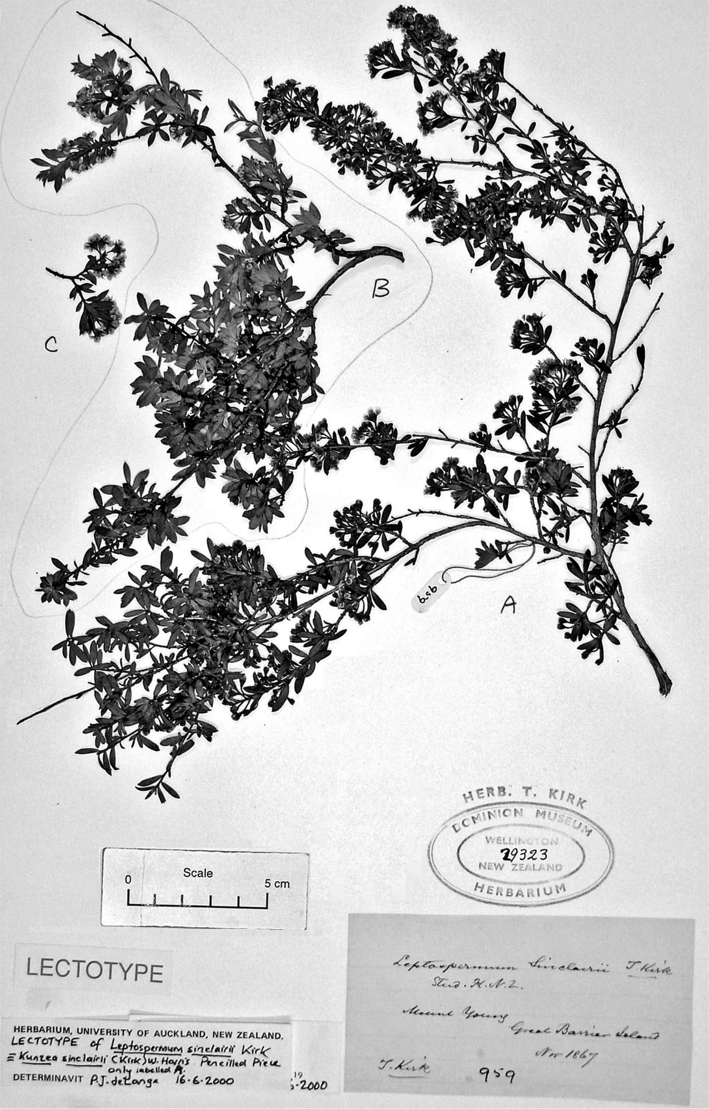
Figure 44.Lectotype of Leptospermum sinclairii Kirk (T. Kirk 959, WELT SP029323 (piece labelled in pencil “A”).
-
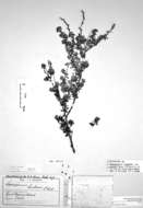
Figure 45.Kunzea sinclairii specimen from the private T. Kirk Herbarium (WELT SP044298) bearing two conflicting labels, one by Hutton and the other by Kirk.
-
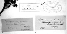
Figure 46.Details of labels on herbarium sheet WELT SP044298. A Pencil label on blue paper in handwriting of Captain F. W. Hutton bearing annotations in Indian ink by T. Kirk—annotations comprise the manuscript name ‘v. Sinclairii’, a change of collection date and the crossing out of Hutton’s name as collector B Second label on herbarium paper bearing the handwriting of T. Kirk in Indian ink. Neither label can be matched with certainty to the specimen mounted on WELT SP044298.
-
Figure 47.Lectotype of Leptospermum ericoides var. pubescens Kirk (T. Kirk s.n., AK 5515).
-
Figure 48.Label details of the lectotype of Leptospermum ericoides var. pubescens Kirk (AK 5515). A Bottom-most label written by T. Kirk and including critical details from the protologue of var. pubescens B Second label from bottom in handwriting of T. Cheeseman C Top most label recording use of specimen AK 5515 for Cheeseman’s Illustrations of the New Zealand Flora (Cheeseman 1914). Arrows indicate position of the preceding labels.
-

Figure 49.Distinguishing features of Kunzea sinclairii. A Flowering branchlets (ex cult. AK 246813) B Vegetative bud, leaf and branchlet indumentum (ex cult. AK 246813) C Three year old seedling (no voucher, ex cult. Aotea (Great Barrier Island), Mt Young) D Adaxial leaf surface (ex cult. AK 246813) E Abaxial leaf surface (ex cult. AK 246813) F Leaf margin indumentum (ex cult. AK 246813) G Leaf variation from seedling to adult (taken from (C) above): (G1) glabrous leaves of seedling (first year of growth), (G2) second year transitional leaves, first three w, next three hairy; (G3) third year adult leaves (no voucher, ex cult. Aotea (Great Barrier Island), Mt Young) H Flower (top view) (ex cult. AK 246813) I Flower and hypanthium (side view) (ex cult. AK 246813) J Flower cross section showing anther, style and ovules (ex cult. AK 246813) K Style and stigma (ex cult. AK 246813) L Stamens (ex cult. AK 246813) M Dehisced fruit (ex cult. AK 246813). Scales bars: (A, C, G) 10 mm; (D, D, E, H–M) 1 mm; (F) 0.5 mm.
-
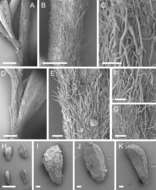
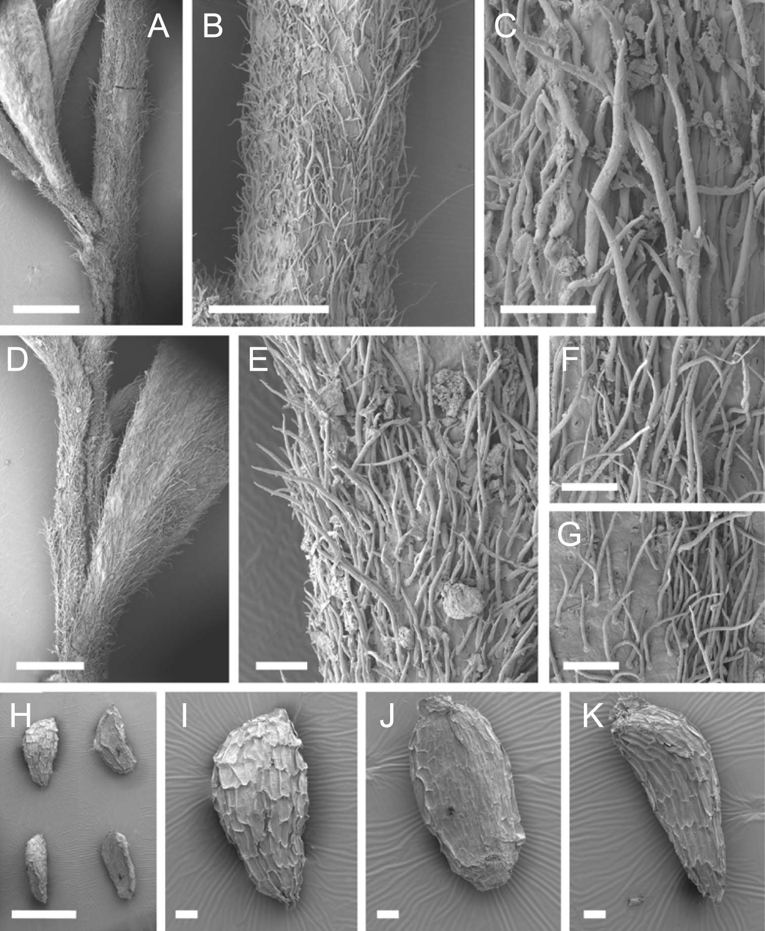
Figure 50.Scanning Electron Micrographs of Kunzea sinclairii. (A–G all AK 140485) Branchlet indumentum H–K Seeds (AK 278809). Scale bars: (A, D, H) 1 mm; (B) 500 μm; (E–G, I–K) 100 μm.
-
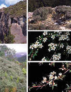
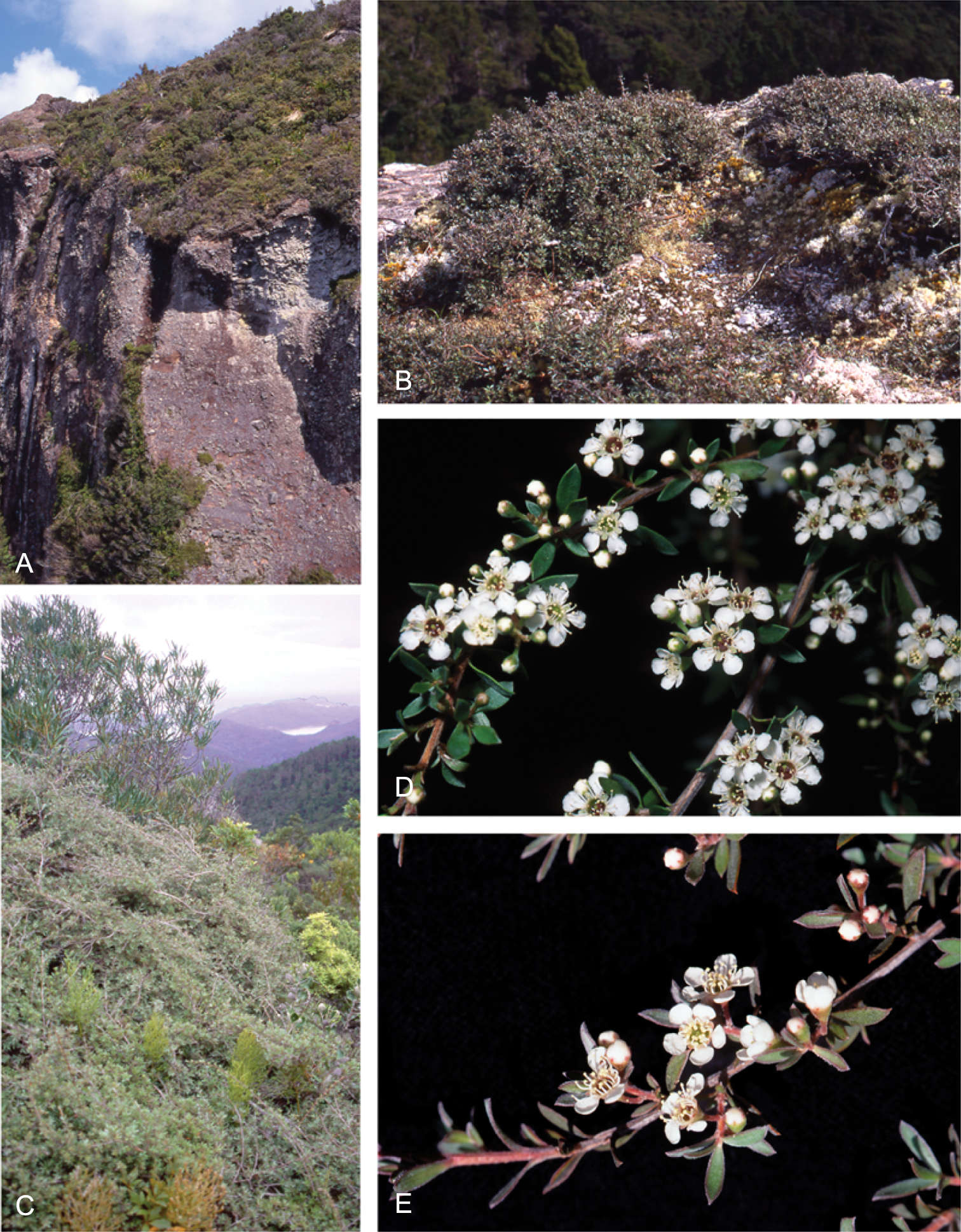
Figure 51.Kunzea sinclairii. A Rhyolite rock canyons on Aotea (Great Barrier Island), providing one of the key habitats for Kunzea sinclairii (which is the dominant shrub in the image), Aotea (Great Barrier Island), Windy Canyon (photo: P. J. de Lange) B Decumbent Kunzea sinclairii shrubs on rhyolitic saprolite at the type locality for the species, Aotea (Great Barrier Island), Mt Young (photo: P. J. de Lange) C Typical long trailing form of Kunzea sinclairii cascading down Rhyolite cliffs; Aotea (Great Barrier Island), near Mt Young D Kunzea sinclairii in full flower, ex cult. Aotea (Great Barrier Island), Mt Young (photo: J. E. Braggins) E Kunzea sinclairii freshly opened flowers, flower buds, and bud just prior to bud burst, Aotea (Great Barrier Island), Mt Heale (photo: G. M. Crowcroft).
-
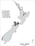
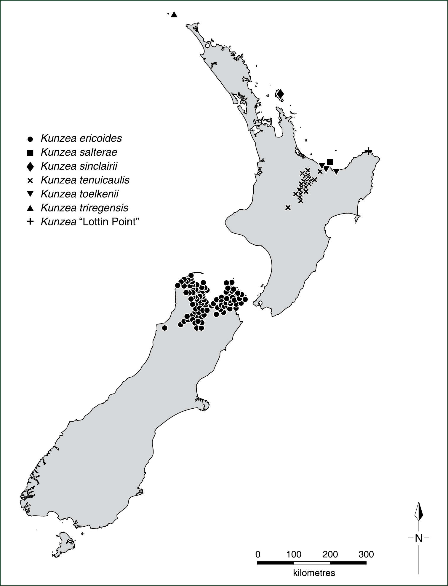
Figure 7.Distribution of Kunzea ericoides, Kunzea salterae, Kunzea sinclairii, Kunzea tenuicaulis, Kunzea toelkenii, Kunzea triregensis and Kunzea “Lottin Point”.
-
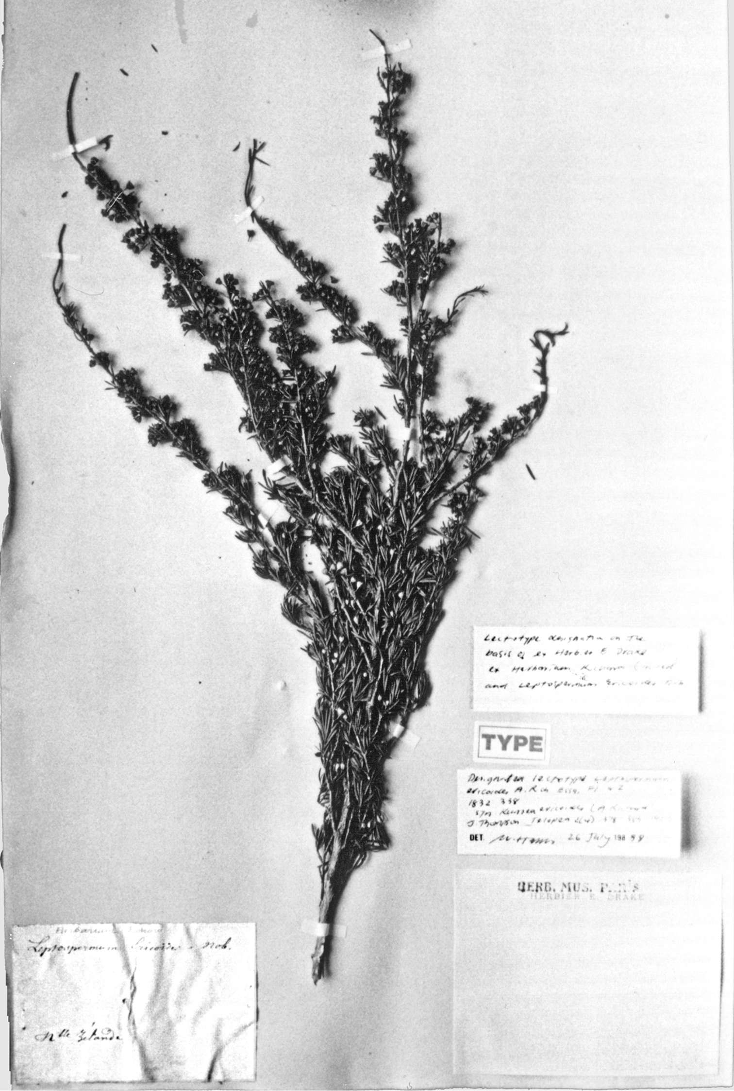
Figure 2.Lectotype of Leptospermum ericoides A.Rich. (held at l’Herbier du Laboratoire de Phanérogamie du Muséum National d’Histoire Naturelle (P)).
-
Figure 3.Paralectotype of A Leptospermum ericoides (held at l’Herbier du Laboratoire de Phanérogamie du Muséum National d’Histoire Naturelle (P)). B Enlargement of paralectotype label showing Achille Richard’s distinctive handwriting.