-
Boonsatien Boonsoong, Dietrich Braasch
Zookeys
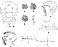
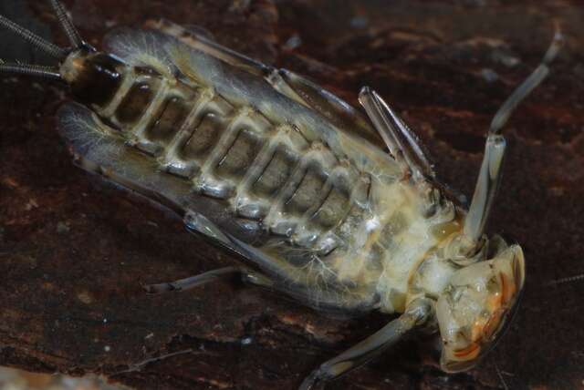
Figure 2.A Ventral view of abdomen of Rhithrogena siamensis Braasch & Boonsoong, 2009 B–E lamellae of gills 1 (B), 3 (C), 5 (D) and 7 (E) of Trichogenia maxillaris Braasch & Soldán, 1988 F ventral view of left maxilla of Trichogenia maxillaris Braasch & Soldán, 1988 G bristles on dorsal face of abdominal terga of Trichogenia maxillaris Braasch & Soldán, 1988 H ventral view of left maxilla of Compsoneuria langensis Braasch & Boonsoong, 2010 I-J abdominal terga (I) and tergum VII (J) of Notacanthurus baei Braasch & Boonsoong, 2009.
-
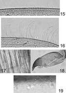
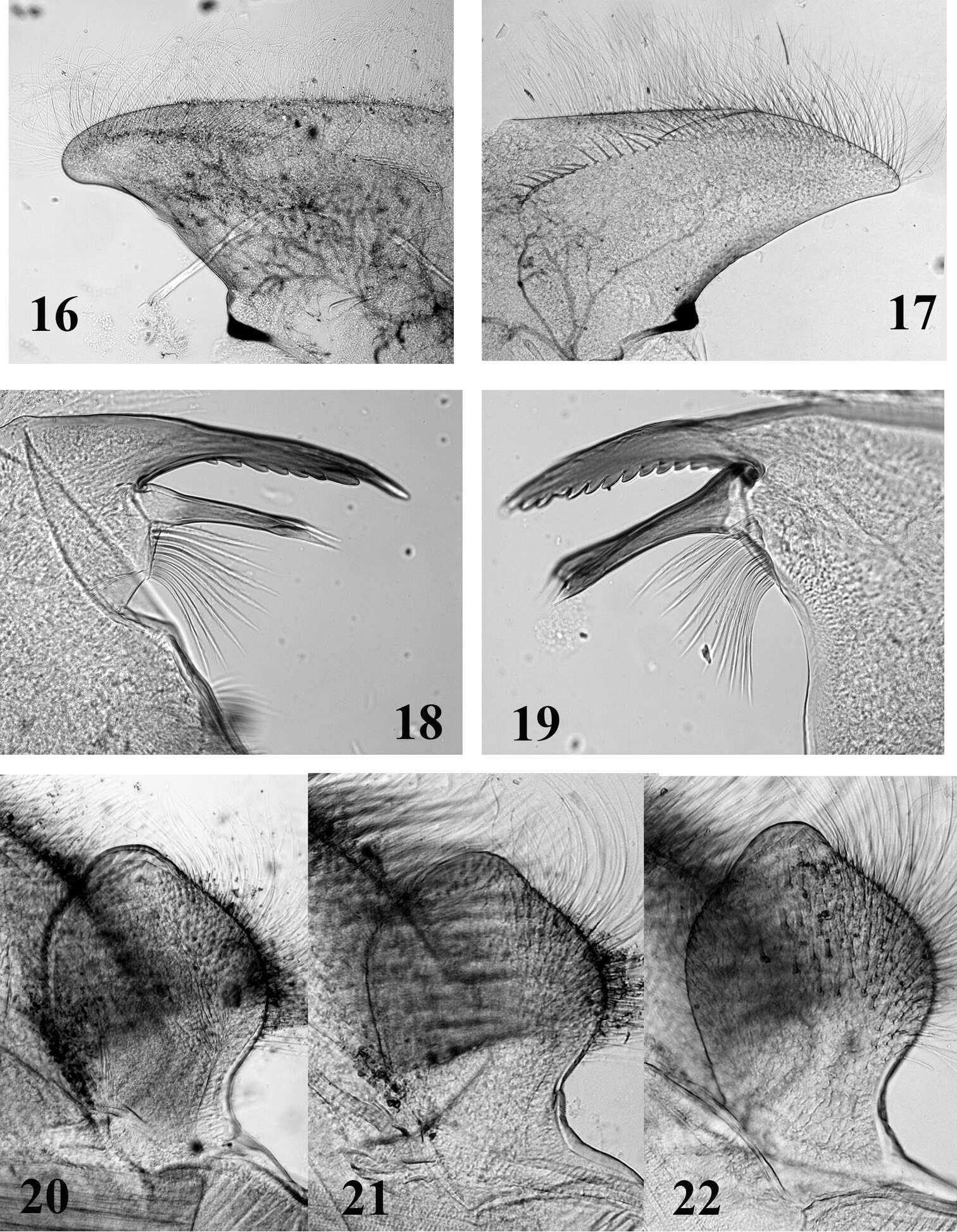
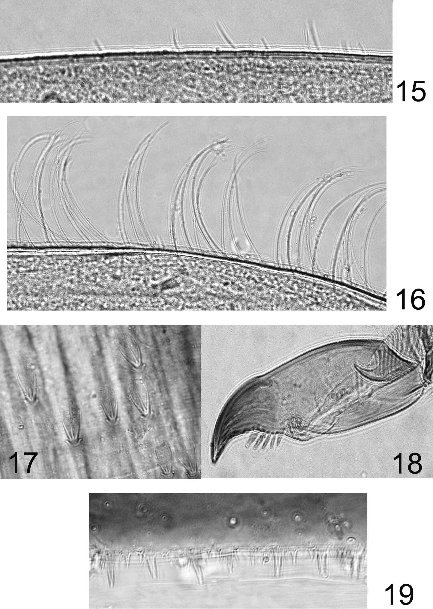
Figures 16–22.Mouthparts structure of Thalerosphyrus determinatus (20), Thalerosphyrus sinuosus (16, 21) and Thalerosphyrus lamuriensis (17, 18, 19, 22). 16–17 Hemi-labrum 18 Left mandible 19 Right mandible 20–22 Labial glossa.
-
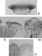
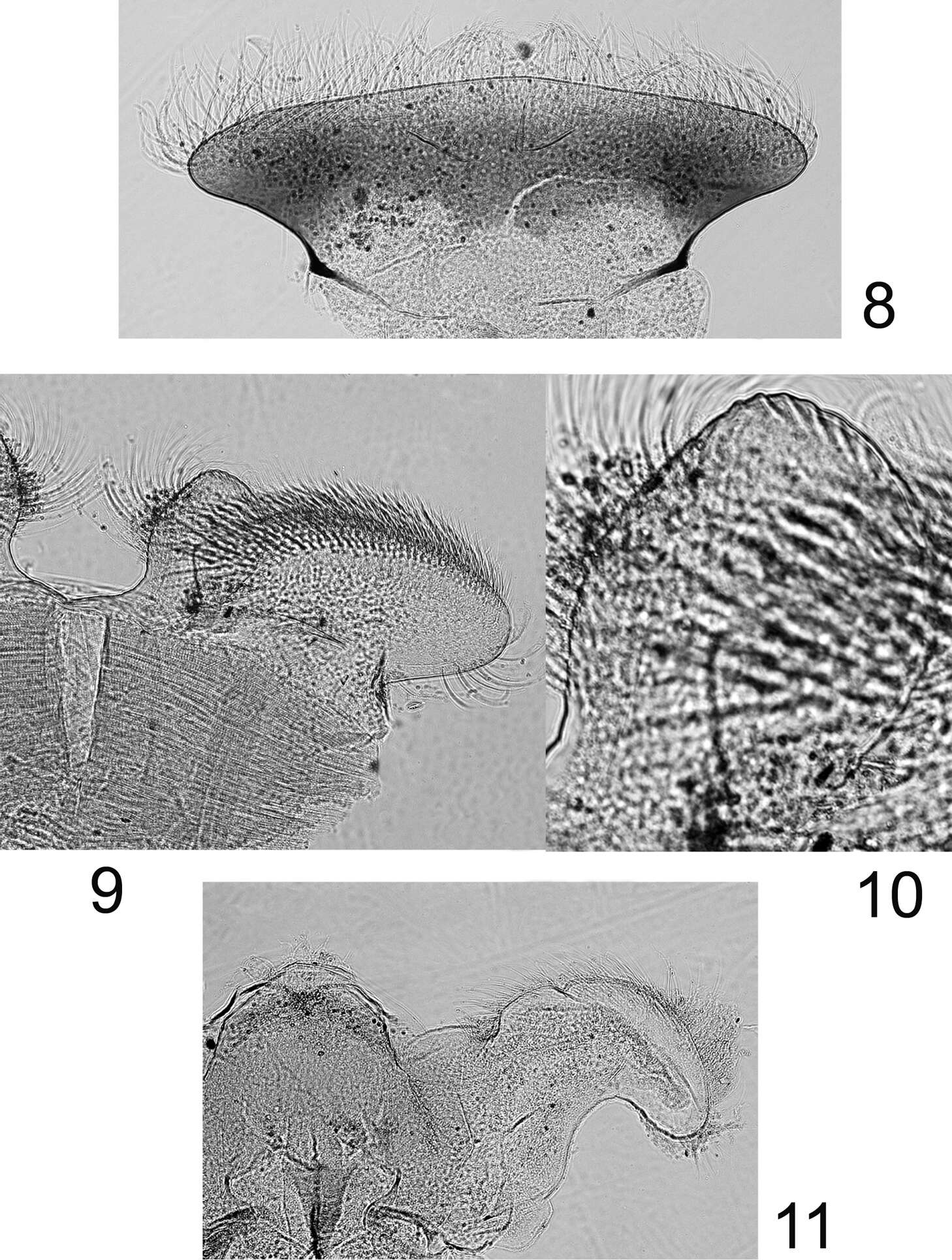
Figures 8–11.Rhithrogeniella ornata Ulmer, 1939, nymphal mouthparts. 8 Labrum in dorsal view 9 Left glossae and paraglossae of the labium 10 Detail of the glossae from 9 11 Hypopharynx, ventral view lingua and left superlingua.
-
Gudenå opstrøms Klostermølle, Mattrup Å ved Stidsmølle
-
Midtjylland
-
Boonsatien Boonsoong, Dietrich Braasch
Zookeys
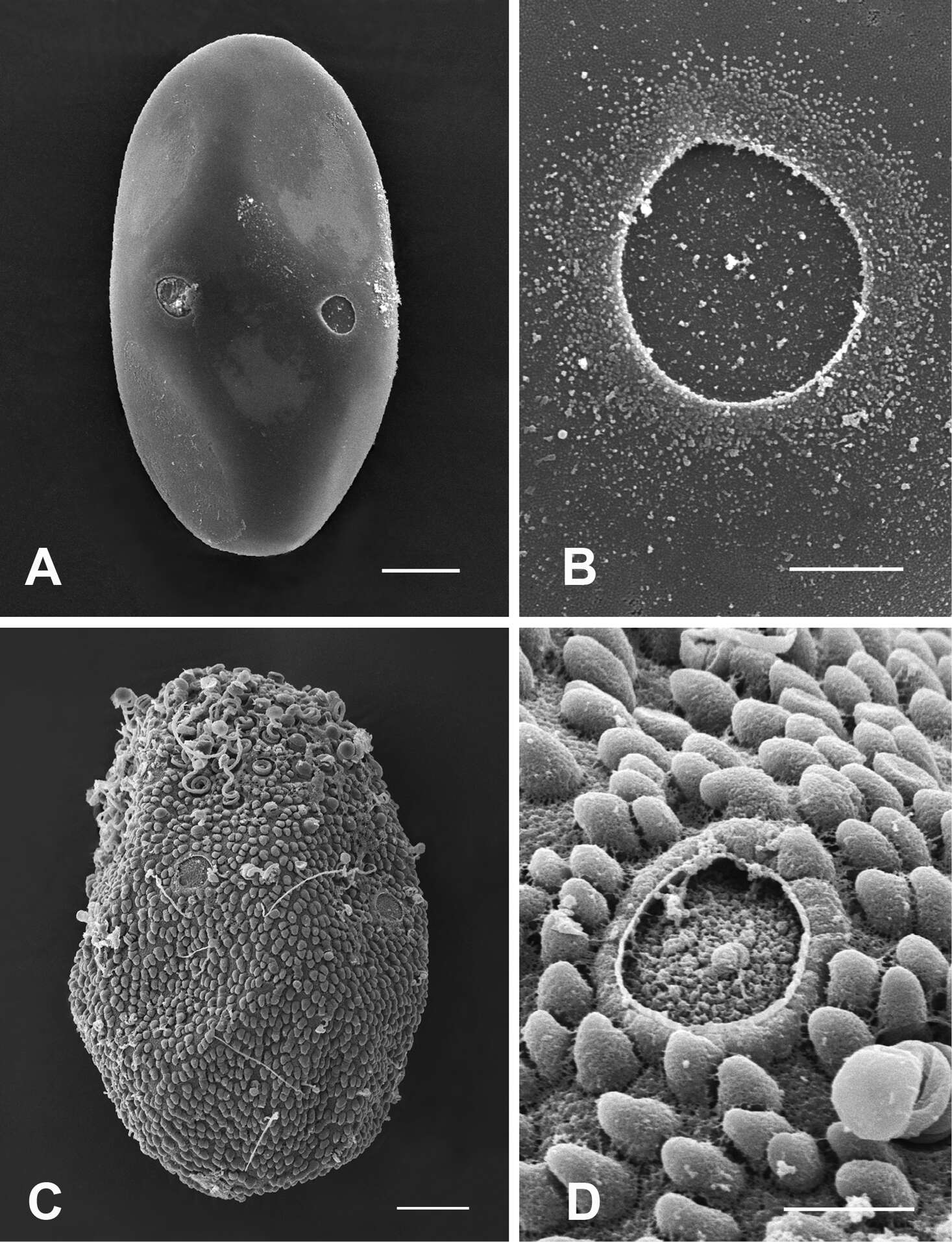
Figure 5.A–B General outline (A) and micropyle (B) of the egg of Epeorus khayengensis Boonsoong & Braasch, 2010 C-D General outline (C) and micropyle (D) of the egg of Rhithrogena siamensis Braasch & Boonsoong, 2009. Scale bars 20 µm for A and C; 5 µm for B and D.
-
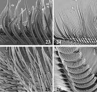
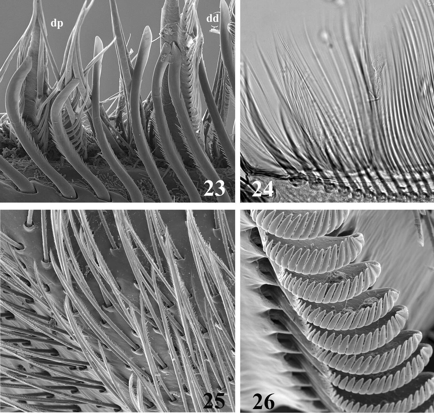
Figures 23–26.SEM (23, 25, 26) and optic (24) pictures of maxillar structure. 23–24 Dentisetae of Thalerosphyrus lamuriensis dp: proximal dentisetae, dd distal dentisetae 25 Scattered setae on the ventral face of the galea-lacinia of Thalerosphyrus sinuosus 26 Comb-shape setae on the crown of the galea-lacinia of Thalerosphyrus sinuosus.
-
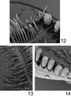
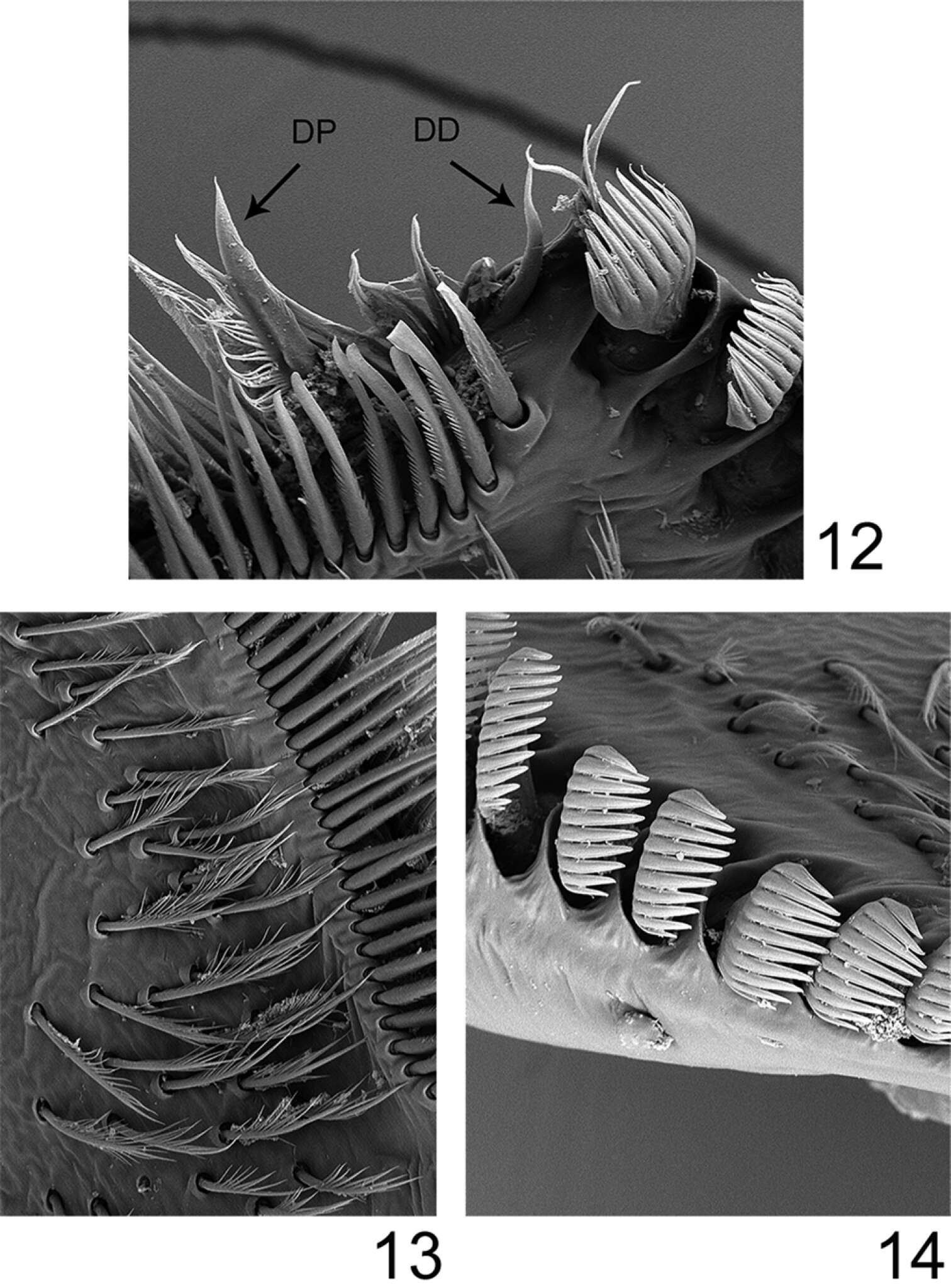
Figures 12–14.Rhithrogeniella ornata Ulmer, 1939, SEM pictures of the maxilla. 12 Dentisetae (DP: proximal dentiseta, DD: distal dentiseta) 13 Fimbriate setae on the ventral surface 14 Comb-shape setae on the crown of the galea-lacinia.
-
Vänern ved Karlstad
-
Midtjylland
-
Boonsatien Boonsoong, Dietrich Braasch
Zookeys
Figure 2.A Ventral view of abdomen of Rhithrogena siamensis Braasch & Boonsoong, 2009 B–E lamellae of gills 1 (B), 3 (C), 5 (D) and 7 (E) of Trichogenia maxillaris Braasch & Soldán, 1988 F ventral view of left maxilla of Trichogenia maxillaris Braasch & Soldán, 1988 G bristles on dorsal face of abdominal terga of Trichogenia maxillaris Braasch & Soldán, 1988 H ventral view of left maxilla of Compsoneuria langensis Braasch & Boonsoong, 2010 I-J abdominal terga (I) and tergum VII (J) of Notacanthurus baei Braasch & Boonsoong, 2009.
-
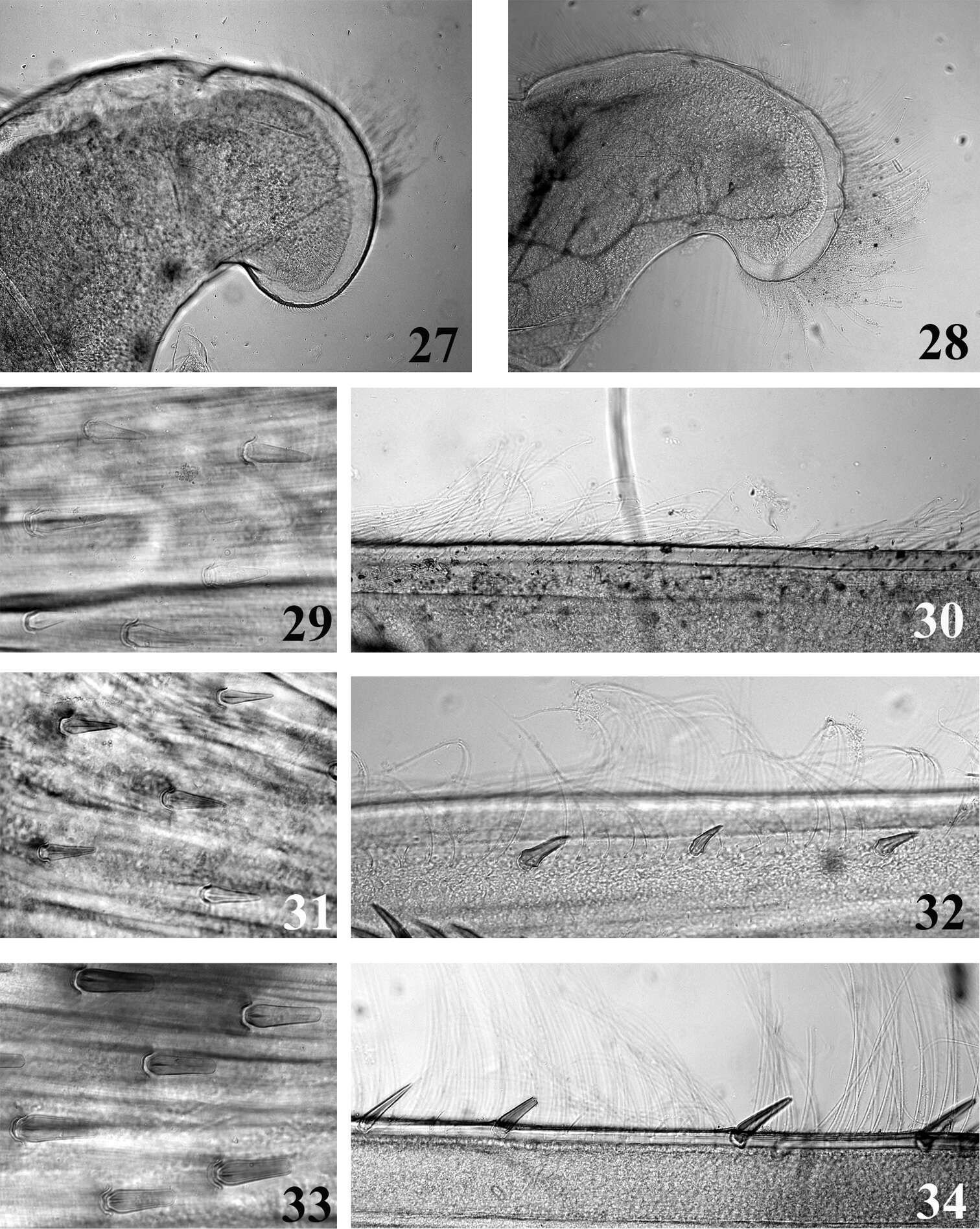
Figures 27–34.Mouthpart (27–28) and thoracic (29–34) structures of Thalerosphyrus determinatus (27, 29, 30), Thalerosphyrus sinuosus (31, 32) and Thalerosphyrus lamuriensis (28, 33, 34). 27–28 Apex of superlingua of hypopharynx 29, 31, 33 Bristles on the dorsal face of hind femur 30, 32, 34 Outer margin of hind tibia.
-
Figures 15–19.Rhithrogeniella ornata Ulmer, 1939. 15 Outer margin of the fore tibia 16 Outer margin of the hind tibia 17 Bristles on the dorsal surface of hind femur 18 Tarsal claw 19 Posterior margin of tergite V.
-
Vänern ved Karlstad
-
Midtjylland
-
Boonsatien Boonsoong, Dietrich Braasch
Zookeys
Figure 2.A Ventral view of abdomen of Rhithrogena siamensis Braasch & Boonsoong, 2009 B–E lamellae of gills 1 (B), 3 (C), 5 (D) and 7 (E) of Trichogenia maxillaris Braasch & Soldán, 1988 F ventral view of left maxilla of Trichogenia maxillaris Braasch & Soldán, 1988 G bristles on dorsal face of abdominal terga of Trichogenia maxillaris Braasch & Soldán, 1988 H ventral view of left maxilla of Compsoneuria langensis Braasch & Boonsoong, 2010 I-J abdominal terga (I) and tergum VII (J) of Notacanthurus baei Braasch & Boonsoong, 2009.
-
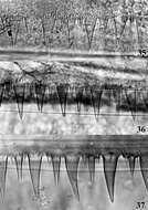
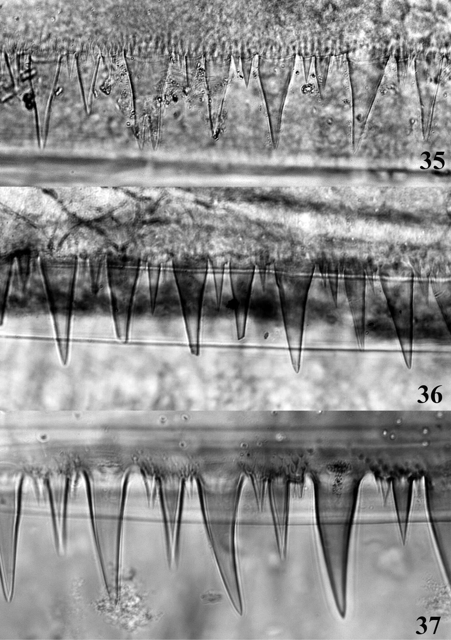
Figures 35–37.Posterior margin of abdominal tergite IV. 35 Thalerosphyrus determinatus 36 Thalerosphyrus sinuosus 37 Thalerosphyrus lamuriensis.
-
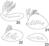
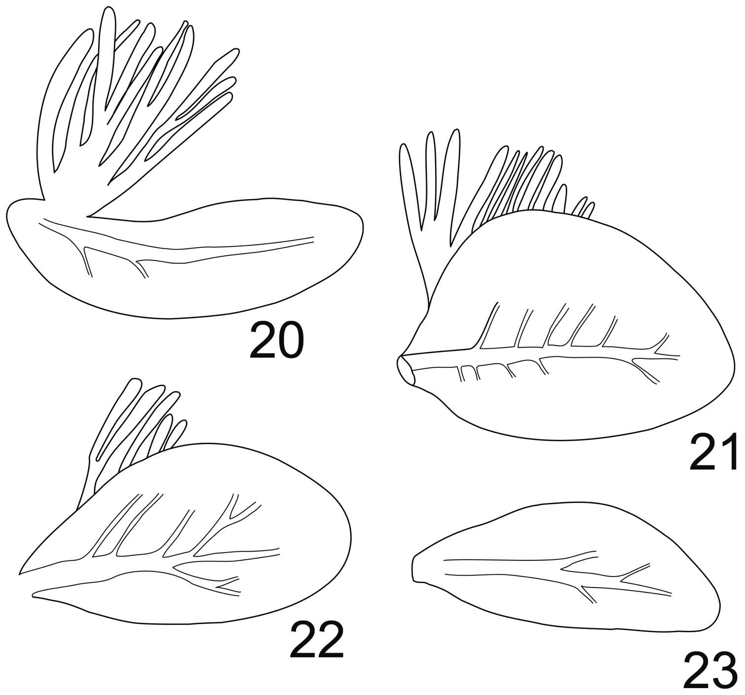
Figures 20–23.Rhithrogeniella ornata Ulmer, 1939. 20 Gill I 21 Gill IV 22 Gill VI 23 Gill VII.
-
Vänern ved Karlstad
-
Midtjylland
-
Boonsatien Boonsoong, Dietrich Braasch
Zookeys
Figure 2.A Ventral view of abdomen of Rhithrogena siamensis Braasch & Boonsoong, 2009 B–E lamellae of gills 1 (B), 3 (C), 5 (D) and 7 (E) of Trichogenia maxillaris Braasch & Soldán, 1988 F ventral view of left maxilla of Trichogenia maxillaris Braasch & Soldán, 1988 G bristles on dorsal face of abdominal terga of Trichogenia maxillaris Braasch & Soldán, 1988 H ventral view of left maxilla of Compsoneuria langensis Braasch & Boonsoong, 2010 I-J abdominal terga (I) and tergum VII (J) of Notacanthurus baei Braasch & Boonsoong, 2009.
-
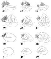
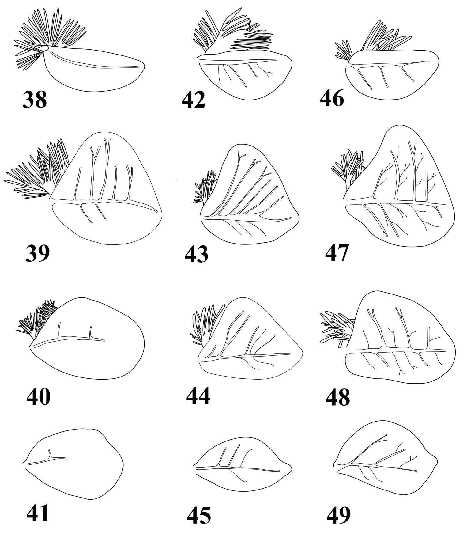
Figures 38–49.Gills of Thalerosphyrus determinatus (38–41), Thalerosphyrus sinuosus (42–45) and Thalerosphyrus lamuriensis (46–49). 38, 42, 46 Gill I 39, 43, 47 Gill IV 40, 44, 48 Gill VI 41, 45, 49 Gill VII.
-
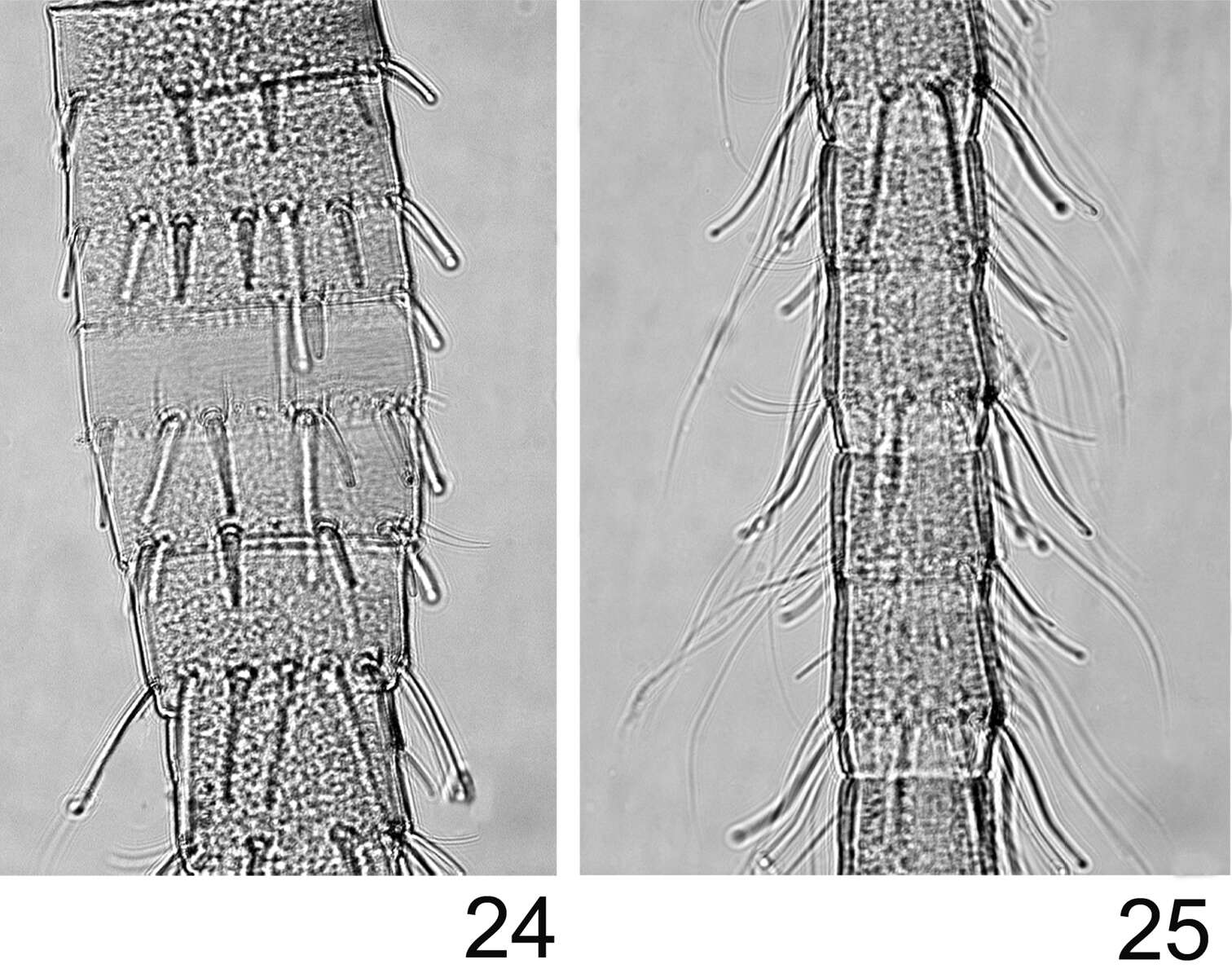
Figures 24–25.Rhithrogeniella ornata Ulmer, 1939. 24 Proximal part of the terminal filament 25 Median part of the terminal filament.
-
Gudenå v. Vilholt Mølle