Unionids use specialized structures to visually attract potential fish hosts. The combination of the statocysts and the statolith aids the mussel in maintaining equilibrium by sensing gravity. They may also be able to detect vibrations (Meglitsch & Schram 1991). Although the function of the osphradia is uncertain, some researchers believe that they detect foreign particles brought in through the inhalant aperture (Smith 2001). Drastic changes in the intensity of the light in the environment can be detected by the mantle border (Smith 2001). Glochidia can usually detect light changes with ocelli, but the eyes are generally lost after metamorphosis (Meglitsch & Schram 1991). Many mussel species also have tactile cells lining the exposed portion of the mantle, which aid in the organism's sense of touch (Meglitsch & Schram 1991). The glochidia are especially sensitive to touch, which helps in the attachment to a host as it comes close to them (Arey 1921).
Communication Channels: visual ; chemical
Other Communication Modes: mimicry
Perception Channels: visual ; tactile ; vibrations ; chemical
Muskrats are probably the most important mammal that preys on freshwater mussels (Cummings & Mayer 1992; Smith 2001). These animals drag the mussels, primarily the thin-shelled species, on the shore and either break the shells open with their teeth or leave them on the banks until the mussel dies and the shell opens (Smith 2001). In active muskrat foraging areas, there are often middens of a variety of shells which have been cleaned by the muskrats. Other common unionid predators include minks, otters, raccoons, turtles, hellbenders, fish, some species of birds, and humans (Cummings & Mayer 1992; Smith 2001; Watters 1998). Some of the common fish species include the freshwater drum, sheepshead, lake sturgeon, spotted suckers, redhorses, and pumpkinseed. Shortnose sturgeon often consume Elliptio juveniles. Catfish and sheepshead have been found preying upon the more soft-shelled species (Smith 2001). In Europe, hooded crows have been known to prey upon mussels. They are able to reach the soft tissue by dropping the mussels to crack the shell open (Watters 1998).
Aside from predators, mussels are often invaded by destructive parasites. Unionicolid mites and monogenean trematodes are often found feeding upon the mantle and branchial tissue. A chironomid midge, Baeoctenus bicolor, feeds and lays its eggs upon the demibranchs (Smith 2001) and has been known to consume up to 50% of the gill tissues (Watters 1998), which interferes with the respiration and reproduction of the mussel. Other trematodes infect the nacre causing the formation of irregular pearls and blisters and often discolor the nacre (Smith 2001). Aspidogastrean trematodes are known to infect the branchial, intestinal, and pericardinal cavities (Smith 2001). Matteson (1955) found a leech attached to the mantle cavity of a female Lampsilis siliquoidia.
To avoid these predators, mussels will bury themselves into the lake or stream sediments. Because adult unionids do not have true siphons, only openings in the mantle, they must leave the posterior margin out of the sediments to allow for sufficient respiration. This exposure leaves the organism vulnerable to predation, desiccation, and temperature extremes (Watters 1998).
Known Predators:
In general, members of the Unionidae are acephalic, have two calcium carbonate/organic shells called "valves" (bivalved) attached at the hinge by an elastic ligament. They have an umbo (beak) along the dorsal margin and slightly anterior to the hinge and are bilaterally symmetrical along a plane running between the two valves. Individuals do not have true siphons. Instead, they have two to three openings in the mantle along the posterior margin that act as the inhalant and exhalant apertures (Smith 2001). These openings are either papillated (bumpy) or crenulated (grooved) along the external margin. Under each mantle is a gill made up of two demibranchs. Each demibranch is composed of two lamellae fused at the ventral surface but open at the dorsal surface forming a “W.” Each lamella is lined vertically with compact water tubes, which are closed at the bottom but open into a larger, shared cavity at the top called the suprabranchial chamber. These water tubes are characteristic of Unionidae but not Margaritiferidae. The ax-shaped foot is found on the anterior end of the organism and between the demibranchs in the two valves. The majority of the median visceral mass in the posterior portion of the organism is primarily dorsal and not as confined in the anterior portion (Smith 2001). Unionids have a simple sensory system. Their nervous system is comprised of three pairs of ganglia: cerebropleural, pedal, and visceral. With one on each side of the esophagus, the cerebropleural ganglia are located on the posterior side of the anterior adductor muscle and are connected by a short commissure. In the foot and fused is the pair of pedal ganglia and anterior to the posterior adductor muscle is the partially fused visceral ganglia. The ganglia are connected by long commissures and each pair is the source of the nerve fibers for the surrounding organs (Smith 2001). Near the pedal ganglia is a pair of statocysts, which are ovid or spherical. These statocysts are filled with fluid and lined with sensory cells. They also contain a solid sphere called a statolith (Smith 2001). These mussels generally have closed statocysts and a single statolith (Meglitsch & Schram 1991). Osphradia are specialized epithelium concentrated in two small regions on the roof of the cloacal chamber (the posterior end of the suprabranchial chamber in the gills where it is fused) (Smith 2001). In some species, there is a spot of pigmentation near the inhalant aperture that may be photoactive (Smith 2001).
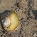
Adult unionids can range anywhere from 30 to 250 mm (Smith 2001) in length, and are just as variable in shape and color. Among the common shapes are triangular, circular, rhomboidal, quadrate, trapezoidal, and elliptical (Burch 1975). Shape is a general description; it cannot be heavily relied upon in the identification of species because it can vary among individuals of the same species. It is not uncommon to have a more inflated, rounded form of a species found in large rivers, while the larger, more compressed form of the same species is found in smaller streams and lakes where currents are not as strong. Many genera in the subfamily Ambleminae, tribe Lampsilini exhibit sexual dimorphism. In these species, the males are usually bluntly pointed or squared along the posterior-ventral margin, while females are broadly truncated. Periostracum colors vary from yellow or tan to shades of green to dark brown or black. Some have solid rays, broken rays, wavy rays, rays composed of chevrons, or even a combination of rays and spots. External shell sculpturing can also vary from one species to another and can be used to distinguish some taxa. Sculptures can be one of several combinations of ridges and bumps called "nodules" or "pustules." Not all mussel species have sculpturing. Nearly the entire Unioninae subfamily has smooth surfaces with the exception of ridges formed from the concentric growth rings. Another exterior sculpturing that is relied upon in identification is beak sculpture. Beak sculptures range from numerous fine concentric ridges to a few distinctive bars to double-looped or v-shaped ridges. In some cases, the difference in beak sculpture is the best way to distinguish between two species. Other exterior shell characteristics may include a prominent posterior ridge extending from beak to posterior-ventral margin, a unique texture to the periostracum, or a wing-like structure extending from the dorsal margin.
Aside from the exterior surface of the shell, researchers involved in identifying mussel species examine various aspects of the interior of the shell as well. In fact because of the high individual variability of the exterior, the interior characteristics are relied more heavily upon in identification. Probably the most important interior features are the size, shape, number, and orientation of the hinge teeth. Pseudocardinal teeth are situated slightly anterior to the beak and are generally short and triangular in shape. These teeth can vary from being broad and triangular to thin and elevated and are generally serrated. Orientation of the pseudocardinals refers to the angle between the pseudocardinals and the lateral teeth, which can be obtuse (angled away from the center of the shell), nearly right (vertical), or acute (angled toward the center of the shell). In some species, the pseudocardinal teeth are practically parallel with the lateral teeth. Lateral teeth are the long, slender, raised ridges posterior to the beak. These teeth can vary in length; can be straight or curved, smooth or serrated, thick or thin, and compressed or elevated. In some species, the teeth are well-developed, while in other species, the teeth are poorly-developed or reduced to a thickening along the hinge. In addition to the characteristics of the hinge teeth, mussels vary in the depth of the beak cavity, the width of the interdentum (the area between the pseudocardinal and lateral teeth along the hinge), the color of the nacre, the degree of impression of the muscle scars and pallial line (the line running parallel to the ventral margin), the thickness of the shell, the degree of inflation of the shell (the width or distance from one beak to the other when the two valves are together), the curvature of each margin, and the height of the umbo. Most species are identified by a combination of several characteristics.
Glochidia are the parasitic stage of the larvae and are generally dependent on a host to survive. Mature glochidia range from 0.05 to 0.5 mm in diameter. They are bivalves, which vary in shape from triangular, circular, oblong, or (in Potamilus only) ax head shaped and are typically attached by a single adductor muscle. Most glochidia have sensory hairs lining their mantle and a larval thread protruding from the open valves, which may allow them to attach to the host. Many species have hook-like structures to allow them to attach to the fins or skin of the fish. Those species without hooks usually attach to the gills.
Measurements are generally taken of the length, height, and width. The length is the distance from the anterior to the posterior margin. The height is the distance from dorsal to ventral margin, usually at the beak. Width is the widest point when the mussel valves are together, which is usually below the beaks. In addition, some identification keys will use the length to height ratio as a way to distinguish some species.
Other Physical Features: ectothermic ; heterothermic ; bilateral symmetry ; polymorphic
Sexual Dimorphism: sexes alike; sexes shaped differently
For small organisms, unionids are long-lived, living an average of 10 or more years (Cummings & Mayer 1992). Some genera live only 8 to 9 years, while others can live up to 10 to 15 years (Smith 2001). Given the proper conditions, many species can live up to 20 or 30 years (Watters 1998). Bauer (2001b) suggested life span is dependent upon metabolic rate. Mussels with a higher metabolic rate tend to have a shorter life span. Those unionids in larger rivers or streams would have a higher metabolic rate due to the abundance of food, and would be expected to have a short life. Unionids that thrive further upstream may have a longer lifespan because they would have adapted to a limited food supply by decreasing their metabolic rate. Although metabolic rate is a key factor affecting longevity in some species, it is not a universal constant. Some species with similar metabolic rates may have very different lifespans.
Unionids are found in various permanent freshwater sources such as lakes, streams, and rivers. The family Unionidae is not found in high mountain lakes, probably due to a lack of proper fish hosts for the glochidia or poor nutrient supply (Smith 2001). Most species are generally found where there are coarse substrates like sand or gravel (Smith 2001) however, the predictive value of substrate has been questioned (Strayer and Ralley, 1993). In Michigan, different mussel distributions may more strongly tied to surface geology in the streams (Strayer, 1983). Constantly shifting substrates or stream basins composed of solid rock have few mussels. Rivers tend to have a more abundant food supply and higher dissolved oxygen content than bodies of water with little or no current. Large rivers tend to contain a wider diversity of mussel species and larger populations than smaller streams (Cummings & Mayer 1992). Watters (1992) found as the area of a drainage basin increases, so does the fish diversity. This relationship is likely due to the increased diversity in habitat for fish. Watters (1992) also found a linear correlation between fish diversity and mussel diversity, likely due to the increase number of host fish species available.
Because the shell is primarily composed of calcium carbonate, mussels prefer an aquatic habitat with an alkaline pH, an abundance of calcium, a bound carbon dioxide content of more than 15 mg/L, and a potassium level less than 7 mg/L. Some species are able to tolerate an acidic pH for a short time, but eventually the acid will dissolve the shell and alter the internal chemistry of the visceral mass. Calcium and carbon dioxide are important for the development of the shell, and potassium appears to be toxic.
Unionids are most abundant in depths less than 2 m, but will populate waters as deep as 7 m (Smith 2001). The record depth for a Unionidae genus was 31 m. At this depth six Pyganodon grandis specimens ranging between 7 and 14 years old and less than 53 mm long were collected from Lake Michigan (Reigle 1967).
Habitat Regions: temperate ; tropical ; freshwater
Aquatic Biomes: benthic ; lakes and ponds; rivers and streams
Other Habitat Features: intertidal or littoral
Members of the family Unionidae occur in North America, Europe, Asia, Africa, and the Indonesian Archipelago (Graf and Cummings, 2002) and can thrive in tropical to temperate climates. The most diversity is in North America, where there are approximately 286 species (Turgeon et al., 1998), mainly east of the Rocky Mountains (Jennings, 2000). The nearly 300 species in North America are grouped into 49 genera, which make up two subfamilies: Unioninae and Ambleminae (Graf, 2002). The genus Elliptio is an example of this broad diversity. Only two species are found in the interior United States (Mississippi River basin), and the majority of the species (36 currently recognized) are found in the rivers of the southeastern Atlantic coastal plain (Watters, 2001a). Historical documentation describes mussels paving the beds of the Ohio and Wabash rivers (Warren, 2000).
Biogeographic Regions: nearctic (Native ); palearctic (Native ); oriental (Native ); australian (Native )
Other Geographic Terms: holarctic
Adult freshwater mussels are filter feeders; they continuously filter food particles out of the water (Watters 1998; Allen 1921). Water is constantly pumped into the inhalant aperture, through the gills, and out the exhalant aperture by cilia. The cilia lining the inner surface of the mantle, demibranchs, and visceral mass create a current by beating in a coordinated manner. Organic and inorganic particles suspended in the water surrounding the inhalant aperture are brought in by the current and caught in the mucus lining the demibranchs. The constant current created by the cilia moves the mucus with any trapped particles to the cilia lining the labial palps. The labial palps remove the inorganic particles and push them toward the ventral margin where they drop off, are move by the cilia backward, and released between the valves just below the inhalant aperture (Smith 2001). The organic particles are separated by size in sorting areas on the labial palps and are then directed into the mouth. From the mouth, particles are moved through a short esophagus to the digestive gland surrounding the stomach. Food particles enter the stomach through the subdivided pores of the large digestive gland (Meglitsch & Schram 1991). Small particles are digested intracellularly as they enter the stomach. The intestinal glands are responsible for phagocytosis, intracellular digestion, food absorption, secretion of enzymes and excretion (Meglitsch & Schram 1991). The intestine coils behind and below the stomach before it extends dorsally and empties into the mantle cavity through the anus located just above the exhalant aperture. At the anterior end of the stomach is a lateral diverticulum (groove) containing a crystalline style (an elongated structure composed of a dense gelatinous material) (Smith 2001). The style extends into the stomach and rotates clockwise by the ciliary epithelium within the style sac (Meglitsch & Schram 1991; Smith 2001). The anterior end, which is in the stomach, erodes as it rubs against the digestive shield. As is erodes, it releases the digestive enzymes, amylase, lipase, and cellulase (Meglitsch & Schram 1991). Because unionids do not ingest large particles, the ciliary action surrounding the style returns the larger particles back to the entrance of the digestive glands to be resorted. During periods of starvation, the crystalline style will disappear, but will regenerate when food is more abundant (Smith 2001). Regeneration is a slow process which occurs during low water temperatures.
The exact type of food consumed by adult freshwater mussels has been debated for some time now. Some researchers have suggested mussels eat algae and diatoms (Allen 1914), while others suggest bacteria, protozoans and other organic particles were ingested (Watters 1998). A few studies have even suggested ingesting silt somehow enhances the survival of the organism (Watters 1998). Generally, unionids feed on the bacteria and microphytoplankton but nothing larger (Smith 2001; Cummings & Mayer 1992).
The phagocytic mantle cells of the glochidia feed off of the host’s tissue (Meglitsch & Schram 1991). Before attachment, glochidia must locate a proper host. In most cases, they end up in the stream or lake sediments with the open end of the valves up awaiting a fish to brush up against the mud allowing the larvae to attach themselves to the fins. The glochidia of other species swim around in the water by clapping the valves together.
Foraging Behavior: filter-feeding
Primary Diet: planktivore ; detritivore
Like all other organisms, freshwater mussels play an important role within their ecosystem. Not only do they provide a food source for muskrats and other predators, but they also aid in the decomposition of detritus and keep the bacterial and planktonic populations under control (Pusch et al 2001; Jorgensen 1990). Dense mussel populations rely on rapid currents for survival. During periods of little or no current, these dense mussel beds can cause a depletion of the dissolved oxygen and food supply, causing a rise in the mortality rate of the mussel and other faunal populations along the basin (Jorgensen 1990).
Researchers have found that the glochidia generally do not cause sufficient enough damage to the host to cause problems. Cases of over 3000 glochidia infecting a fish without apparent harm have been reported. However, there have also been cases where 30 mm fingerling trout have died of secondary bacterial infections caused by a little more than 100 glochidia (Smith 2001). Some fish species are able to develop an immune response to resist the glochidia causing them to pre-maturely drop off the fish.
Ecosystem Impact: parasite
Species Used as Host:
Commensal/Parasitic Species:
Humans have used freshwater mussels and their shells for food, jewelry, tools, utensils, and pottery temper for thousands of years (Cummings & Mayer 1992). Native Americans have been carving shells for implements and ornamentation for at least 3000 years. Around 1000 years ago, people in North America discovered that tempering their pottery with crushed shells rather than sand or gravel allowed them to create a smoother, thinner vessel. During this same period, people were creating beads, hoes and spoons with freshwater mussel shells (Wiant 2000).
Before 1890, freshwater mussels were used for only a few decorative items such as pistol grips, brush handles, and jewelry. Both U.S. tariffs on imported goods (including buttons) and the rise of the new ready-to-wear clothing industry created high demand for buttons. The pearl button industry began in 1891 with the start of a new fashion trend to use shell buttons to fasten clothes. With Muscatine, Iowa as the center of the industry, pearl button production became a major industry for hundreds of river towns along the Mississippi and other Midwestern rivers. Thousands of tons of mussels were harvested. The demand was so high that by 1900 the Illinois and Wabash rivers were depleted of mussels. The peak of the industry occurred in 1909 with a record of 2600 boats on the Mississippi River alone. By the 1940s and 1950s, cheaper plastic buttons became widely available and replaced the shell buttons, causing the collapse of this industry and the recovery of many depleted mussel populations.
In the 1950s, the Japanese pearl growers developed another use for freshwater mussel shells (Cummings & Mayer 1992). They discovered that small beads could be carved out of the shells of freshwater mussels and inserted into oysters to artificially form pearls. They also found that certain species, particularly the threeridge and the washboard, were easier to manipulate and created a better nucleus. These discoveries were the beginning of the cultured pearl industry. Today, thousands of tons of freshwater mussel shells from North America are exported to Japan to support the pearl industry (Cummings & Mayer 1992).
In addition to the many products, freshwater mussels act as water quality indicators. Because they are filter-feeders, pollutants in the water will accumulate in the tissue of mussels until they reach a toxic level killing the organism. A drastic drop in the mussel population is an indication of poor water quality.
Positive Impacts: food ; body parts are source of valuable material; research and education
There are no known adverse effects of unionid mussels on humans.
The family Unionidae is commonly referred to as pearly mussels, naiads, or unionids. Although no full accounts for the family Unionidae exist and the exact number is currently disputed, researchers agree that the order Unionoida includes around 1,000 species worldwide (Bauer 2001a). Charles Torrey Simpson described 1,172 species in 1900 and 1,337 in 1914. A more recent account by Fritz Haas (1969) combined over 4,000 names into just 837 recognized species (Graf & Cummings 2002).
The Unionidae are acephalic (no head), bivalved mollusks usually with the beak (the elevated portion of the dorsal margin) slightly anterior. When present, the pseudocardinal teeth are generally anterior to the beak. The lateral teeth, generally posterior to the beak, are parallel to the hinge line. The species in this family have a foot rather than a byssus, fibrous structures found in other mussel families. Along with Margaritiferidae, another family included in the order Unionoida, Unionidae does not have true siphons (True siphons are formed when tissues between the inhalent and exhalent openings are fused and mantle aperatures are elongated). Unlike the family Margaritiferidae, the inhalant aperture (opening in the posterior end of the mantle border where water enters the mussel) of Unionidae has unbranched papillae (bumps). Individuals vary in shape, size and coloration. Adult individuals can range from 30 to 250 mm.
Embryonic unionids develop within the marsupia, or specialized portions of the gills, of the female. Once fully developed, they are released from the female and must attach to the gills or fins of a fish host within a few days or they will die. Strophitus undulatus and Utterbackia imbecillis are the only two species capable of direct development without a host (Watters 1994c). Only one species, Simpsonaias ambigua, metamorphoses on a salamander, Necturus maculosus. Many unionids are species-specific, requiring one or a narrow range of species. If attached to the wrong species, the glochidia will die as a result of the fish's immune system response (Watters 1998). Within a couple of days, the hosts’ dermal tissue will encapsulate each glochidium forming a nodular cyst. While encysted, the glochidia will metamorphose, allowing the organs to develop more like an adult’s organs (Meglitsch & Schram 1991). There is a mortality rate of over 99.99% from the time the glochidia are released from the mother to the time in which the metamorphosed juveniles settle in the sediments (Jansen et al 2001).
After an average of 10-30 days (the record is 190 days), the metamorphosis will be complete and the glochidia will break from the cysts and drop from the host. The third and final stage of development occurs in the sediments of the stream or lake and may last anywhere from one to eight years before the juvenile is sexually mature. In this juvenile stage, the young mussel will complete its internal development, create the adult shell, and begin to live independently in the stream or lake.
As in most bivalves, the shell is composed of three layers: the periostracum, the prismatic layer, and the nacre. The periostracum is the outermost layer and is composed of an organic material. The prismatic layer is the middle layer and is composed of thin blocks of a prism-like calcium carbonate, which are oriented perpendicular to the mantle and the other two layers. The nacre, or mother of pearl, is the innermost layer, which is composed of thin, alternating, laminae (flakes or sheets) of calcium carbonate and an organic material (Smith 2001). The mantle is responsible for generating new shell as the mussel ages. A mantle flap is pressed against the interior of each valve and ends in three folds. The periostracum forms at the outer margin and the prismatic layer forms at the outer border. The nacre forms along the entire surface of the mantle. Muscle scars form where the muscle attaches to the shell, disrupting the formation of the nacre. Instead of the shell forming along the dorsal edge where the hinge is located, an elastic hinge ligament composed of conchiolin (a protein-rich substance) forms, binding the two valves together (Meglitsch & Schram 1991).
Growth of the mussel begins at the elevated portion called the umbo or beak. Because new shell is added along the entire edge of the mantle, concentric rings form around the beak. In some species, these rings may be grouped closer together in some areas than others, forming ridges. These ridges indicate the period of diapause during the winter or unfavorable environmental conditions, such as lower water level or lack of food. The period of growth in northern populations is typically from April to September. The growth rate depends mostly on environmental conditions such as water temperature, food supply, and the chemical composition of the water. Many mussel species are capable of growing 30 to 80 mm every two growing seasons.
Development - Life Cycle: metamorphosis
Worldwide, freshwater mussels are one of the most endangered groups with significant population declines documented in recent surveys. In the United States alone, nearly 70 species are either endangered or threatened (USFWS 2003). Reasons for the past decline include the effects of the pearl button industry of the late 19th and early 20th centuries and the cultured pearl industry of the past 50 years. Today, siltation from agriculture, forestry, and construction smothers the organisms inhibiting feeding and respiration. Impoundments alter the habitat, killing first the mussels that thrive in rapid currents. Dams cause an increase in silt as well as a constant cold water temperature. Since many mussel species are temperature sensitive, the cold will slow the growth and may inhibit the reproduction of the mussels that survived the initial shock of the construction. In-stream sand and gravel mining often buries, crushes, or removes the mussels in the substrate and releases silt, which affects the species downstream. Agricultural runoff is another threat to mussel populations. Many species cannot tolerate pollutants introduced in the water from pesticides, herbicides, and fertilizers. At sub-lethal concentrations these chemicals inhibit respiration and accumulate in the tissues of the organism. Mussels are also sensitive to heavy metals which accumulate in the tissues. Mine runoff creates an acidic pH in the water, which many mussel species cannot tolerate for long periods of time. Salinity from road salt runoff is lethal to glochidia.
In addition to industrial wastes and depletion, mussels now compete for resources with introduced species. The Asian clam and the zebra mussel are probably the two most common exotic species, which have been introduced to North American freshwaters.
The study of freshwater mussels as a group is made more complicated by a particularly high rate of duplicate species names. Many 19th century biologists didn't realize that members of the same mussel species grow somewhat differently-shaped shells depending on the conditions of the stream or lake they are growing in. The biologists described new species based on shell shape, and consequently there are many hundreds of duplicate names for the same species.
A few species are occasionally or permanently simultaneous hermaphrodites (Bauer 1987), but in most cases, unionid sexes are separate. Bauer (1987) suggested that hermaphroditism occurs when the population density is low or gene flow is limited. In these cases, the female is the only one of the two sexes that can become hermaphroditic. Despite the dioecious nature of most mussels, males and females do not make contact with each other. Males produce sperm year round and release during the time of year when females ovulate (Matteson 1948). This simultaneous release of gametes may be triggered by a change in the water temperature and the intensity of light in the environment. The male’s sperm leaves the suprabranchial chamber of each demibranch and exits the organism through the exhalant aperture to be carried by the water current to a nearby female. Because sperm cannot swim against the current, the receiving female must be downstream (Watters 1994a). The sperm enters the female through the inhalant aperture and fertilizes the eggs stored in the water tubes of the demibranch’s lamellae (Smith 2001).
Mating System: polygynandrous (promiscuous)
Depending on the species, sexual maturity is reached between one and eight years (Smith 2001). Gamete production is initiated by a change in the water temperature surrounding the mussel (Watters 1998). Annual gametogenesis and gravidity may occur throughout the year or during certain seasons depending on latitude. The more northern populations tend to be gravid for a few months or all winter long and release the glochidia in the spring. There are a few species that release the glochidia in the fall. In many cases, southern populations are not restricted to reproducing during certain seasons. The number of larvae developing in one female at a time may range from several thousand in some of the smaller Unionidae genera to possibly over 1 million. The maximum amount of glochidia in one female is unknown, but Tankersley and Dimock (1992) recorded nearly 1 million in a Pyganodon cataracta female. The closest related family, Margaritiferidae, contains species which have produced more than 3 million per individual (Smith 2001). Bradytictic (long term) breeders will maintain the glochidia within the marsupia, the specialized portions of the gills, until the following spring or summer before releasing them into the water. Tachytictic (short term) breeders will release the glochidia in the same year, usually by July or August (Watters 1998). Matteson (1948) was convinced that the membrane surrounding the developing embryos provides all of the necessary nutrients, rather than the female transferring food to the developing young. His conclusion was based on a lack of connective structure from the gills to the young and that the fertilization membrane surrounding each embryo, which prevents the passing of any materials, remains until development is complete.
Key Reproductive Features: iteroparous ; seasonal breeding ; year-round breeding ; gonochoric/gonochoristic/dioecious (sexes separate); simultaneous hermaphrodite; sexual ; fertilization (Internal ); ovoviviparous
Unionid embryos spend the first stage of development in the marsupial portion of the female unionid's gills, where they develop into glochidia, the parasitic stage. Once the first stage is complete, usually in the spring, the female will release the glochidia into the water to begin the second stage as a parasite. Because glochidial mortality is high, many unionids have developed specialized methods of attracting fish to the mother before the glochidia are released, increasing the chances the larvae can attach to a host. Some of these species extend the glochidia encapsulated in conglutinates (Chamberlain 1934). These conglutinates (sacs) are attached to the parent organism and move in the current like worms. This encapsulated appendage acts as a lure to attract the host fish, which then eats the glochidia freeing them from the capsule and allowing them to attach to the gills of the fish. Other species use a modified mantle flap to attract the fish. This flap mimics the prey of the potential host fish. The glochidia are sensitive enough to attach themselves to the fish as soon as contact is made.
Parental Investment: pre-fertilization (Provisioning, Protecting: Female); pre-hatching/birth (Provisioning: Female, Protecting: Female)
The Unionidae are a family of freshwater mussels, the largest in the order Unionida, the bivalve molluscs sometimes known as river mussels, or simply as unionids.[1][2]
The range of distribution for this family is world-wide. It is at its most diverse in North America, with about 297 recognised taxa,[3][4][5] but China and Southeast Asia also support very diverse faunas.
Freshwater mussels occupy a wide range of habitats, but most often occupy lotic waters, i.e. flowing water such as rivers, streams and creeks.
The recent phylogenetic study reveals that the Unionidae most likely originated in Southeast and East Asia in the Jurassic, with the earliest expansions into North America and Africa (since the mid-Cretaceous) followed by the colonization of Europe and India (since the Paleocene).[6]
Unionidae burrow into the substrate, with their posterior margins exposed. They pump water through the incurrent aperture, obtaining oxygen and food. They remove phytoplankton and zooplankton, as well as suspended bacteria, fungal spores, and dissolved organic matter.[7][8][9][10][11][12][13][14][15][16] Despite extensive laboratory studies, which of these filtrates unionoids actually process remains uncertain. In high densities, they have the ability to influence water clarity [17][18] but filtration rates are dependent on water temperature, current velocity, and particle size and concentration. In addition, gill morphology can determine particle size filtered, as well as the rate.[11]
Unionidae are distinguished by a unique and complex lifecycle. Most unionids are of separate sex, although some species, such as Elliptio complanata, are known to be hermaphroditic.[19]
The sperm is ejected from the mantle cavity through the male's excurrent aperture and taken into the female's mantle cavity through the incurrent aperture. Fertilised eggs move from the gonads to the gills (marsupia) where they further ripen and metamorph into glochidia, the first larval stage. Mature glochidia are released by the female and then attach to the gills, fins, or skin of a host fish. A cyst is quickly formed around the glochidia, and they stay on the fish for several weeks or months before they fall off as juvenile mussels, which then bury themselves in the sediment.
Some of the species in the Unionidae, commonly known as pocketbook mussels, have evolved a remarkable reproductive strategy. The edge of the female's body that protrudes from the valves of the shell develops into an imitation of a small fish complete with markings and false eyes. This decoy moves in the current and attracts the attention of real fish. Some fish see the decoy as prey, while others see a conspecific, i.e. a member of their own species. Whatever they see, they approach for a closer look and the mussel releases huge numbers of larvae from her gills, dousing the inquisitive fish with her tiny, parasitic young. These glochidial larvae are drawn into the fish's gills, where they attach and trigger a tissue response that forms a small cyst in which the young mussel resides. It feeds by breaking down and digesting the tissue of the fish within the cyst.[20]
Sex is determined by a region located on the mitochondrial DNA, the male open reading frame (M-ORF) and female open-reading frame (F-ORF). Hermaphroditic mussels lack these regions and contain a female-like open-reading frame dubbed hermaphroditic open-reading frame (H-ORF). In many mussels, the hermaphroditic state is ancestral and the male sex evolved later. This region of the mitochondria also may be responsible for the evolution of doubly uniparental inheritance seen in freshwater mussels.[21]
The following classification is based on MolluscaBase and the MUSSEL Project database:[22][23]
In large enough quantities, unionid shells can have enough of an impact on environmental conditions to affect the ability of organic remains in the local environment to fossilize.[24] For example, in the Dinosaur Park Formation, fossil hadrosaur eggshell is rare[24] because the breakdown of tannins from local coniferous vegetation would have caused the ancient waters to become acidic.[24] Eggshell fragments are present in only two microfossil sites, both of which are dominated by the preserved shells of invertebrate life, including unionids.[24] The slow dissolution of these shells releasing calcium carbonate into the water raised the water's pH high enough to prevent the eggshell fragments from dissolving before they could be fossilized.[24]
{{cite journal}}: CS1 maint: multiple names: authors list (link) The Unionidae are a family of freshwater mussels, the largest in the order Unionida, the bivalve molluscs sometimes known as river mussels, or simply as unionids.
The range of distribution for this family is world-wide. It is at its most diverse in North America, with about 297 recognised taxa, but China and Southeast Asia also support very diverse faunas.
Freshwater mussels occupy a wide range of habitats, but most often occupy lotic waters, i.e. flowing water such as rivers, streams and creeks.