-
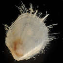
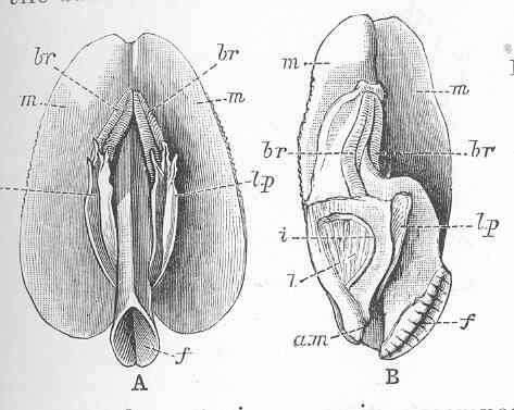
Yoldia limatula Say, Greenland, showing the short plumed branchiae (br,br), the gasteropodous foot (f'), and the large labial palpa (l,p, l,p).
-
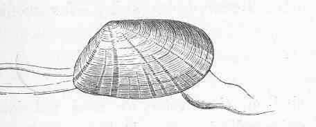
Donax trunculus (Linnaeus).
-
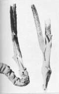
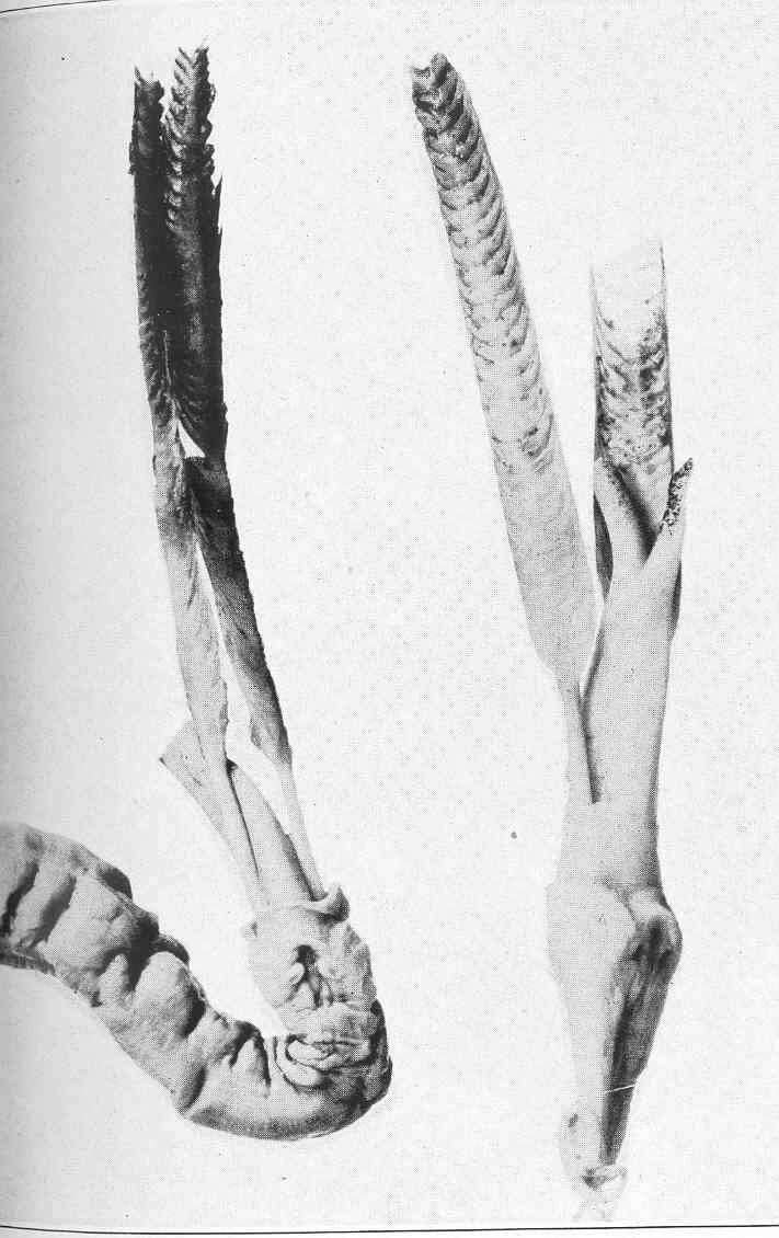
Pallets and siphons of Xylotrya sciacca Tryon.
-
Shells of Xylotrya sciacca Tryon.
-
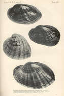
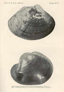
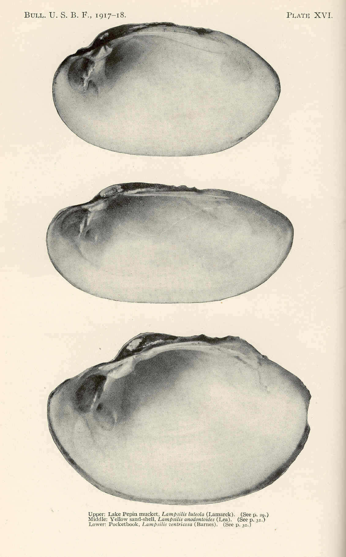
Lake Pepin mucket, Lampsilis luteola (Lamarck), upper; Yellow sand-shell, Lampsilis anodontoides (Lea), middle; Pocketbook, Lampsilis ventricosa (Barnes), lower.
-
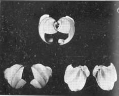
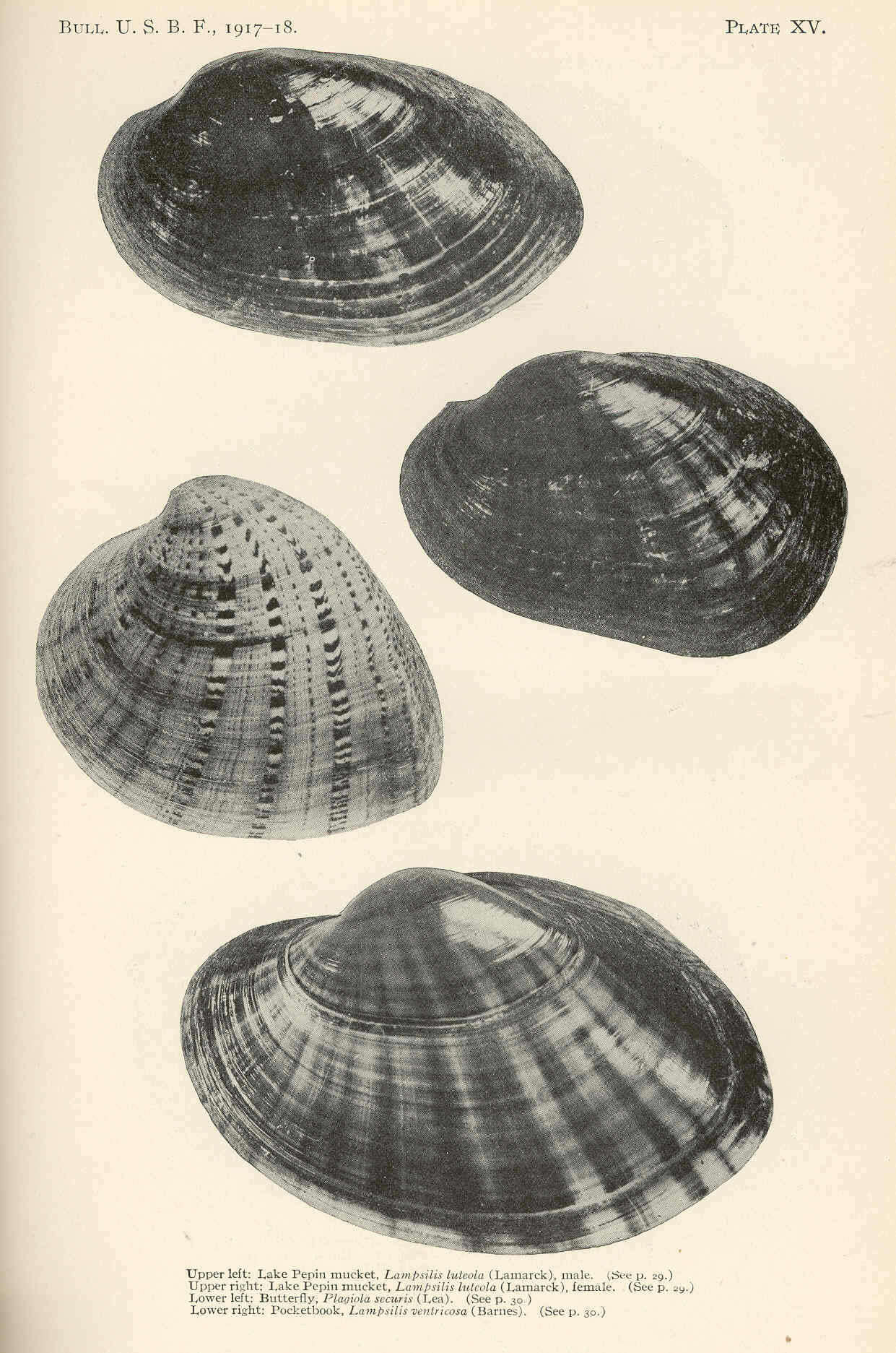
Lape Pepin mucket, Lampsilis luteola (Lamarck), male, upper left; Lake Pepin mucket, Lampsilis luteola (Lamarck), female, upper right; Butterfly, Plagiola securis (Lea), lower left); Pocketbook, Lampsilis ventricosa (Barnes), lower right. 1921. Lampsilis;
cc-publicdomain
Lape Pepin mucket, Lampsilis luteola (Lamarck), male, upper left; Lake Pepin mucket, Lampsilis luteola (Lamarck), female, upper right; Butterfly, Plagiola securis (Lea), lower left); Pocketbook, Lampsilis ventricosa (Barnes), lower right.
-
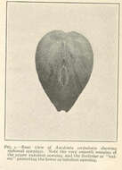
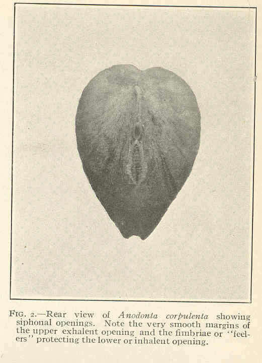
Rear view of Anodonta corpulenta showing siphonal openings. Note the very smooth margins of the upper exhalent opening and the fimbriae or feelers protecting the lower or inhalent opening.
-
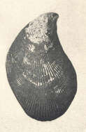
Modiola nigra.
-
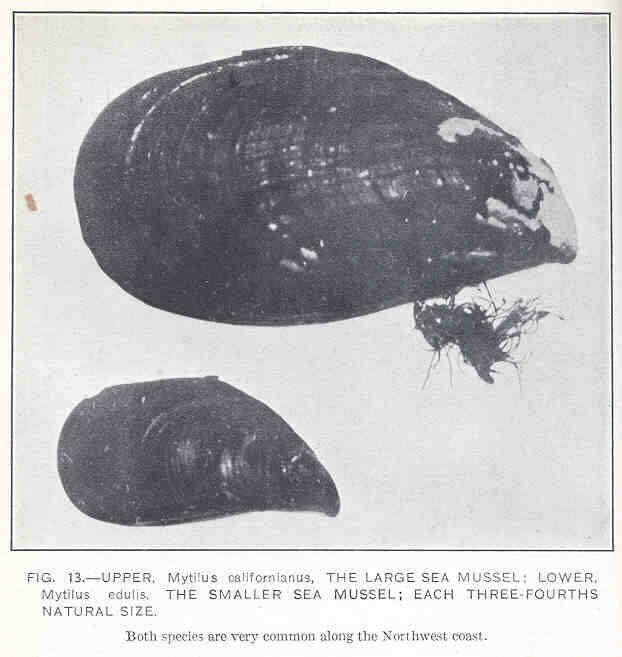
Mytilus californianus, the Large Sea Mussel (Upper); Mytilus edulis, the Smaller Sea Mussel (Lower).
-
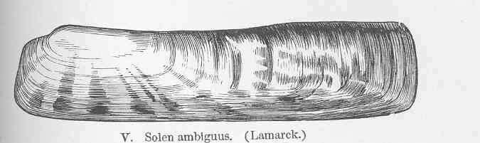
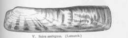
Solen ambiguns (Lamarck).
-
Mytilus hamatus.
-
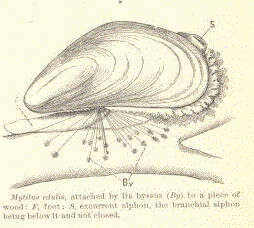
Mytilus edulis, attached by its byssus (By) to a piece of wood: F, food; S, exeurrent siphon, the branchial siphon being below it and not closed.
-
Grandma, Lampsilis ovata (Say), from Cumberland River, upper; Pocketbook, Lampsilis capax (Green), from Mississippi River, lower.
-
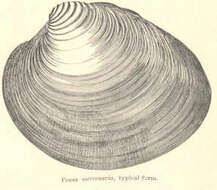
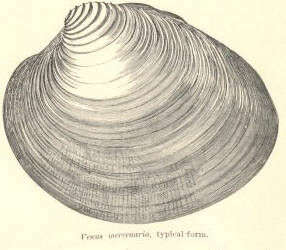
Venus mercenaria.
-
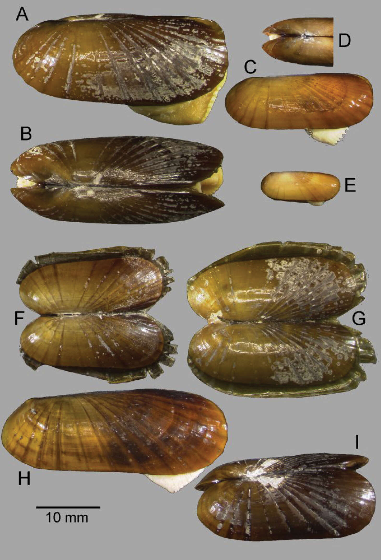
Graham Olive, Clara F. Rodrigues, Marina R. Cunha
Zookeys
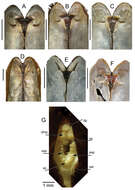
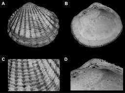
Figure 2.Solemya (Petrasma) elarraichensis sp. n. A–E from Kidd MV; A–B lateral and dorsal views of holotype C–D lateral and dorsal views of medium sized paratype E lateral view of small paratype. F paired valves from Pen Duick Escarpment G paired valves from Mercator MV H lateral view of specimen from Meknès MV I lateral view of shell from Yuma MV.
-
Graham Olive, Clara F. Rodrigues, Marina R. Cunha
Zookeys
Figure 3. A–FInternal views of ligament, scale bars = 5mm. A–D Solemya (Petrasma) elarraichensis sp. n. from A Kidd MV B Pen Duick Escarpment C Mercator MV D Yuma MV. E Solemya togata, Mediterranean F Solemya (Petrasma) velum, Rhode Island (from Taylor et al. 2009). c, chondrophore; cr chondrophore ridge; pa, posterior adductor scar; r, resilium. G posterior siphon of Solemya (Petrasma) elarraichensis. dp, dorsal papilla; dmp, dorsal marginal papillae; psp, primary siphonal papillae; sa, smooth area; ssp, secondary siphonal papillae; ssr, subsiphonal ridge; vp, ventral papilla.
-
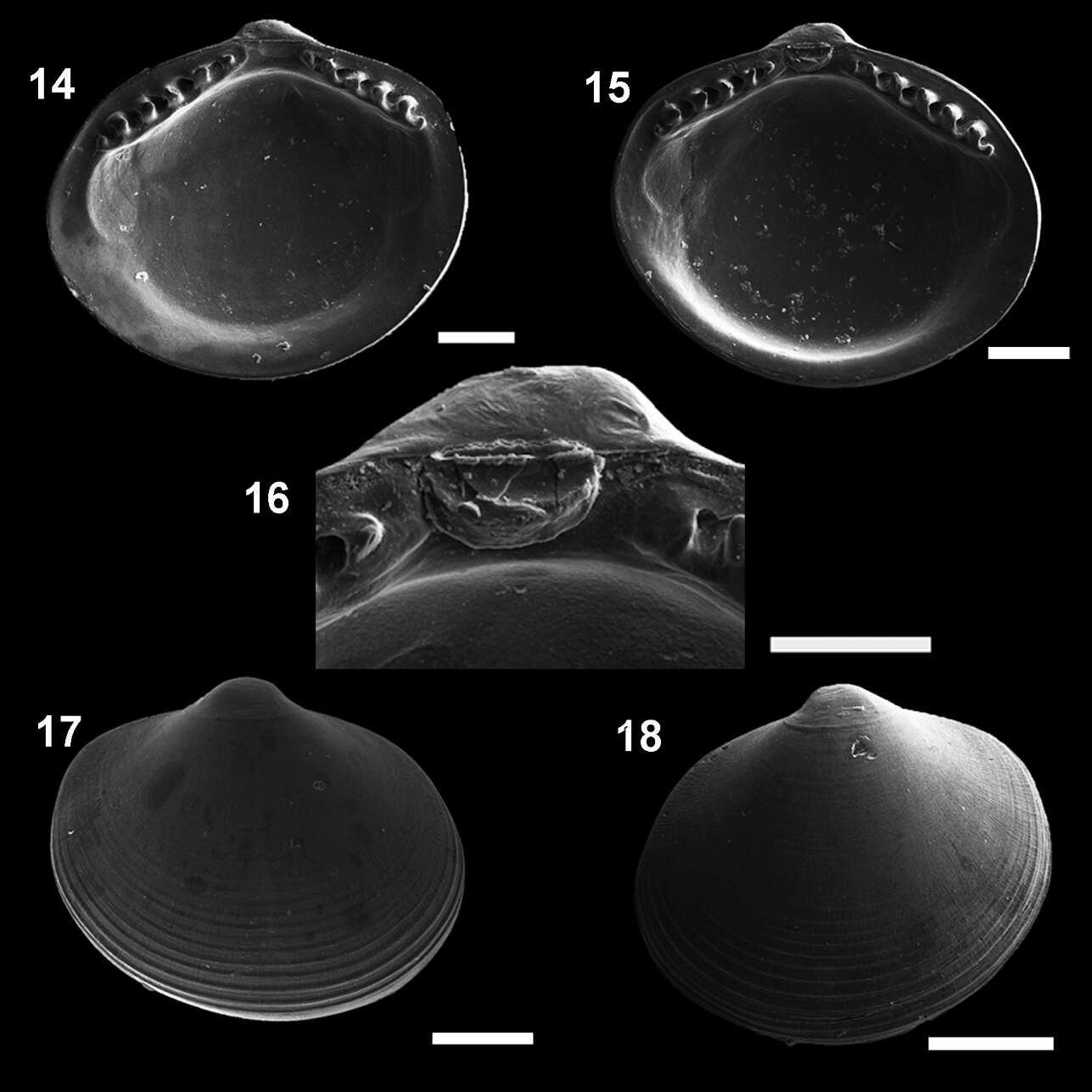
Natalia Pereira Benaim, Ricardo Silva Absalão
Zookeys
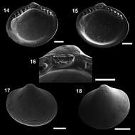
Figures 14–18.Microgloma macaron sp. n. Internal view, right valve 14 left valve 15 detail of the hinge plate and ligament 16 External view, right valve 17 left valve 18 Holotype MNRJ 19112 (14,16,18). Paratype IBUFRJ 15297 15, 17 Scale bars: 14, 16= 200 µm; 15, 18 = 300 µm; 17 = 250 µm.
-
Natalia Pereira Benaim, Ricardo Silva Absalão
Zookeys
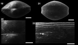
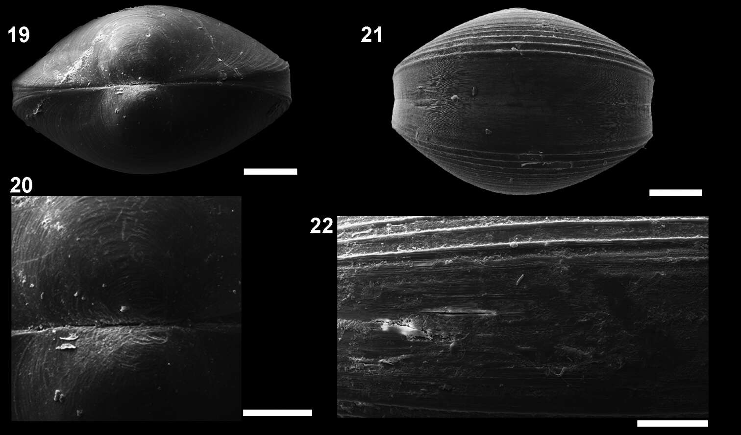
Figures 19–22.Microgloma macaron sp. n. Dorsal view 19 prodissoconch 20 (IBUFRJ 15297). Ventral margin view, extended margin 21 periostracum of the ventral margin 22 (IBUFRJ 19177). Scale bars: 19, 21 = 200 µm; 20, 22 = 100 µm.
-
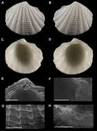
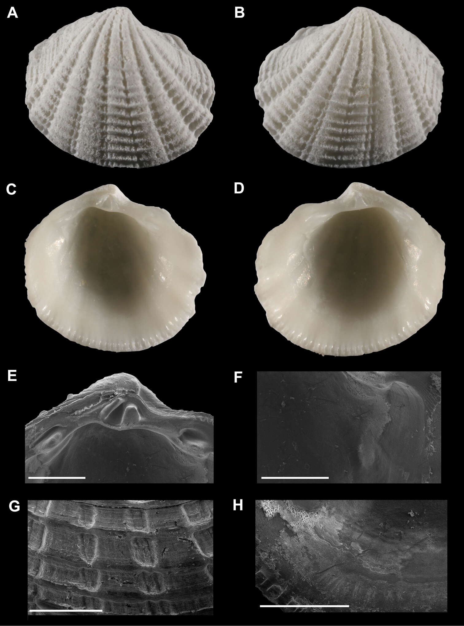
Figure 1.A–D Radiolucina amianta (SBMNH 357639, USA, Florida, Santa Petersburg, Tampa Bay) length = 5.4 mm A Exterior of right valve B Exterior of left valve C Interior of left valve D Interior of right valve E Close up of hinge of left valve F Close up of anterior adductor muscle scar of left valve G Close up of ribs of right valve H Close up of pallial line of left valve. E–H scale bar = 1 mm.
-
Figure 4.A–D Phacoides (Bellucina) amiantus Dall, 1901lectotype herein(USNM 64276, Mexico, Yucatan Strait) length = 6 mm A Exterior of right valve B Interior of right valve C Close up of ribs of right valve D Close up of hinge of right valve.
-
Bárbara L. V. Romera, Luiz R. L. Simone, Carlo M. Cunha
Zookeys
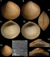
Figures 1–6.Diplodonta portesiana. Holotype (NHMUK 1854.12.4.770; L: 20 mm; H: 17 mm). 1 Left valve, external view 2 Right valve, external view 3 Left valve, internal view 4 Right valve, internal view 5 Left hinge detail 6 Right hinge detail. Scale: 2 mm.
-
Bárbara L. V. Romera, Luiz R. L. Simone, Carlo M. Cunha
Zookeys
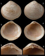
Figures 7–14.Specimen of Diplodonta portesiana (MZSP 22747, L: 13.4 mm, H: 13.1 mm, width: 7.7 mm). 7 Left valve, external view 8 Right valve, external view 9 Left valve, internal view 10 Right valve, internal view 11 Dorsal view 12 Left hinge detail 13 Right hinge detail 14 External surface of shell under SEM; Scale: 2 mm, except 14: 200 µm.
-
Paul Valentich-Scott, Diarmaid Ó Foighil, Jingchun Li
Zookeys
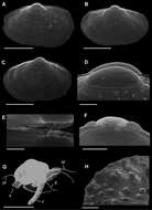
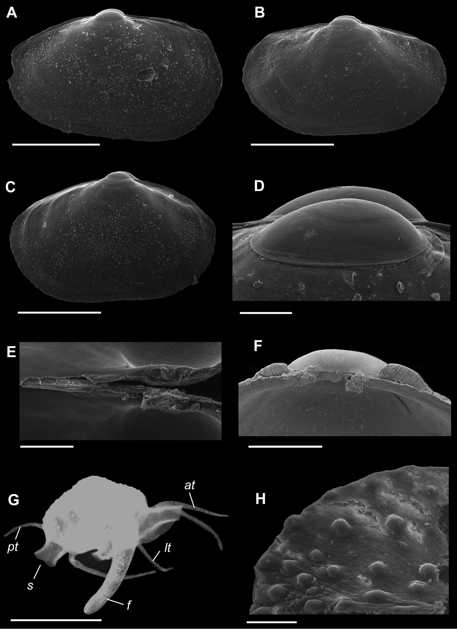
Figure 1.A–H Waldo arthuri new species A–E paratypes, SBMNH 149934 A–C Exterior of left valve D Prodissoconch E Close up of hinge of both valves F Close up of hinge of right valve G Live animal with extended mantle and mantle tentacles; posterior mantle tentacle (pt); siphon (s), foot (f), lateral mantle tentacle (lt), anterior mantle tentacle (at) H Detail of mantle papillae. A–C, G scale bar = 1 mm; D–F, H scale bar = 100 µm.
-
Paul Valentich-Scott, Diarmaid Ó Foighil, Jingchun Li
Zookeys
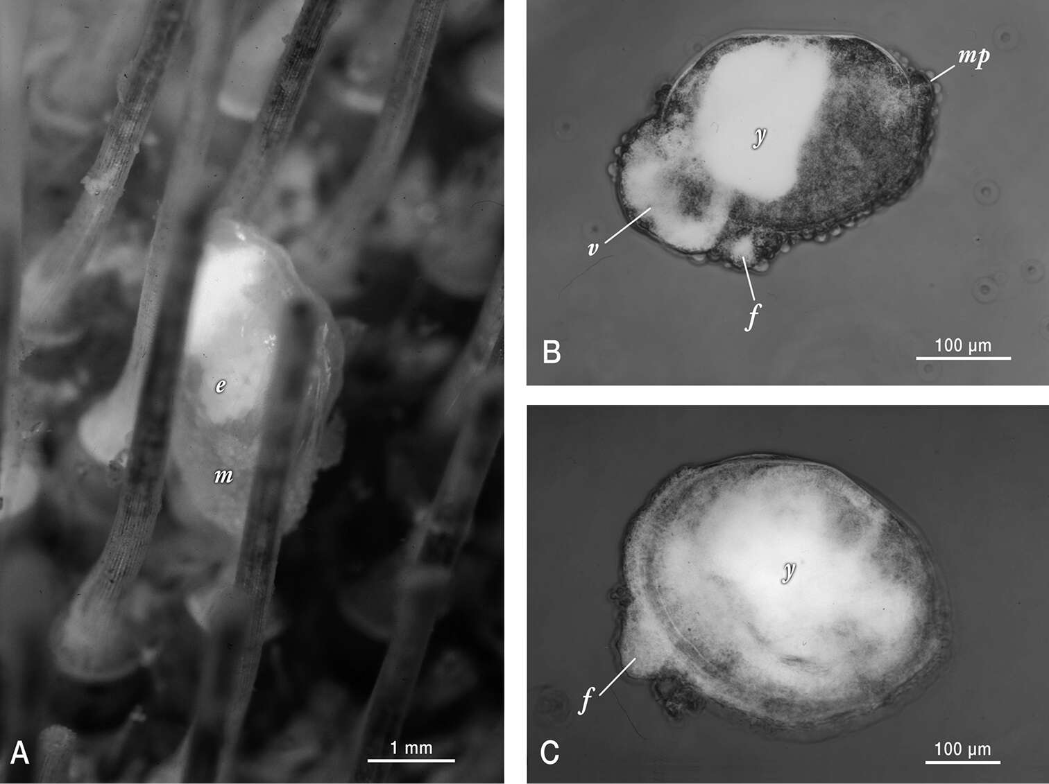
Figure 2.Photographs of live Waldo arthuri material sampled in Barkeley Sound in 1989. A Brooding adult attached to its host. Note the papillated mantle (m) that is partially retracted and the presence of ~ 200 µm diameter white yolky early embryos (e) in its ctenidia, visible through the transparent shell B Micrograph of mid-late development embryo (equivalent to the pediveliger stage in pelagic developing bivalves) that was dissected from its brooding parent’s ctenidia. Labels indicate protruding foot (f), modified non-ciliated velum (v) with partially consumed yolk reserves (white areas) and mantle papillae (mp) in addition to a dense mass of yolk (y) sequestered in the anterior shelled half of the embryo C Micrograph of smallest/youngest (20 µm of dissoconch growth) specimen observed attached to an urchin host. Note the protruding foot (f) and the apparent presence of persistent yolk reserves (y) dispersed throughout much of the juvenile’s visceral mass.