-
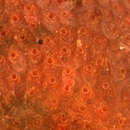
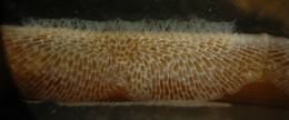
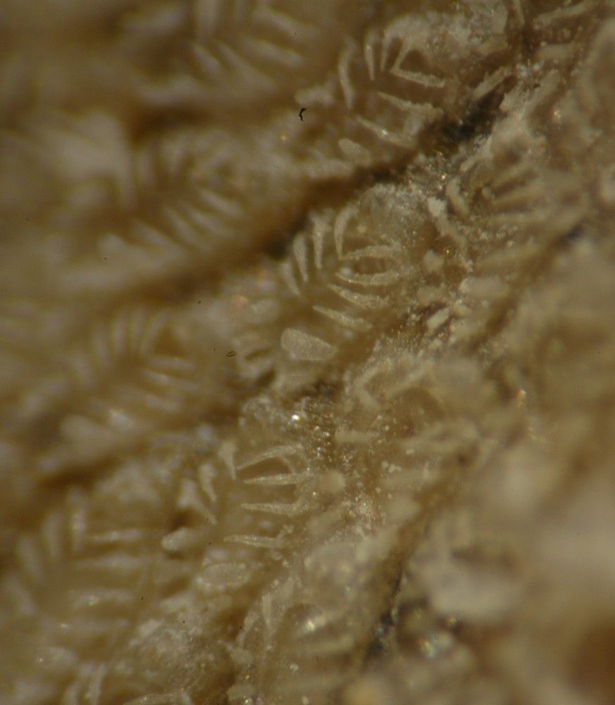
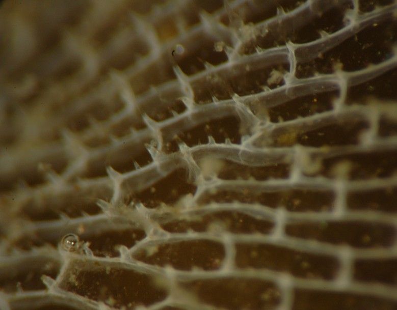
This closeup shows the individual zooids. Note that the frontal area of the zooids is soft and membranous but it is protected by a sheath of spines around the margin. Here and there among the spines are thicker, stalked clublike avicularia. There are no large grooves between the zooids.
-
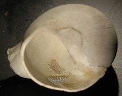
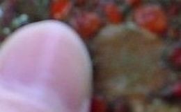
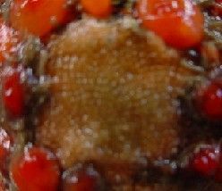
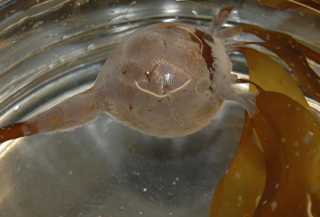
Here is a full view of the moon snail shell, which has a diameter of 2.6 cm in this view. Cauloramphus spiniferum is the bryozoan colony farthest inside the shell mouth (the topmost colony in this view). The other two colonies are Lichenopora verrucaria (middle left) and Lichenopora novae-zelandiae (bottom right).
-
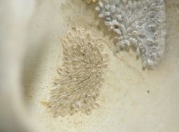
Cauloramphus spiniferum is seen above as the bryozoan patch to the left within the mouth of a small moon snail collected at 120 m depth west of Yellow Island, San Juan Channel, WA. The patch is 5 mm long vertically. The bryozoan to the right is Lichenopora verrucaria. (Photo by: Dave Cowles, July 2007)
-
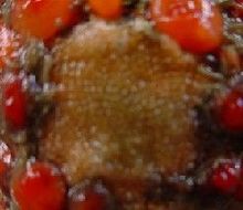
Callopora horrida Slender-spined Bryozoan Callapora horrida collected at Rosario Marine Station, WA Photo by: Anna Dyer, 2002
-
Callopora horrida Slender-spined Bryozoan Callapora horrida collected at Rosario Marine Station, WA Photo by: Anna Dyer, 2002
-
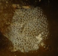
A closer view of one of the colonies shows the lacelike latticework of the boxlike zooecia.
-
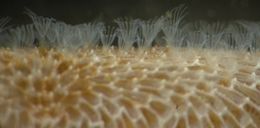
This closer view of the same colony as above shows the small spines present around some of the zooids. Perhaps this colony has been attacked by nudibranchs lately.
-
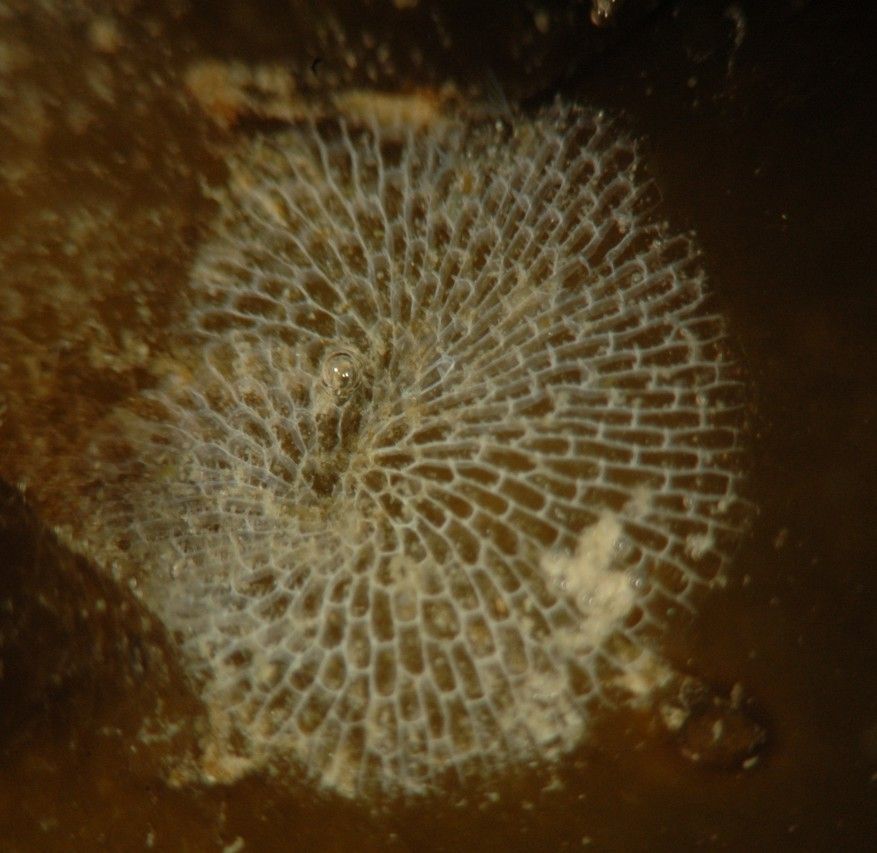
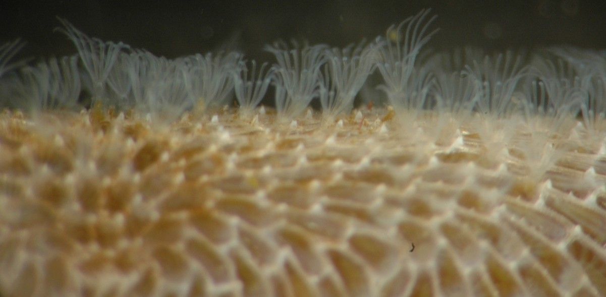
This closeup view of a colony wrapped around a tiny Nereocystis stipe shows the feeding zooids with their lophophores extended in the feeding position.
-
Here is a closer view of the lophophores.
-
A view of the open lophophores from above. Photo 2011 by Brianna Payne
-
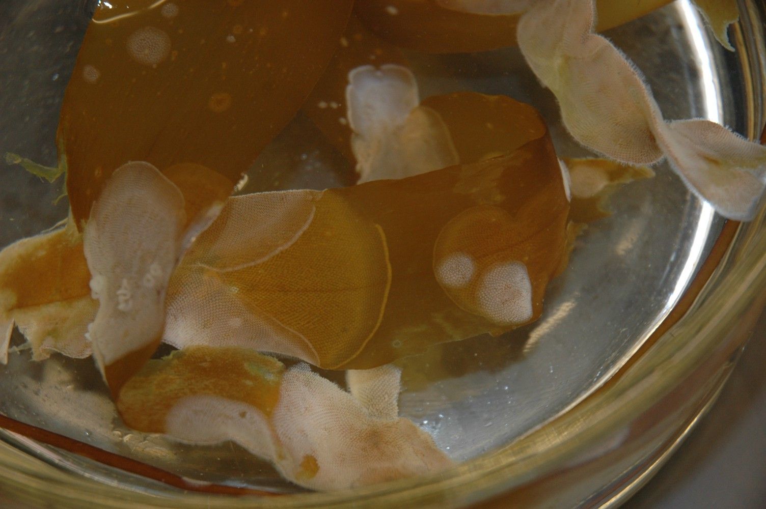
Membranipora membranacea bryozoans on a Nereocystis float and blades. (Photo by: Dave Cowles, July 2007)
-
Membranipora membranacea bryozoans on a Nereocystis float and blades. (Photo by: Dave Cowles, July 2007)