About
Education
Discuss
TraitBank
Sign In
Sign Up
Language
Deutsch
English
Español
français
italiano
Nederlands
Piemontèis
Português do Brasil
suomi
Türkçe
čeština
Ελληνικά
македонски
Українська
العربية
简体中文
繁體中文
names in breadcrumbs
vernacular
scientific
About
Education
Discuss
TraitBank
Sign In
Sign Up
en
Deutsch
English
Español
français
italiano
Nederlands
Piemontèis
Português do Brasil
suomi
Türkçe
čeština
Ελληνικά
македонски
Українська
العربية
简体中文
繁體中文
names in breadcrumbs
vernacular
scientific
Creatures
»
…
»
Animal
»
…
»
Segmented Worms
»
…
Creatures
»
Cellular Organisms
»
Eukaryotes
»
Opisthokonts
»
Animal
»
Bilateria
»
Protostomes
»
Spiralians
»
Segmented Worms
»
Pleistoannelida
»
Sedentaria
«
Aeolosomatidae
collect
overview
data
media
articles
maps
names
CC-BY-NC-SA
any license
CC-BY
CC-BY-NC-SA
CC-BY-SA
type
any type
image
video
Wikimedia Commons
any provider
Wikimedia Commons
Barcode of Life Data Systems
Flickr Group
BioImages, the virtual fieldguide, UK
vimeo
cc-by-3.0
trusted
cc-by-sa-3.0
trusted
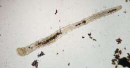
Aeolosoma hemprichi 165891952
cc-by-3.0
Nathan Jones
Wikimedia Commons
Description: English: Aeolosoma Worm (Aeolosoma hemprichi). Date: 26 October 2021. Source:
https://www.inaturalist.org/photos/165891952
. Author:
Nathan Jones
. Camera location
37° 56′ 47.49″ N, 75° 30′ 02.68″ W
View all coordinates using:
OpenStreetMap
37.946524; -75.500744.
: This media file is part of an observation on
iNaturalist
:
inaturalist.org/observations/99506647
This tag does not indicate the copyright status of the attached work. A normal
copyright tag
is still required. See
Commons:Licensing
. : This image was originally posted to iNaturalist by
blastcat
at
https://www.inaturalist.org/photos/165891952
. It was reviewed on 17 May 2022 by
INaturalistReviewBot
and found to be published under the terms of the Cc-by-4.0 license. Reason: sha1.
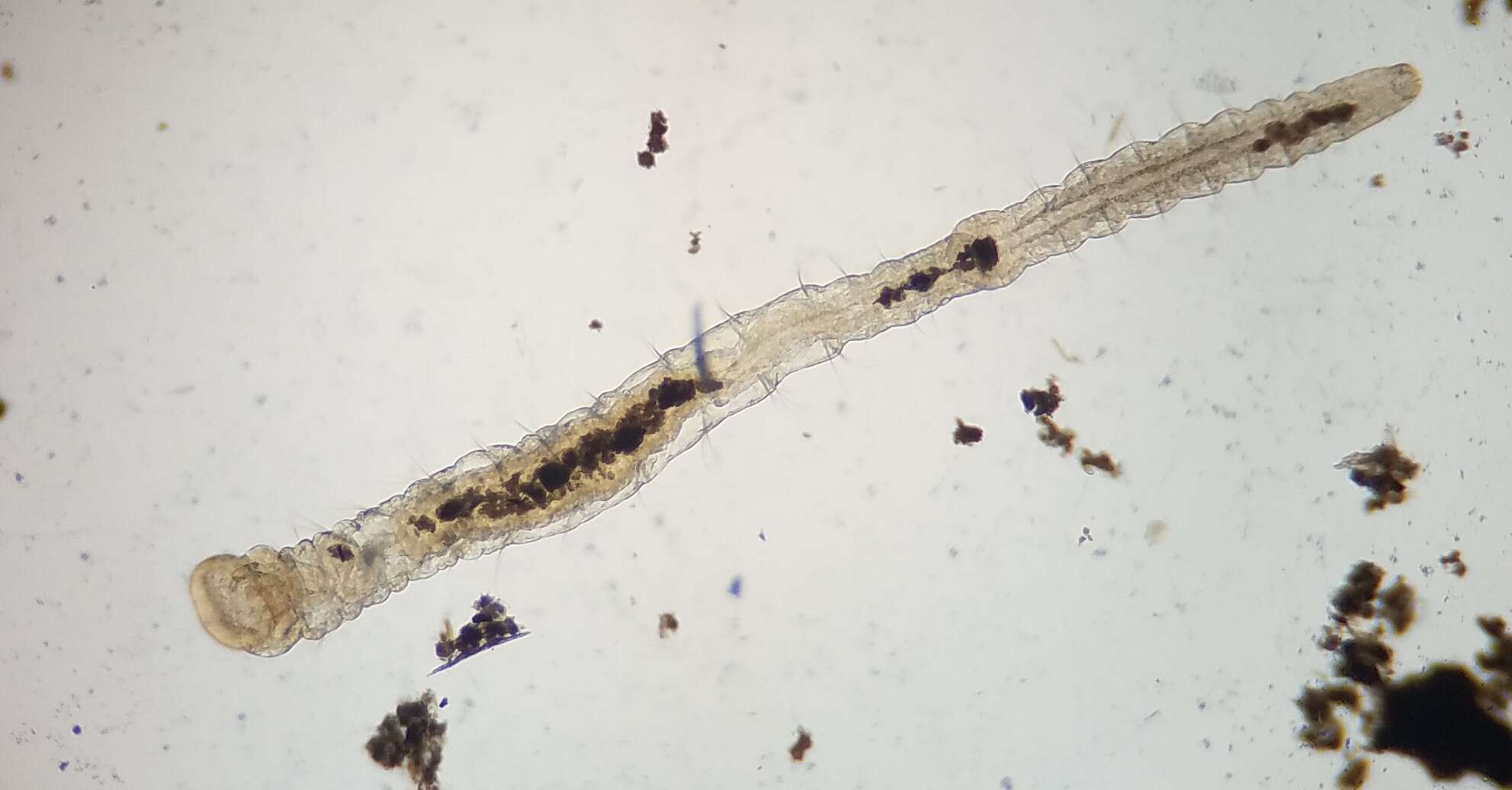
20200520aeolosoma
cc-by-sa-3.0
Ikjbagl
Wikimedia Commons
Description: English: Darkfield microscope capture of aeolosoma at 100x magnification; shows motor function and internal structure. Date: 20 May 2020. Source: I took this video with my phone through my microscope. Author:
Ikjbagl
.
