-
Under a magnification of 44x, this scanning electron micrograph (SEM) depicted the hairy surface of a number of legs of a venomous brown recluse spider, Loxosceles reclusa, found inhabiting a Kentucky farm. The jointed nature of the spiders legs places it in the Phylum, Arthropoda, i.e., Arthro = jointed, and poda = legs, and the fact that this creature has 8 legs places it in the Class, Arachnida. The most proximal leg segment, i.e., nearest to the cephalothorax, is known as the coxa, followed by a trochanter, then a femur, patella. tibia, metatarsus, a tarsus, and finally the claws. Depicted in this particular view are the proximal segments of the spiders first and second right legs. In the lower right you can see the origin of the spiders right fang (far right), and its right pedipalp. The hairs are known as setae, and are sensorial in nature.Created: 2007
-
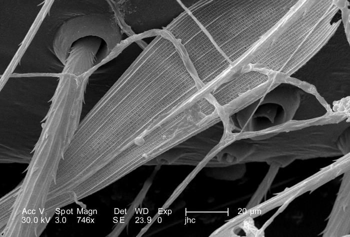
Under a high magnification of 746x, 4X greater than PHIL 10082, this scanning electron micrograph (SEM) depicted the hairy dorsal surface of the anterior cephalothorax of a venomous brown recluse spider, Loxosceles reclusa, found inhabiting a Kentucky farm. These are not really hairs at all in the mammalian sense, which are composed of keratin, but are composed of chitin, and are known as setae, which are sensorial in nature, supplying the spider with information about changes in its environment such as changes in temperature, wind direction, and chemical queues such as pheromones and poisons.In this particular view what appears to be a insect wing scale is entrapped amongst other environmental particulates, which may have represented the remains of one of the spiders previous meals?Created: 2007
-
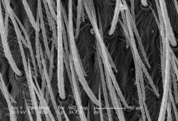
Under a magnification of 186x, 7X greater than PHIL 10075, this scanning electron micrograph (SEM) depicted the hairy dorsal surface of the anterior cephalothorax of a venomous brown recluse spider, Loxosceles reclusa, found inhabiting a Kentucky farm. These are not really hairs at all in the mammalian sense, which are composed of keratin, but are composed of chitin, and are known as setae, which are sensorial in nature, supplying the spider with information about changes in its environment such as changes in temperature, wind direction, and chemical queues such as pheromones and poisons. L. reclusa is sometimes referred to as the violin or fiddle spider, for on its cephalothorax one will see what appears to be coloration in the shape of these stringed instruments, which is quite evident in the color photograph PHIL 1125, depicting a live specimen. Also see PHIL 2224, and 6268 for additional brown recluse images.Created: 2007
-
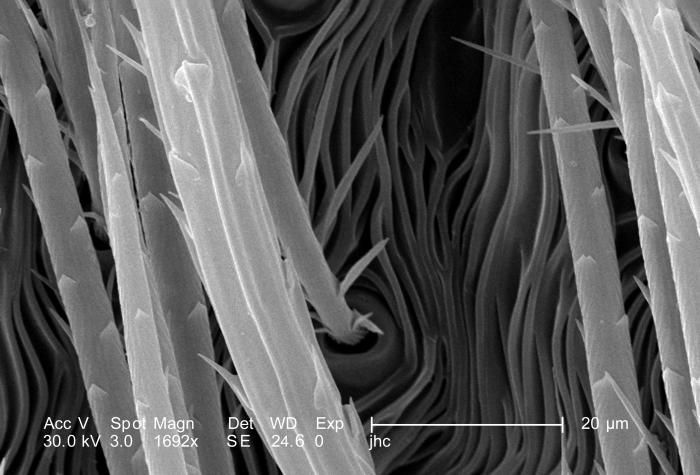
Under a high magnification of 1692x, 4X greater than PHIL 10080, this scanning electron micrograph (SEM) depicted the hairy dorsal surface of the abdominal exoskeleton of a venomous brown recluse spider, Loxosceles reclusa, found inhabiting a Kentucky farm. These are not really hairs at all, in the mammalian sense, which are composed of keratin, but are composed of chitin, and are known as setae, which are sensorial in nature, supplying the spider with information about changes in its environment such as changes in temperature, wind direction, and chemical queues such as pheromones and poisons. L. reclusa is sometimes referred to as the violin or fiddle spider, for on its cephalothorax one will see what appears to be coloration in the shape of these stringed instruments, which is quite evident in the color photograph PHIL 1125, depicting a live specimen. Also see PHIL 2224, and 6268 for additional brown recluse images.Created: 2007
-
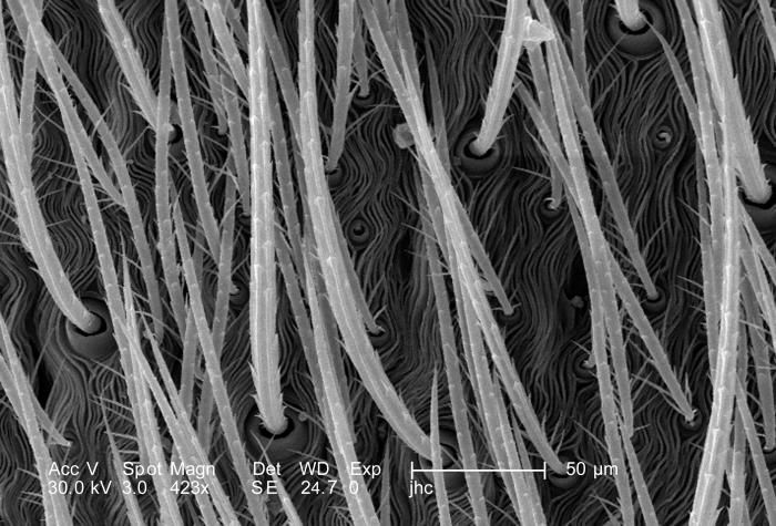
Under a magnification of 423x, 8X greater than PHIL 10079, this scanning electron micrograph (SEM) depicted the hairy dorsal surface of the abdominal exoskeleton of a venomous brown recluse spider, Loxosceles reclusa, found inhabiting a Kentucky farm. These are not really hairs at all, in the mammalian sense, which are composed of keratin, but are composed of chitin, and are known as setae, which are sensorial in nature, supplying the spider with information about changes in its environment such as changes in temperature, wind direction, and chemical queues such as pheromones and poisons. L. reclusa is sometimes referred to as the violin or fiddle spider, for on its cephalothorax one will see what appears to be coloration in the shape of these stringed instruments, which is quite evident in the color photograph PHIL 1125, depicting a live specimen. Also see PHIL 2224, and 6268 for additional brown recluse images.Created: 2007
-
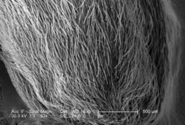
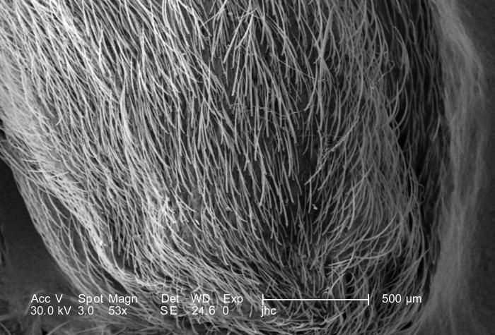
Under a very low magnification of 53x, this scanning electron micrograph (SEM) depicted the hairy dorsal surface of the abdominal exoskeleton of a venomous brown recluse spider, Loxosceles reclusa, found inhabiting a Kentucky farm. These are not really hairs at all, in the mammalian sense, which are composed of keratin, but are composed of chitin, and are known as setae. Chitin is a molecule made up of bound units of acetylglucosamine, joined in such a way as to allow for increased points at which hydrogen bonding can occur. In this way chitin provides increased strength, and durability as an exoskeletal foundation. L. reclusa is sometimes referred to as the violin or fiddle spider, for on its cephalothorax one will see what appears to be coloration in the shape of these stringed instruments, which is quite evident in the color photograph PHIL 1125, depicting a live specimen. Also see PHIL 2224, and 6268 for additional brown recluse images.Created: 2007
-
Under a high magnification of 6922x this scanning electron micrograph (SEM) depicted the striated texture found on the exoskeletal surface of a venomous brown recluse spider, Loxosceles reclusa, found inhabiting a Kentucky farm. As arthropods, spiders possess an exoskeleton composed of chitin, which is a molecule made up of bound units of acetylglucosamine, joined in such a way as to allow for increased points at which hydrogen bonding can occur. In this way chitin provides increased strength, and durability as an exoskeletal foundation. L. reclusa is sometimes referred to as the violin or fiddle spider, for on its cephalothorax one will see what appears to be coloration in the shape of these stringed instruments, which is quite evident in the color photograph PHIL 1125, depicting a live specimen. Also see PHIL 2224, and 6268 for additional brown recluse images.Created: 2007
-
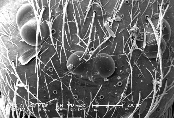
Under a magnification of 108x, approximately twice that of PHIL 10076, this scanning electron micrograph (SEM) depicted the dorsal cephalothorax, i.e., a combination of its head and thoracic regions, of a venomous brown recluse spider, Loxosceles reclusa, found inhabiting a Kentucky farm. Most spiders possess eight eyes (4 pairs), however, as is evidenced in this image, recluse spiders only possess 3 pairs. L. reclusa is sometimes referred to as the violin or fiddle spider, for on its cephalothorax one will see what appears to be coloration in the shape of these stringed instruments, which is quite evident in the color photograph PHIL 1125, depicting a live specimen. Youll also note this spiders four pairs of jointed legs, which places it in the Phylum, Arthropoda, and the Class, Arachnida. Also see PHIL 2224, and 6268 for additional brown recluse images.Created: 2007
-
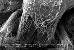
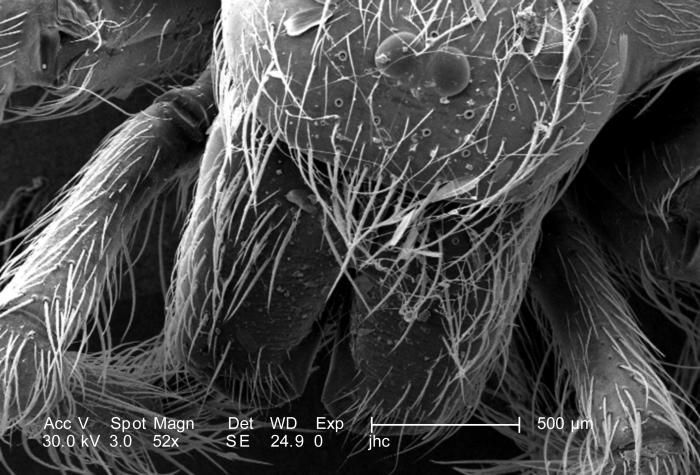
Under a low magnification of only 52x, approximately twice that of PHIL 10075, this scanning electron micrograph (SEM) depicted the dorsal cephalothorax, i.e., a combination of its head and thoracic regions, of a venomous brown recluse spider, Loxosceles reclusa, found inhabiting a Kentucky farm. Most spiders possess eight eyes (4 pairs), however, recluse spiders only possess six. L. reclusa is sometimes referred to as the violin or fiddle spider, for on its cephalothorax one will see what appears to be coloration in the shape of these stringed instruments, which is quite evident in the color photograph PHIL 1125, depicting a live specimen. Youll also note this spiders four pairs of jointed legs, which places it in the Phylum, Arthropoda, and the Class, Arachnida. Also see PHIL 2224, and 6268 for additional brown recluse images.Created: 2007
-
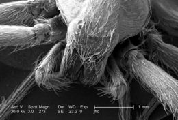
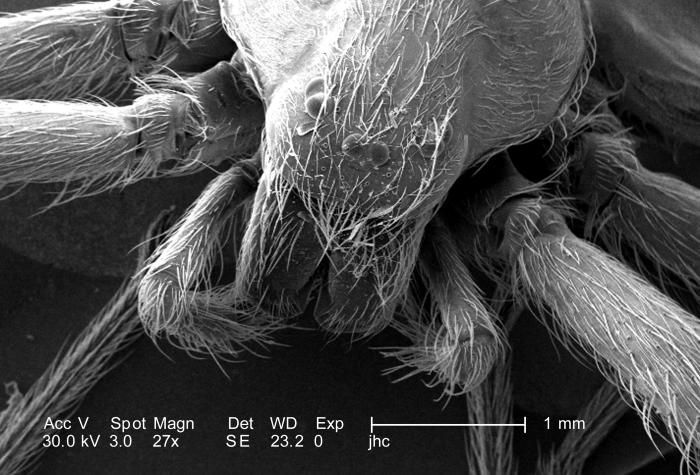
Under a low magnification of only 27x, twice that of PHIL 10074, this scanning electron micrograph (SEM) depicted the dorsal cephalothorax, i.e., a combination of its head and thoracic regions, of a venomous brown recluse spider, Loxosceles reclusa found inhabiting a Kentucky farm. Most spiders possess eight eyes (4 pairs), however, as is evidenced in this image, recluse spiders only possess six. L. reclusa is sometimes referred to as the violin or fiddle spider, for on its cephalothorax one will see what appears to be coloration in the shape of these stringed instruments, which is quite evident in the color photograph PHIL 1125, depicting a live specimen. Youll also note this spiders four pairs of jointed legs, which places it in the Phylum, Arthropoda, and the Class, Arachnida. Also see PHIL 2224, and 6268 for additional brown recluse images.Created: 2007
-
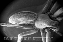
Under a very low magnification of only 14x, this scanning electron micrograph (SEM) depicted the dorsal surface of a venomous brown recluse spider, Loxosceles reclusa, found inhabiting a Kentucky farm. Most spiders possess eight eyes (4 pairs), however, recluse spiders only possess 3 pairs. L. reclusa is sometimes referred to as the violin or fiddle spider, for on its cephalothorax, i.e., a combination of its head and thoracic regions, one will see what appears to be coloration in the shape of these stringed instruments, which is quite evident in the color photograph PHIL 1125, depicting a live specimen. Youll also note this spiders four pairs of jointed legs, which places it in the Phylum, Arthropoda, and the Class, Arachnida. Also see PHIL 2224, and 6268 for additional brown recluse images.Created: 2007
-
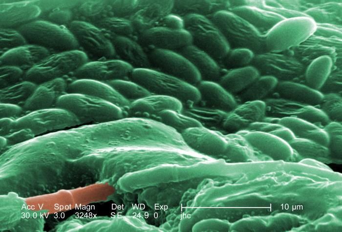
Under three increasingly greater magnifications, this being the highest at 3248X (see PHIL 10089, 10090), what is depicted here is an unidentified pore located on the dorsal abdomen of a venomous brown recluse spider, Loxosceles reclusa, found inhabiting a Kentucky farm. Note the material surrounding the pores orifice, and as the magnification increases, it becomes evident that the material is composed of an unidentified bacterial biofilm. It is not known if these were existing symbiotically upon the spiders exoskeleton, or if they were pathologic in nature, signifying manifestations of a progressive disease process? See PHIL 10088 for a black and white version of this image.L. reclusa is sometimes referred to as the violin or fiddle spider, for on its cephalothorax one will see what appears to be coloration in the shape of these stringed instruments, which is quite evident in the color photograph PHIL 1125, depicting a live specimen.Created: 2007
-
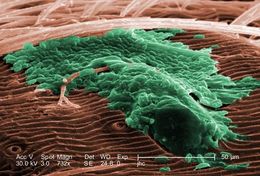
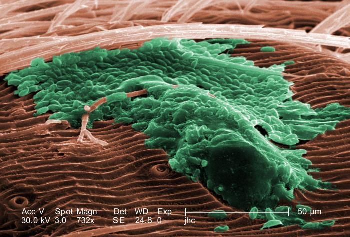
Under three increasingly greater magnifications, this being midway at 732X (see PHIL 10089, 10091), what is depicted here is an unidentified pore located on the dorsal abdomen of a venomous brown recluse spider, Loxosceles reclusa, found inhabiting a Kentucky farm. Note the material surrounding the pores orifice, and as the magnification increases, it becomes evident that the material is composed of an unidentified bacterial biofilm. It is not known if these were existing symbiotically upon the spiders exoskeleton, or if they were pathologic in nature, signifying manifestations of a progressive disease process? See PHIL 10087 for a black and white version of this image.L. reclusa is sometimes referred to as the violin or fiddle spider, for on its cephalothorax one will see what appears to be coloration in the shape of these stringed instruments, which is quite evident in the color photograph PHIL 1125, depicting a live specimen.Created: 2007
-
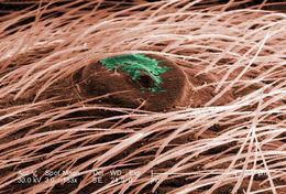
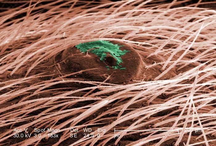
Under three increasingly greater magnifications, this being the lowest at 183X (see PHIL 10089, 10090), what is depicted here is an unidentified pore located on the dorsal abdomen of a venomous brown recluse spider, Loxosceles reclusa, found inhabiting a Kentucky farm. Note the material surrounding the pores orifice, and as the magnification increases, it becomes evident that the material is composed of an unidentified bacterial biofilm. It is not known if these were existing symbiotically upon the spiders exoskeleton, or if they were pathologic in nature, signifying manifestations of a progressive disease process? See PHIL 10086 for a black and white version of this image.L. reclusa is sometimes referred to as the violin or fiddle spider, for on its cephalothorax one will see what appears to be coloration in the shape of these stringed instruments, which is quite evident in the color photograph PHIL 1125, depicting a live specimen.Created: 2007
-
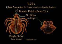
This ventral view illustration depicts a female Rhipicephalus tick showing its unridged palpi, and deeply indented fore coxae.Created: 1976
-
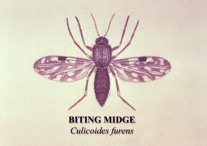
This is an illustration of a sandfly, or the biting midge, Culicoides furens.Created: 1976
-
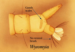
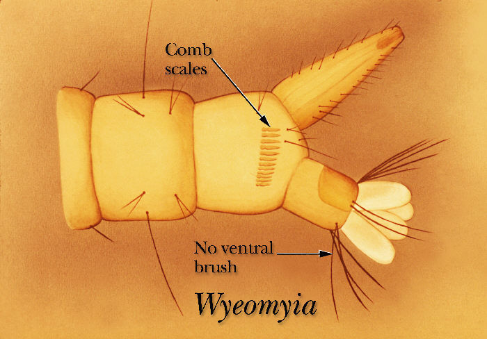
An illustration of the ventral surface of the terminal segment of a Wyeomyia mosquito larva.Created: 1975
-
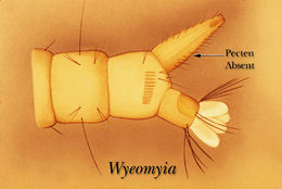
A drawing showing that the Wyeomyia mosquito larva has no pectin row on the ventral surface of its siphon tube.Created: 1975
-
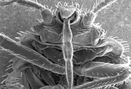
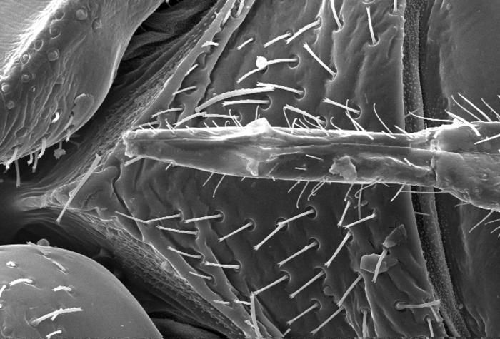
This highly-magnified scanning electron micrograph (SEM) revealed some of the ultrastructural morphology displayed on the ventral surface of a bedbugs, Cimex lectularius, head and thorax. Note the distal anatomical relationships of the insects skin piercing mouthparts that it uses to obtain its blood meal.Clinical Features:Created: 2009
-
This scanning electron micrograph (SEM) revealed some of the ultrastructural morphology displayed on the ventral surface of a bedbugs, Cimex lectularius, head and thorax. Note the anatomical relationships of the insects skin piercing mouthparts that it uses to obtain its blood meal.Clinical Features:Created: 2009
-
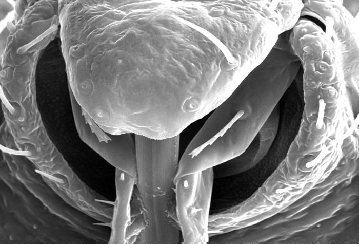
This highly-magnified scanning electron micrograph (SEM) revealed some of the ultrastructural morphology displayed on the rostral head region of a bedbug, Cimex lectularius. Note the proximal anatomical relationships of the insects skin piercing mouthparts it uses to obtain its blood meal.Clinical Features:Created: 2009
-
This highly-magnified scanning electron micrograph (SEM) revealed some of the ultrastructural morphology displayed on the rostral head region of a bedbug, Cimex lectularius. Note the distal anatomical relationships of the insects skin piercing mouthparts used to obtain its blood meal. For a colorized version of this image see PHIL 11140.Clinical Features:Created: 2009
-
This highly-magnified, scanning electron micrograph (SEM) revealed some of the ultrastructural morphology displayed on the rostral head region of a bedbug, Cimex lectularius. Note the proximal anatomical relationships the insects skin piercing mouthparts it uses to obtain its blood meal, and how they join the head. For a colorized version of this image see PHIL 11738.Clinical Features:Although bedbugs have been found naturally-infected with blood-borne pathogens, they are not effective vectors of disease. The primary medical importance is inflammation associated with their bites (due to allergic reactions to components in their saliva).Created: 2009
-
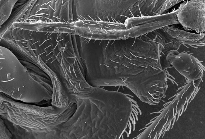
This scanning electron micrograph (SEM) revealed some of the ultrastructural morphology displayed on the ventral surface of a bedbug, Cimex lectularius. From this view you can see the insects skin piercing mouthparts it uses to obtain its blood meal, as well as a number of its six jointed legs. For a colorized version of this image see PHIL 11739.Clinical Features:Created: 2009