-
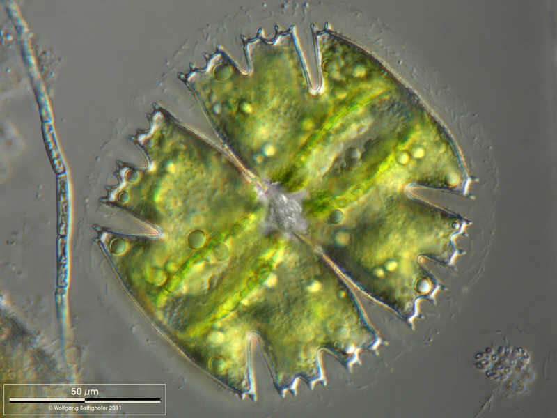
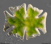
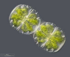
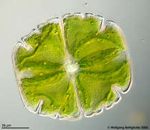
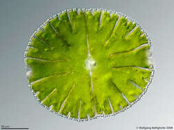
Micrasterias americana Scale bar indicates 50 µm. Sample from a pond near Großostheim, Germany. Sampling date 04/2021. The image was built up using several photomicrographic frames with manual stacking technique. Images were taken using Zeiss Axioplan with Olympus OM-D M5 MKII. Image under Creative Commons License V 3.0 (CC BY-NC-SA). Place name: Pond near Großostheim (Germany) Latitude: 49.88482168 Longitude: 9.09980822 Multiebenen-Abbildung, manuell gestapelt. Der Messbalken markiert eine Länge von 50 µm. Probe aus einem Waldteich bei Großostheim. Datum der Aufsammlung: 04/2021. Mikrotechnik: Zeiss Axioplan, Kamera: Olympus OM-D M5 MKII. Creative Commons License V 3.0 (CC BY-NC-SA). For permission to use of (high-resolution) images please contact postmaster@protisten.de.
-

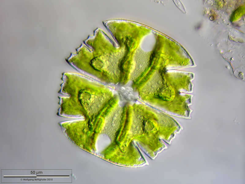
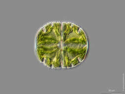
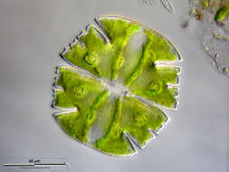
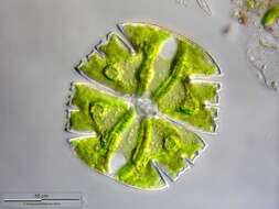
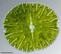
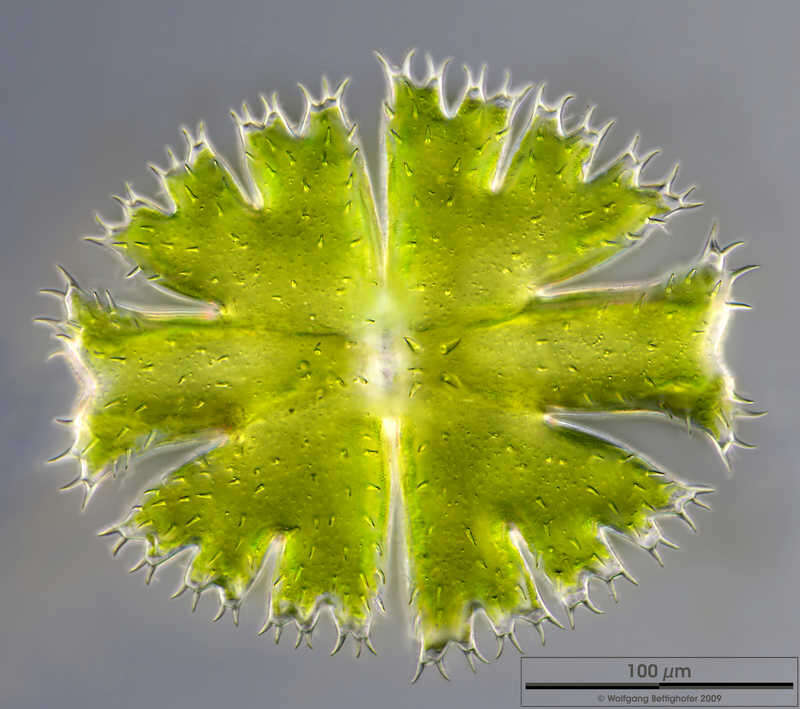
Micrasterias americana Micrasterias americana shows a very special morphological phenomenon. The four minor appendices of apical lobes are optional. But if some of them are formed they show rotational symmetry according to direction of the appendices. This depth of focus picture assembling 14 high resolution shots showes a specimen with one appendix formed Sample from sphagnum pond situated in the northern alpine region of Austria near Salzburg. Images were taken using Zeiss Universal with Olympus C7070 CCD camera.Image under Creative Commons License V 3.0 (CC BY-NC-SA). Place name: Bogs near Salzburg (Austria) Latitude: 48.068516 Longitude: 12.954134 Zelle von Micrasterias americana, die lediglich eine der üblichen vier kleineren Fortsätze der apikalen Lappen ausgebildet hat. Tiefenschärfe durch Multiebenenabbildung aus 14 Bildebenen, manuell gestapelt. Probe aus einem Moor in den nördlichen Kalkalpen von Österreich in der Nähe von Salzburg. Mikrotechnik: Zeiss Universal, Kamera: Olympus C7070. Creative Commons License V 3.0 (CC BY-NC-SA). For permission to use of (high-resolution) images please contact postmaster@protisten.de.
-
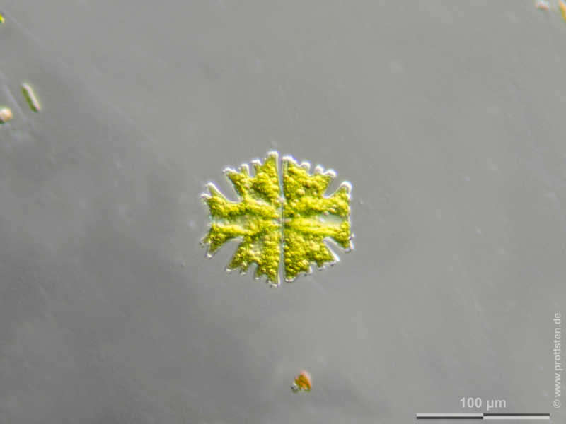
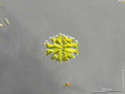
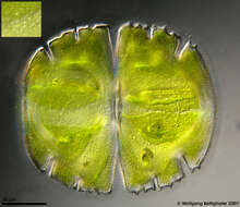
Micrasterias americana Scale bar indicates 100 µm. Sample from a pond near Großostheim, Germany. Sampling date 04/2021. . Images were taken using Olympus stereomicroscope SZX16 with Olympus OM-D M5 MKII. Image under Creative Commons License V 3.0 (CC BY-NC-SA). Place name: Pond near Großostheim (Germany) Latitude: 49.88482168 Longitude: 9.09980822 Multiebenen-Abbildung, manuell gestapelt. Der Messbalken markiert eine Länge von 100 µm. Probe aus einem Waldteich bei Großostheim. Datum der Aufsammlung: 04/2021. Mikrotechnik: Olympus Stereomikroskop SZX16, Kamera: Olympus OM-D M5 MKII. Creative Commons License V 3.0 (CC BY-NC-SA). For permission to use of (high-resolution) images please contact postmaster@protisten.de.
-
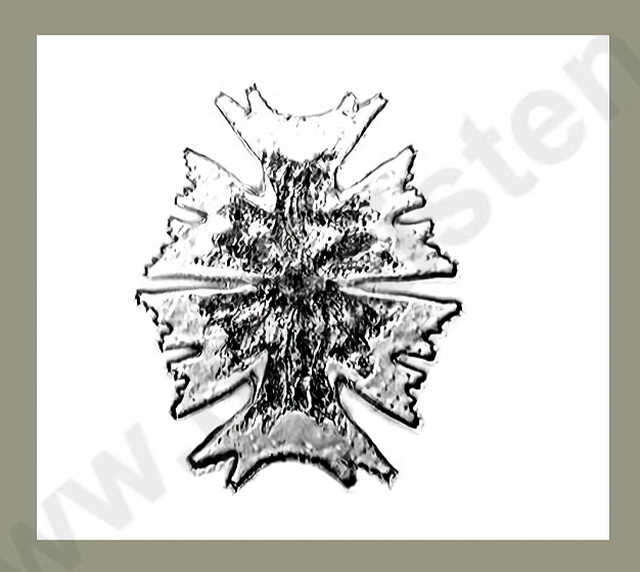
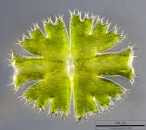
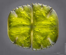
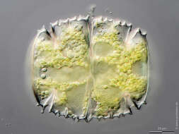
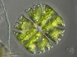
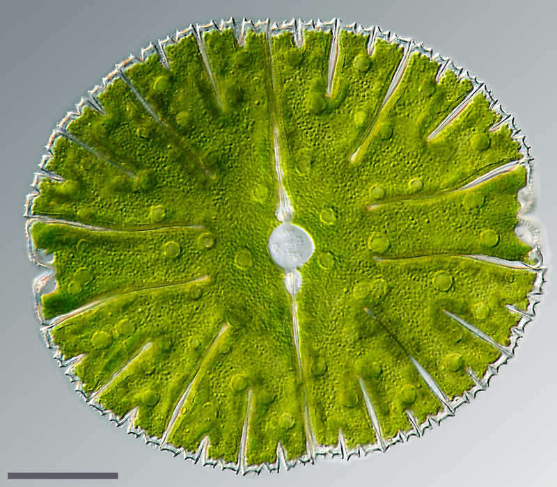
Micrasterias apiculata The picture shows the outline and the surface texture of the cell wall with the numerous spines on the cell wall and was built up using 38 high resolution DIC frames with manual stacking technique. The scale bar indicates 100 µm. Sample from sphagnum pond situated in the northern alpine region of Austria near Salzburg. Images were taken using Zeiss Universal with Olympus C7070 CCD camera.Image under Creative Commons License V 3.0 (CC BY-NC-SA). Place name: Bogs near Salzburg (Austria) Latitude: 48.068516 Longitude: 12.954134 Das Bild zeigt den Umriss und Musterung der Zellwand mit den zahlreichen Stacheln auf der Zellwand. Tiefenschärfe durch Multiebenenabbildung aus 38 Bildebenen, manuell gestapelt. Der Messbalken markiert eine Länge von 100 µm. Probe aus einem Moor in den nördlichen Kalkalpen von Österreich in der Nähe von Salzburg. Mikrotechnik: Zeiss Universal, Kamera: Olympus C7070. Creative Commons License V 3.0 (CC BY-NC-SA). For permission to use of (high-resolution) images please contact postmaster@protisten.de.
-
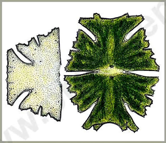
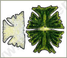
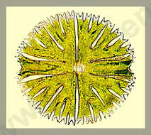
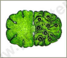
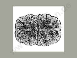
Micrasterias americana (EHR.) RALFS Dimension: Length 130 - 150 µm, width 100 120 µm.Not rare both in lowlands and in alpine waters, sometimes abundant.Copyright by Prof. Rupert Lenzenweger, Ried im Innkreis, Austria. Place name: n. a. Latitude: 0 Longitude: 0 Dimensionen Länge 130 150 µm, Breite 100 120 µm.Sowohl im Flachland als auch in alpinen Gewässern nicht selten, mitunter massenhaft. Copyright by Prof. Rupert Lenzenweger, Ried im Innkreis, Österreich. For permission to use of (high-resolution) images please contact postmaster@protisten.de.
-
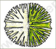
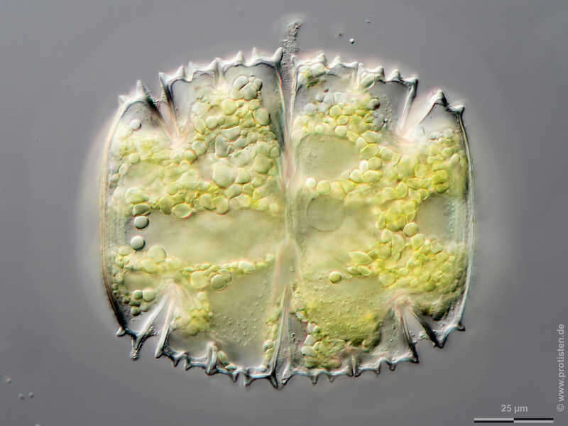
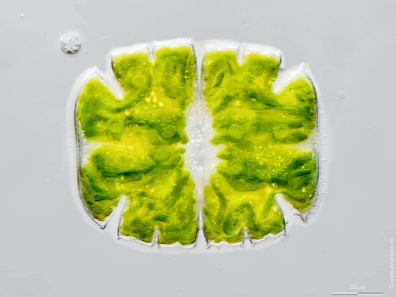
Micrasterias truncata Scale bar indicates 50 µm. Sample from Bog Waasenmoos Pass Thurn near Mittersil, Tyrol, Austria. Sampling date 10/2019. The image was built up using several photomicrographic frames with manual stacking technique. Images were taken using Zeiss Axioplan with Olympus OM-D M5 MKII. Image under Creative Commons License V 3.0 (CC BY-NC-SA). Place name: Bog Waasenmoos Pass Thurn near Mittersil (Tyrol, Austria) Latitude: 47.30234117 Longitude: 12.41751194 Multiebenen-Abbildung, manuell gestapelt. Der Messbalken markiert eine Länge von 50 µm. Probe aus dem Waasenmoos bei Pass Thurn Nähe Mittersil, Tirol. Datum der Aufsammlung: 10/2019. Mikrotechnik: Zeiss Axioplan, Kamera: Olympus OM-D M5 MKII. Creative Commons License V 3.0 (CC BY-NC-SA). For permission to use of (high-resolution) images please contact postmaster@protisten.de.
-
Micrasterias truncata Scale bar indicates 10 µm. Sample from the bog Mittermoos near Pillersee (Tyrol, Austria). The image was built up using several photomicrographic frames with manual stacking technique. Images were taken using Zeiss Universal with Canon EOS 600D.Image under Creative Commons License V 3.0 (CC BY-NC-SA). Place name: Wetland Mittermoos near Fieberbrunn (Tyrol, Austria) Latitude: 47.47998695 Longitude: 12.5240922 Der Messbalken markiert eine Länge von 10 µm. Probe aus dem Mittermoos Nähe Pillersee in Tirol. Mikrotechnik: Zeiss Universal, Kamera: Canon EOS 600D. Creative Commons License V 3.0 (CC BY-NC-SA). For permission to use of (high-resolution) images please contact postmaster@protisten.de.
-
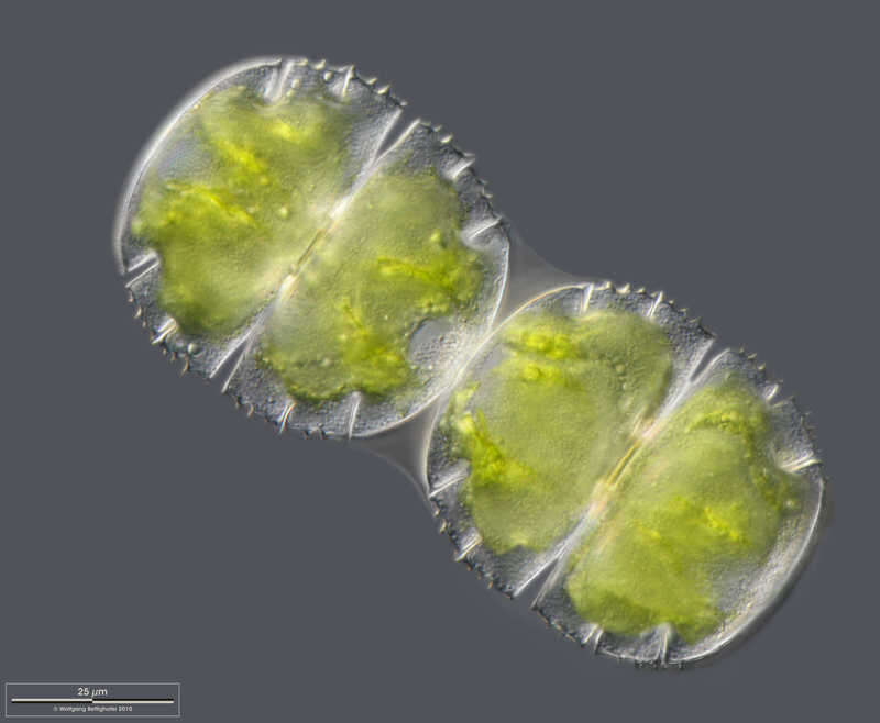
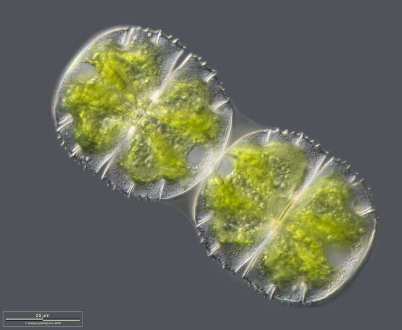
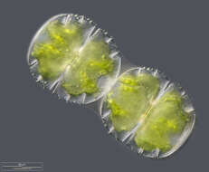
Micrasterias truncata Two cells shortly after binary fission. The connecting sheath of the primary cell wall is visible. Scale bar indicates 25 µm. Sample from a small wetland near Schladming (northern alpine region of Austria near Salzburg). Images were taken using Zeiss Universal with Olympus C7070 CCD camera.Image under Creative Commons License V 3.0 (CC BY-NC-SA). Place name: Wetland near Schladming (Austria) Latitude: 47.37386 Longitude: 13.823016 Zwei Zellen kurz nach der Teilung. Die primäre Zellwand als gemeinsame Hülle ist im Verbindungsbereich der beiden Zellen sichtbar. Der Messbalken markiert eine Länge von 25 µm. Probe aus einer Wiesenvernässung nahe Schladming/Österreich. Mikrotechnik: Zeiss Universal, Kamera: Olympus C7070. Creative Commons License V 3.0 (CC BY-NC-SA). For permission to use of (high-resolution) images please contact postmaster@protisten.de.
-
Micrasterias truncata Scale bar indicates 25 µm. Sample from Bog Waasenmoos Pass Thurn near Mittersil, Tyrol, Austria. Sampling date 10/2019. The image was built up using several photomicrographic frames with manual stacking technique. Images were taken using Zeiss Axioplan with Olympus OM-D M5 MKII. Image under Creative Commons License V 3.0 (CC BY-NC-SA). Place name: Bog Waasenmoos Pass Thurn near Mittersil (Tyrol, Austria) Latitude: 47.30234117 Longitude: 12.41751194 Multiebenen-Abbildung, manuell gestapelt. Der Messbalken markiert eine Länge von 25 µm. Probe aus dem Waasenmoos bei Pass Thurn Nähe Mittersil, Tirol. Datum der Aufsammlung: 10/2019. Mikrotechnik: Zeiss Axioplan, Kamera: Olympus OM-D M5 MKII. Creative Commons License V 3.0 (CC BY-NC-SA). For permission to use of (high-resolution) images please contact postmaster@protisten.de.
-
Micrasterias truncata Two cells shortly after binary fission. Optical transversal section. The connecting sheath of the primary cell wall is visible. Scale bar indicates 25 µm. Sample from a small wetland near Schladming (northern alpine region of Austria near Salzburg). Images were taken using Zeiss Universal with Olympus C7070 CCD camera.Image under Creative Commons License V 3.0 (CC BY-NC-SA). Place name: Wetland near Schladming (Austria) Latitude: 47.37386 Longitude: 13.823016 Zwei Zellen kurz nach der Teilung; optischer Querschnitt. Die primäre Zellwand als gemeinsame Hülle ist im Verbindungsbereich der beiden Zellen sichtbar. Der Messbalken markiert eine Länge von 25 µm. Probe aus einer Wiesenvernässung nahe Schladming/Österreich. Mikrotechnik: Zeiss Universal, Kamera: Olympus C7070. Creative Commons License V 3.0 (CC BY-NC-SA). For permission to use of (high-resolution) images please contact postmaster@protisten.de.
-
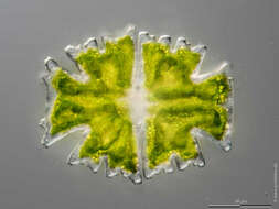
Micrasterias truncata Desmid alga from a bog. Scale bar indicates 50 µm. Sample from a small wetland near Schladming (northern alpine region of Austria near Salzburg). Images were taken using Zeiss Universal with Olympus C7070 CCD camera.Image under Creative Commons License V 3.0 (CC BY-NC-SA). Place name: Wetland near Schladming (Austria) Latitude: 47.37386 Longitude: 13.823016 Zieralge aus einem Moorteich. Der Messbalken markiert eine Länge von 50 µm. Probe aus einer Wiesenvernässung nahe Schladming/Österreich. Mikrotechnik: Zeiss Universal, Kamera: Olympus C7070. Creative Commons License V 3.0 (CC BY-NC-SA). For permission to use of (high-resolution) images please contact postmaster@protisten.de.
-

Micrasterias truncata Non-filamentous desmids have the ability to move slowly by means of directed mucilage secretion. Depht of focus approach can show cell surface together with folded chloroplasts, cell contour and nucleus. In zip archive there are more DOF pictures of this object. Picture generated from 9 shots using CombineZ by Alan Hadley, MicroPicS by Bernhard Wiedemann and Photoshop. Sample from spagnum pond Dosenmoor near Neumuenster (Schleswig-Holstein, Germany). Images were taken using Zeiss Universal with Olympus C7070 CCD camera.Image under Creative Commons License V 3.0 (CC BY-NC-SA). Place name: Bog Dosenmoor near Neumuenster (Schleswig-Holstein, Germany) Latitude: 54.136219 Longitude: 10.026433 Nicht-fädigen Desmidiaceen haben die Fähigkeit, sich langsam mittels gerichteter Schleim-Sekretion zu bewegen. Mittels Multiebenenabbildung kann die Zelloberfläche zusammen mit gefalteten Chloroplasten, dem Zellumriss und dem Kern in hoher Auflösung wiedergegeben werden. Tiefenschärfe durch Multiebenenabbildung aus 9 Bildebenen. Probe aus dem Dosenmoor in der Nähe von Neumünster. Mikrotechnik: Zeiss Universal, Kamera: Olympus C7070. Creative Commons License V 3.0 (CC BY-NC-SA). For permission to use of (high-resolution) images please contact postmaster@protisten.de.
-
Micrasterias truncata The optical transversal section shows the nucleus and the pyrenoid bodies. Scale bar indicates 50 µm. Sample from a small wetland near Schladming (northern alpine region of Austria near Salzburg). Images were taken using Zeiss Universal with Olympus C7070 CCD camera.Image under Creative Commons License V 3.0 (CC BY-NC-SA). Place name: Wetland near Schladming (Austria) Latitude: 47.37386 Longitude: 13.823016 Der optische Querschnitt zeigt den Kern und die Pyrenoid-Körper. Der Messbalken markiert eine Länge von 50 µm. Probe aus einer Wiesenvernässung nahe Schladming/Österreich. Mikrotechnik: Zeiss Universal, Kamera: Olympus C7070. Creative Commons License V 3.0 (CC BY-NC-SA). For permission to use of (high-resolution) images please contact postmaster@protisten.de.
-
Micrasterias truncata Scale bar indicates 50 µm.Sample from the pond Hegne Moor situated in the vicinity of Lake Constance. The image was built up using several photomicrographic frames with manual stacking technique. Images were taken using Zeiss Universal with Olympus C7070 CCD camera.Image under Creative Commons License V 3.0 (CC BY-NC-SA). Place name: Bog Hegne Moor near Lake Constance (Germany) Latitude: 47.718106 Longitude: 9.093974 Multiebenen-Abbildung, manuell gestapelt. Der Messbalken markiert eine Länge von 50 µm. Probe aus dem Simmelried nahe Konstanz. Mikrotechnik: Zeiss Universal, Kamera: Olympus C7070. Creative Commons License V 3.0 (CC BY-NC-SA). For permission to use of (high-resolution) images please contact postmaster@protisten.de.
-

Micrasterias truncata In photosynthesis the enzyme RuBisCO (Ribulose-1,5-Biphosphat- Carboxylase/Oxygenase) plays a very important role. Many algae concentrate RuBisCO in special grains named pyrenoids. The depth of focus picture shows the structured chloroplasts with their pyrenoids. Micrasterias truncata has a mucilaginous envelope. The genus have two types of pores, small ones situated all over the cell surface for building their mucilaginous envelope and special ones with bigger lumen to excrete mucilage for movement. Deph of focus technique was used to gather details from over 40 shots. Sample from spagnum pond situated in the northern alpine region of Austria near Salzburg. Images were taken using Zeiss Universal with Olympus C7070 CCD camera.Image under Creative Commons License V 3.0 (CC BY-NC-SA). Place name: Bog Dosenmoor near Neumuenster (Schleswig-Holstein, Germany) Latitude: 54.136219 Longitude: 10.026433 Das Enzym Rubisco (Ribulose-1 ,5-Biphosphat-Carboxylase / Oxygenase) spielt in der Photosynthese eine zentrale Rolle. Viele Algen konzentrieren RuBisCO in speziellen Körper, die Pyrenoide genannt werden. Die Multiebenenabbildung zeigt die strukturierten Chloroplasten mit ihren Pyrenoiden. Wie bei Desmidiaceen üblich, hat Micrasterias truncata eine schleimige Hülle. Die Gattung hat zwei Arten von Poren, kleine auf der ganzen Zelloberfläche für den Aufbau ihrer schleimigen Hülle und spezielle mit größerem Durchmesser, an den Spitzen der beiden Zell-Lappen sitzend, die zur Absonderung des für die Bewegung notwendigen Schleims dienen. Tiefenschärfe durch Multiebenenabbildung aus 40 Bildebenen, manuell gestapelt. Probe aus einem Moor in den nördlichen Kalkalpen von Österreich in der Nähe von Salzburg. Mikrotechnik: Zeiss Universal, Kamera: Olympus C7070. Creative Commons License V 3.0 (CC BY-NC-SA). For permission to use of (high-resolution) images please contact postmaster@protisten.de.
-
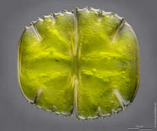
Micrasterias thomasiana var. notata The picture shows the outline and the surface texture of the cell wall and was built up using 34 high resolution DIC frames with manual stacking technique. The scale bar indicates 50 µm. Sample from sphagnum pond situated in the northern alpine region of Austria near Salzburg. Images were taken using Zeiss Universal with Olympus C7070 CCD camera.Image under Creative Commons License V 3.0 (CC BY-NC-SA). Place name: Bogs near Salzburg (Austria) Latitude: 48.068516 Longitude: 12.954134 Das Bild zeigt den Umriss und die Musterung der Zellwand. Tiefenschärfe durch Multiebenenabbildung aus 34 Bildebenen, manuell gestapelt. Der Messbalken markiert eine Länge von 50 µm. Probe aus einem Moor in den nördlichen Kalkalpen von Österreich in der Nähe von Salzburg. Mikrotechnik: Zeiss Universal, Kamera: Olympus C7070. Creative Commons License V 3.0 (CC BY-NC-SA). For permission to use of (high-resolution) images please contact postmaster@protisten.de.
-
Micrasterias thomasiana var. notata The variety notata is very more common than the type variety. This optical transversal section of the desmid cell shows the outline, the surface texture of the chloroplast with many pyrenoids and the nucleus at the center of the cell. This picture was built up using appr. 20 high resolution DIC frames with manual stacking technique. The scale bar indicates 50 µm. Sample from sphagnum pond situated in the northern alpine region of Austria near Salzburg. Images were taken using Zeiss Universal with Olympus C7070 CCD camera.Image under Creative Commons License V 3.0 (CC BY-NC-SA). Place name: Bogs near Salzburg (Austria) Latitude: 48.068516 Longitude: 12.954134 Die Varietät notata ist erstaunlicherweise viel häufiger als die Typusvarietät Micrasterias thomasiana. Der optische Querschnitt zeigt den Umriss und die Oberflächenstruktur der Chloroplasten mit vielen Pyrenoiden und den Kern in der Zellmitte. Tiefenschärfe durch Multiebenenabbildung aus 20 Bildebenen, manuell gestapelt. Der Messbalken markiert eine Länge von 50 µm. Probe aus einem Moor in den nördlichen Kalkalpen von Österreich in der Nähe von Salzburg. Mikrotechnik: Zeiss Universal, Kamera: Olympus C7070. Creative Commons License V 3.0 (CC BY-NC-SA). For permission to use of (high-resolution) images please contact postmaster@protisten.de.
-
Micrasterias truncata var. bahusiensis Scale bar indicates 25 µm. Sample from Kaltenhof Bog in the north of Kiel, Germany. Sampling date 9/2018. The image was built up using several photomicrographic frames with manual stacking technique. Images were taken using Zeiss Axioplan with Olympus OM-D M5 MKII. Image under Creative Commons License V 3.0 (CC BY-NC-SA). Place name: Bog Kaltenhof near Kiel (Schleswig-Holstein, Germany) Latitude: 54.42102744 Longitude: 10.07686615 Multiebenen-Abbildung, manuell gestapelt. Der Messbalken markiert eine Länge von 25 µm. Probe aus dem Kaltenhofer Moor nördlich von Kiel. Datum der Aufsammlung: 9/2018. Mikrotechnik: Zeiss Axioplan, Kamera: Olympus OM-D M5 MKII. Creative Commons License V 3.0 (CC BY-NC-SA). For permission to use of (high-resolution) images please contact postmaster@protisten.de.
-
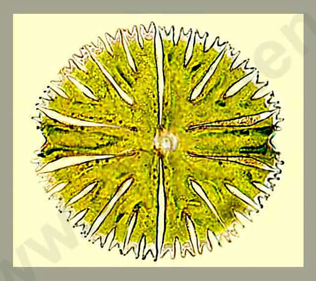
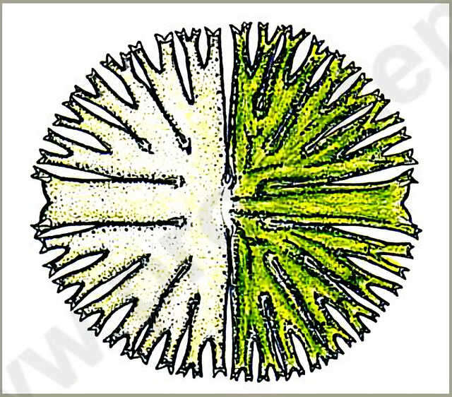
Micrasterias radiosa RALFS Dimension: Length 170 - 180 µm, width 165 - 175 µm.Occurrence: .Copyright by Prof. Rupert Lenzenweger, Ried im Innkreis, Austria. Place name: n. a. Latitude: 0 Longitude: 0 Dimensionen: Länge 170 180 µm, Breite 165 175 µm.Vorkommen: In mäßig sauren Gewässern, mitunter auch im Plankton, in Mitteleuropa selten. Copyright by Prof. Rupert Lenzenweger, Ried im Innkreis, Österreich. For permission to use of (high-resolution) images please contact postmaster@protisten.de.
-

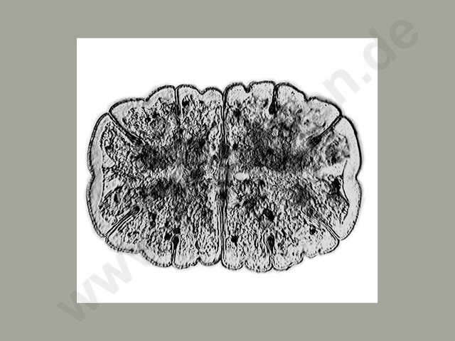
Micrasterias jenneri RALFS The cells are appr. 1.5 times longer than wide, rounded off rectangular in shape. The cells have five lobes, the lateral lobes are broadened towards the periphery and emaginated in the center, the cuts between them are closed and short. The lateral lobes arent spread apart, they are strongly widened peripherally and rounded with shallow emarginations in the center. The central cut is deep, closed, and not peripherally widened. Dimension: Length 150 - 180 µm, width 100 130 µm.Ecology: Acidophilic alga, lives in sphagnum ponds together with other algae which prefer upland moor waters.Occurrence: Probably ubiquitous, mainly in colder zones.Copyright by Prof. Rupert Lenzenweger, Ried im Innkreis, Austria. Place name: n. a. Latitude: 0 Longitude: 0 Die Zellen sind ungefähr 1,5 mal länger als breit, im groben Umriss abgerundet-rechteckig. Die Zellhälften sind 5-lappig, die Seitenlappen sind nach außen verbreitert, in der Mitte eingebuchtet, die Einschnitte dazwischen sind geschlossen und kurz. Die Scheitellappen sind nicht abgesetzt, nach außen stark verbreitert und abgerundet mit einer seichten Einbuchtung in der Mitte. Die Mitteleinschnitte sind tief, geschlossen, nach außen nicht erweitert. Dimensionen: Länge 150 180 µm, Breite 100 130 µm.Ökologie: Acidophile Alge, kommt daher in Hochmoorgewässern gemeinsam mit anderen Hochmooralgen vor.Verbreitung: Wahrscheinlich weltweit, hautsächlich in kälteren Zonen. Copyright by Prof. Rupert Lenzenweger, Ried im Innkreis, Österreich. For permission to use of (high-resolution) images please contact postmaster@protisten.de.
-
Micrasterias jenneri RALFS The cells are appr. 1.5 times longer than wide, rounded off rectangular in shape. The cells have five lobes, the lateral lobes are broadened towards the periphery and emaginated in the center, the cuts between them are closed and short. The lateral lobes arent spread apart, they are strongly widened peripherally and rounded with shallow emarginations in the center. The central cut is deep, closed, and not peripherally widened. Dimension: Length 150 - 180 µm, width 100 130 µm.Ecology: Acidophilic alga, lives in sphagnum ponds together with other algae which prefer upland moor waters.Occurrence: Probably ubiquitous, mainly in colder zones.Copyright by Prof. Rupert Lenzenweger, Ried im Innkreis, Austria. Place name: n. a. Latitude: 0 Longitude: 0 Die Zellen sind ungefähr 1,5 mal länger als breit, im groben Umriss abgerundet-rechteckig. Die Zellhälften sind 5-lappig, die Seitenlappen sind nach außen verbreitert, in der Mitte eingebuchtet, die Einschnitte dazwischen sind geschlossen und kurz. Die Scheitellappen sind nicht abgesetzt, nach außen stark verbreitert und abgerundet mit einer seichten Einbuchtung in der Mitte. Die Mitteleinschnitte sind tief, geschlossen, nach außen nicht erweitert. Dimensionen: Länge 150 180 µm, Breite 100 130 µm.Ökologie: Acidophile Alge, kommt daher in Hochmoorgewässern gemeinsam mit anderen Hochmooralgen vor.Verbreitung: Wahrscheinlich weltweit, hautsächlich in kälteren Zonen. Copyright by Prof. Rupert Lenzenweger, Ried im Innkreis, Österreich. For permission to use of (high-resolution) images please contact postmaster@protisten.de.