-
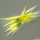
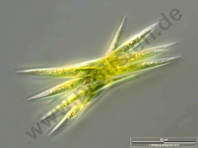
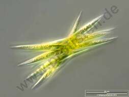
Scale bar indicates 25 µm.Sample from a wetland at the Pillersee (Tyrol, Austria). The image was built up using several photomicrographic frames with manual stacking technique. Images were taken using Zeiss Universal with Olympus C7070 CCD camera.Image under Creative Commons License V 3.0 (CC BY-NC-SA).
-
Villar del Pedroso, Extremadura, Spain
-
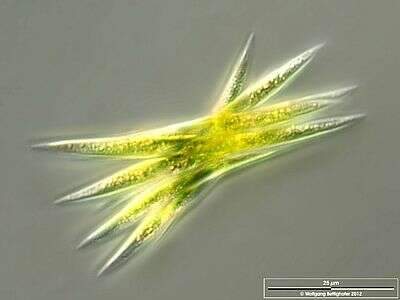
Scale bar indicates 25 m.Sample from a wetland at the Pillersee (Tyrol, Austria). The image was built up using several photomicrographic frames with manual stacking technique. Images were taken using Zeiss Universal with Olympus C7070 CCD camera.For permission to use of (high-resolution) images please contact postmaster@protisten.de.
-
San Martin De Castaneda, Castille and Leon, Spain
-
Melgar de Tera, Castille and Leon, Spain
-
Villar del Pedroso, Extremadura, Spain
-
Neila, Castille and Leon, Spain
-
Villar del Pedroso, Extremadura, Spain
-
Neila, Castille and Leon, Spain
-
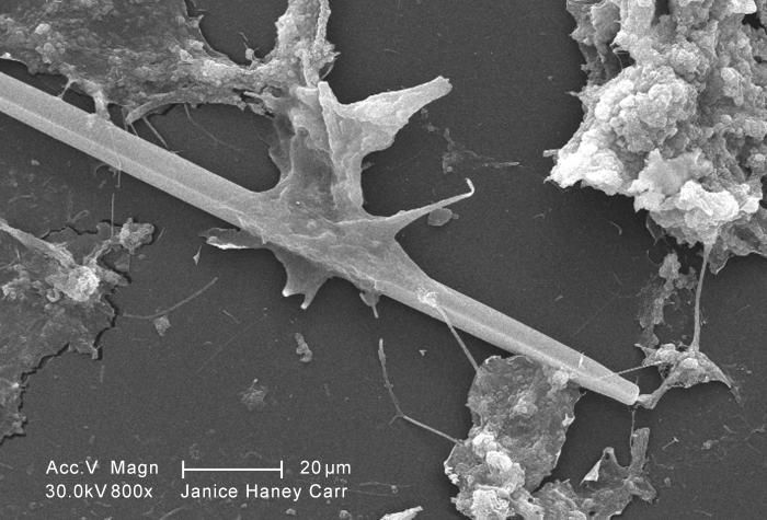
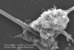
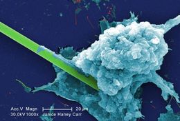
At a magnification of 1000X, this scanning electron micrograph (SEM) of an untreated water specimen extracted from a wild stream mainly used to control flooding during inclement weather, revealed the presence of unidentified organisms, which included bacteria, protozoa, and algae. In this particular image, a needle-shaped structure appeared to be caught up in an amorphous gelatinous biofilm, which though unidentified, appeared to be the green algae, Ankistrodesmus. See PHIL 11664 for another view of this spear-shaped organism. For a colorized version of this image see PHIL 11716.Created: 2009
-
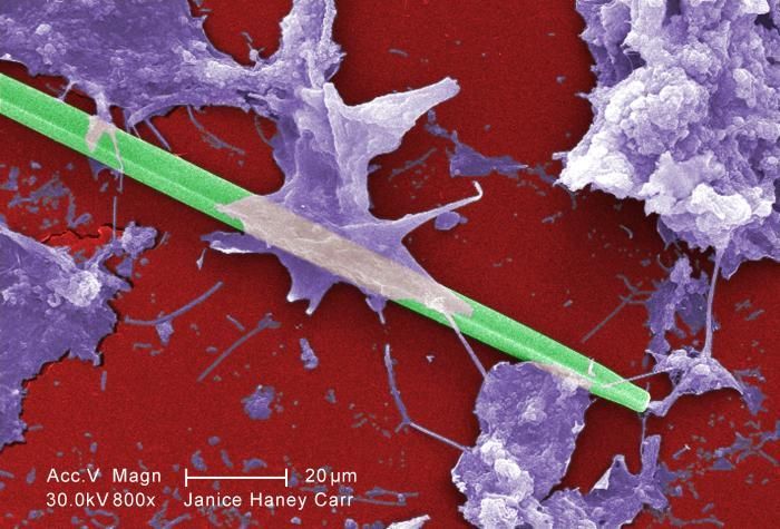
This scanning electron micrograph (SEM) of an untreated water specimen extracted from a wild stream mainly used to control flooding during inclement weather, revealed the presence of unidentified organisms, which included bacteria, protozoa, and algae. In this particular image, a needle-shaped structure appeared to be caught up in an amorphous gelatinous biofilm, which though unidentified, appeared to be the green algae, Ankistrodesmus. See PHIL 11697 for a colorized version of this image.Created: 2009
-
At a magnification of 1000X, this digitally-colorized scanning electron micrograph (SEM) of an untreated water specimen extracted from a wild stream mainly used to control flooding during inclement weather, revealed the presence of unidentified organisms, which included bacteria, protozoa, and algae. In this particular image, a needle-shaped structure appeared to be caught up in an amorphous gelatinous biofilm, which though unidentified, appeared to be the green algae, Ankistrodesmus. See PHIL 11664 for another view of this spear-shaped organism.Created: 2009
-
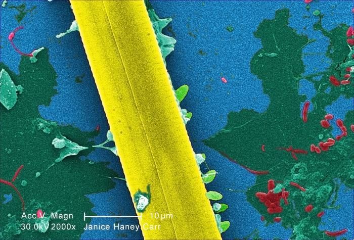
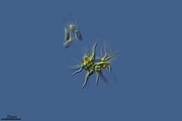
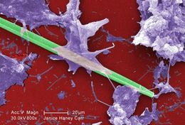
At a magnification of 2000X, this digitally-colorized scanning electron micrograph (SEM) of an untreated water specimen extracted from a wild stream mainly used to control flooding during inclement weather, revealed the presence of unidentified organisms, which included bacteria, protozoa, and algae. Though unidentified, this view depicts a section of a suspected green algae, Ankistrodesmus. Note the bacteria inhabiting the biofilm coating in the bachground.Created: 2009
-
This digitally-colorized scanning electron micrograph (SEM) of an untreated water specimen extracted from a wild stream mainly used to control flooding during inclement weather, revealed the presence of unidentified organisms, which included bacteria, protozoa, and algae. In this particular image, a needle-shaped structure appeared to be caught up in an amorphous gelatinous biofilm, which though unidentified, appeared to be the green algae, Ankistrodesmus.Created: 2009
-
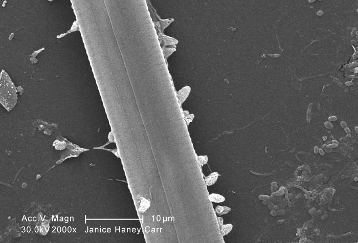
At a magnification of 2000X, this scanning electron micrograph (SEM) of an untreated water specimen extracted from a wild stream mainly used to control flooding during inclement weather, revealed the presence of unidentified organisms, which included bacteria, protozoa, and algae. Though unidentified, this view depicts a section of a suspected green algae, Ankistrodesmus. Note the bacteria inhabiting the biofilm coating in the bachground. See PHIL 11710 for a colorized version of this image.Created: 2009
-
At a magnification of 1000X, this digitally-colorized scanning electron micrograph (SEM) of an untreated water specimen extracted from a wild stream mainly used to control flooding during inclement weather, revealed the presence of unidentified organisms, which included bacteria, protozoa, and algae. In this particular image, a needle-shaped structure appeared to be caught up in an amorphous gelatinous biofilm, which though unidentified, appeared to be the green algae, Ankistrodesmus. See PHIL 11664 for another view of this spear-shaped organism.Created: 2009
-
At a magnification of 2000X, this digitally-colorized scanning electron micrograph (SEM) of an untreated water specimen extracted from a wild stream mainly used to control flooding during inclement weather, revealed the presence of unidentified organisms, which included bacteria, protozoa, and algae. Though unidentified, this view depicts a section of a suspected green algae, Ankistrodesmus. Note the bacteria inhabiting the biofilm coating in the bachground.Created: 2009
-
This digitally-colorized scanning electron micrograph (SEM) of an untreated water specimen extracted from a wild stream mainly used to control flooding during inclement weather, revealed the presence of unidentified organisms, which included bacteria, protozoa, and algae. In this particular image, a needle-shaped structure appeared to be caught up in an amorphous gelatinous biofilm, which though unidentified, appeared to be the green algae, Ankistrodesmus.Created: 2009
-
At a magnification of 2000X, this scanning electron micrograph (SEM) of an untreated water specimen extracted from a wild stream mainly used to control flooding during inclement weather, revealed the presence of unidentified organisms, which included bacteria, protozoa, and algae. Though unidentified, this view depicts a section of a suspected green algae, Ankistrodesmus. Note the bacteria inhabiting the biofilm coating in the bachground. See PHIL 11710 for a colorized version of this image.Created: 2009
-
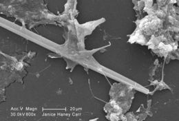
At a magnification of 1000X, this scanning electron micrograph (SEM) of an untreated water specimen extracted from a wild stream mainly used to control flooding during inclement weather, revealed the presence of unidentified organisms, which included bacteria, protozoa, and algae. In this particular image, a needle-shaped structure appeared to be caught up in an amorphous gelatinous biofilm, which though unidentified, appeared to be the green algae, Ankistrodesmus. See PHIL 11664 for another view of this spear-shaped organism. For a colorized version of this image see PHIL 11716.Created: 2009
-
This scanning electron micrograph (SEM) of an untreated water specimen extracted from a wild stream mainly used to control flooding during inclement weather, revealed the presence of unidentified organisms, which included bacteria, protozoa, and algae. In this particular image, a needle-shaped structure appeared to be caught up in an amorphous gelatinous biofilm, which though unidentified, appeared to be the green algae, Ankistrodesmus. See PHIL 11697 for a colorized version of this image.Created: 2009
-
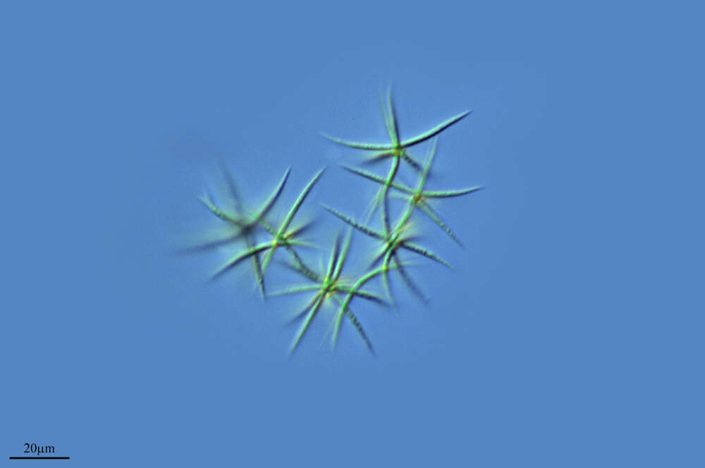
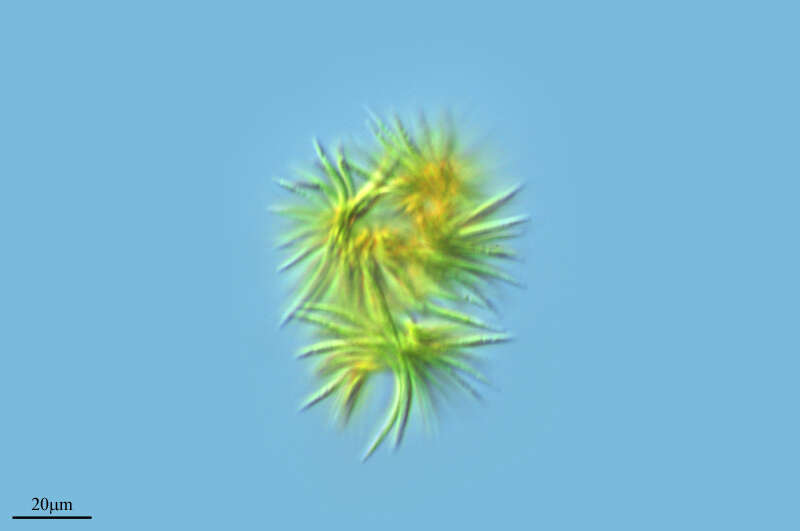
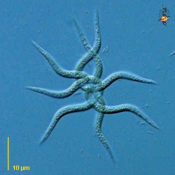
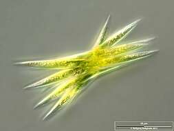
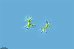
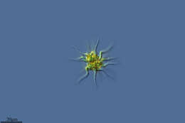
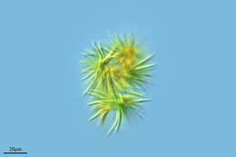
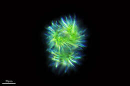
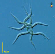
Ankistrodesmus (anne-kissed-ro-des-muss) is a green alga, small tangled clusters of twisted cells. Four cells in this tangle. Not uncommon in freshwater habitats. Differential interference contrast.
-
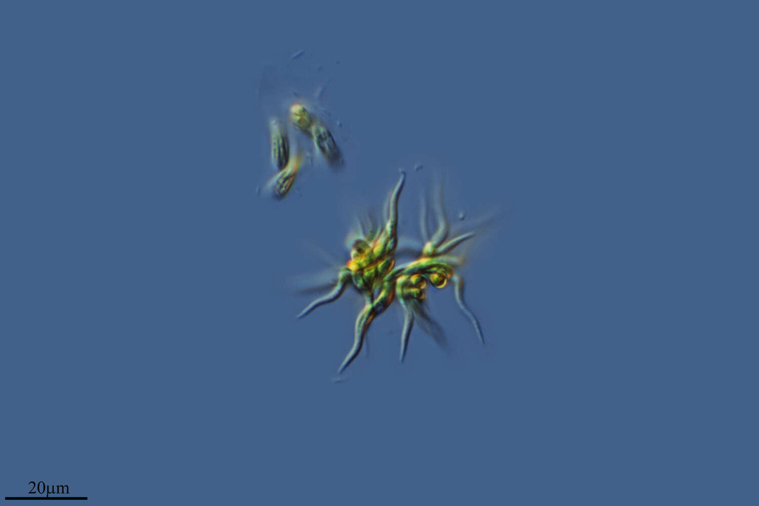
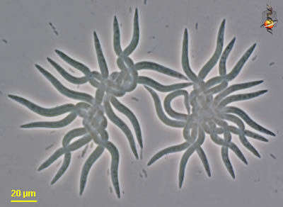
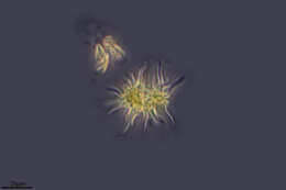
Ankistrodesmus (anne-kissed-ro-des-muss) is a green alga, small tangled clusters of twisted cells. Two tangles seen here making a spectacle of themselves. Not uncommon in freshwater habitats. Phase contrast.
-
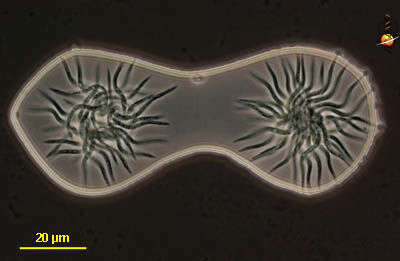
Ankistrodesmus (anne-kissed-ro-des-muss) is a green alga, small tangled clusters of twisted cells. Two tangles seen here Not uncommon in freshwater habitats. Phase contrast.