-
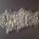
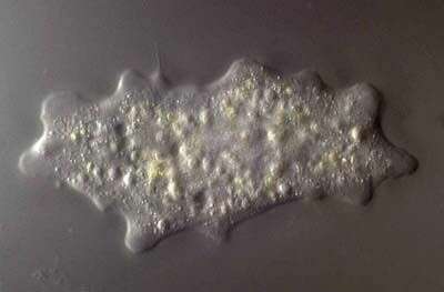
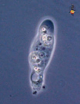
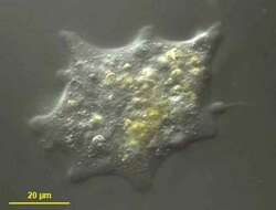
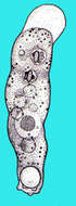
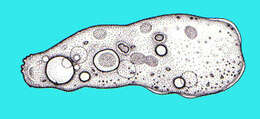
Trichospaherium is a marine amoeba that can occur in a spicule-covered form or as a naked form - as is illustrated here. the cone-shaped pseudopodia with terminal filaments are distinctive.
-
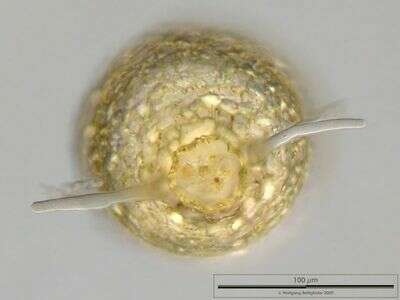
The lateral view shows an elliptical profile whereas the shape is circular in topview. Pseudopods appear as tapered protuberances of protoplasma. The aperture is almost as wide as the shell diameter, the rim is visible. Dimensions of specimen: 15µm x 20 µm Sample from a pond on the island of Hiddensee (Baltic Sea, Germany). This image was taken using Zeiss Universal with Olympus C7070 CCD camera.
-
Portrait of Netzelia tuberculata (Wallich,1864) test. The test or shell is composed of quartz particles arranged in small knob-like aggregates on its surface. The approximately circular shell aperture has a narrow collar of quartz particles. Pseudopodia (not seen in this image) are lobose. Collected from freshwater irrigation canal near Boise, Idaho in November 2003. Brightfield illumination.
-
-
Marine amoeba, exists as a spicule-covered form and as a naked form. the second is illustrated here. The naked form used to be called Pontifex maximum before it became clear that the two forms are of the same species.
-
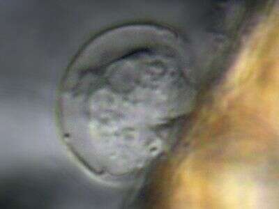
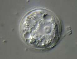
Cell lived on the neuston, the biofilm on the water surface. The test, the two contractile vacuoles and the nucleus with nucleolus are visible. Dimensions: Diameter 20 µm Sample from a pond on the island of Hiddensee (Baltic Sea, Germany). This image was taken using Zeiss Universal with Olympus C7070 CCD camera.
-
This species has been moved from genus Difflugia to Netzelia (family Lesqueruesiidae). Most of the agglutinated particles are silicious idiosoms, all xenosomes are covered with silica.This multi layer image was built up using 4 brightfield frames with a manual stacking technique using Corel Photopaint. The specimen was gathered in a tiny freshwater pond at the island of Hiddensee (Baltic Sea, Germany) which shows a fascinating biodiversity of naked and testate amoebae. Images were taken using Zeiss Standard with Olympus C7070 CCD camera.
-
-
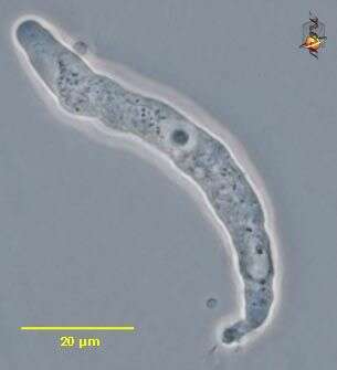
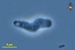
Hartmannella (heart-man-ella), a naked amoeba, limax (slug-like) body form, well developed hyaline cap, central nucleus and scrunched up uroidal region. Phase contrast.
-
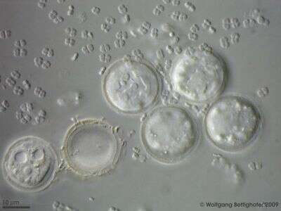
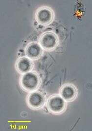
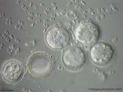
A group of Pyxidicula living on Hyponeuston, the aquatic area closely attached to the water surface, together with bacteria, probably their prey. On the left the nucleus and the two contractile vacuoles are visible, right to this specimen lies an empty test. Neuston has enough tension so that this testate amoebae can walk on it. Scale bar indicates 10 µm. Sample from a freshwater pond on the island of Hiddensee (Baltic Sea, Germany). This image was taken using Zeiss Universal with Olympus C7070 CCD camera.
-
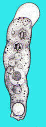
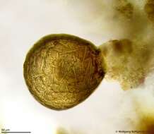
This species has been moved from genus Difflugia to Netzelia (family Lesqueruesiidae). Most of the agglutinated particles are silicious idiosoms, all xenosomes are covered with silica. This multi layer image of Netzelia tuberculata was built up using 12 brightfield frames with a manual stacking technique using Corel Photopaint. The scale bar indicates 50 µm. The specimen was gathered in a tiny freshwater pond at the island of Hiddensee (Baltic Sea, Germany) which shows a fascinating biodiversity of naked and testate amoebae. Images were taken using Zeiss Standard with Olympus C7070 CCD camera.
-
Hartmannella (heart-man-ella), a naked amoeba, limax (slug-like) body form, cysts. Phase contrast.
-
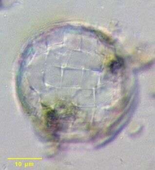
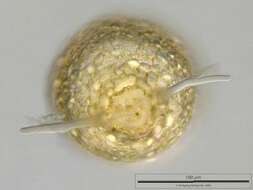
Paraquadrula irregularis test. This amoeba is a member of the Arcellinida. The test is round when viewed en-face (the view in these images) and ovoid in the lateral view. The test is composed of secreted rectangular calcite plates cemented into slightly irregular rows. The pseudostome is a broad oval shape (located towards the bottom in in these images). Although the test plates are similar in appearance to those of Quadrulella the plates are siliceous in the latter and the test of Quadrulella is pyriform with a distinct neck. Feeds on algae. From freshwater pond near Boise, Idaho. Oblique illumination.
-
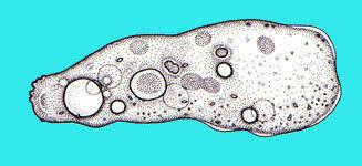
This species has been moved from genus Difflugia to Netzelia (family Lesqueruesiidae). Most of the agglutinated particles are silicious idiosoms, all xenosomes are covered with silica. The texture of the test and the outline of the pseudostome collar of Netzelia tuberculata are (according to R. Meisterfeld) very variable. This multi layer image was built up using 17 brightfield frames with a manual stacking technique using Corel Photopaint. The specimen was gathered in a tiny freshwater pond at the island of Hiddensee (Baltic Sea, Germany) which shows a fascinating biodiversity of naked and testate amoebae. Images were taken using Zeiss Standard with Olympus C7070 CCD camera.
-
Hartmannella (heart-man-ella), a naked amoeba, limax (slug-like) body form, well developed hyaline cap, central nucleus and scrunched up uroidal region. Phase contrast.
-
Paraquadrula irregularis - a member of the Arcellinida. The test is round when viewed en-face (the view in these images) and ovoid in the lateral view. The test is composed of secreted rectangular calcite plates cemented into slightly irregular rows. The pseudostome is a broad oval shape (located atowards the bottom in in these images). Although the test plates are similar in appearance to those of Quadrulella the plates are siliceous in the latter and the test of Quadrulella is pyriform with a distinct neck. Feeds on algae. From freshwater pond near Boise, Idaho. Phase contrast.
-
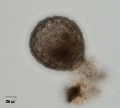
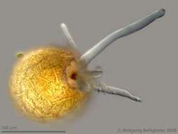
This species has been moved from genus Difflugia to Netzelia (family Lesqueruesiidae). Most of the agglutinated particles are silicious idiosoms, all xenosomes are covered with silica. Multi layer image of Netzelia specimen using 10 frames. Stack assembled with Corel Photopaint. The red dot is garbage waiting for defecation. Sample from sphagnum pond near Bergenhusen (Schleswig-Holstein, Germany). This image was taken using Zeiss Universal with Olympus C7070 CCD camera.
-
-
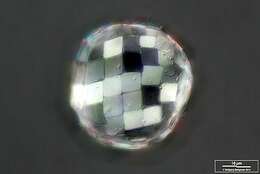
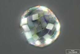
Paraquadrula builds its scales from calcite, which is birefringent. This is the reason why in DIC (polarized light) they show such different magnitudes. Scale bar indicates 10 µm. Sample from a bog near Reith/Pillersee (Tyrol, Austria). The image was built up using several photomicrographic frames with manual stacking technique. Images were taken using Zeiss Universal with Olympus C7070 CCD camera.Image under Creative Commons License V 3.0 (CC BY-NC-SA).
-
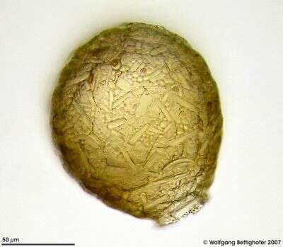
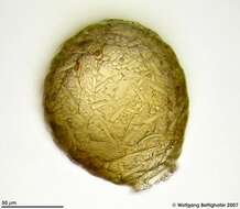
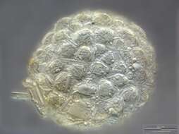
The test of Netzelia tuberculata shows its mulberry surface which gave the name (tuberculata). As all Lesquereusiidae Netzelia builds up the test with self-made siliceous pads (called idiosomes). A few xenosomes (mostly parts of frustule from pennate diatoms) are also visible. All xenosomes are covered with a siliceous coating. Scale bar indicates 25µm Sample from a freshwater pond on the island of Hiddensee (Baltic Sea, Germany). This image was taken using Zeiss Universal with Olympus C7070 CCD camera.
-
Cashia (cash-ee-a) - tentative identification - small limax (slug-shaped) amoeba, hyaline cap to right lacks inclusions, contractile vacuole is associated with the posterior end of the cell. Phase contrast.
-
Paraquadrula builds its scales from calcite, which is birefringent. This is the reason why in DIC (polarized light) they show such different magnitudes. Scale bar indicates 10 µm. Sample from a bog near Reith/Pillersee (Tyrol, Austria). The image was built up using several photomicrographic frames with manual stacking technique. Images were taken using Zeiss Universal with Olympus C7070 CCD camera.Image under Creative Commons License V 3.0 (CC BY-NC-SA).
-
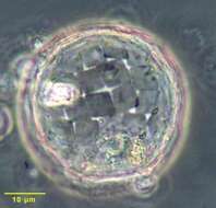
Apertural view on Netzelia tuberculata depicting the pseudostome collar, pseudopods and defecation vacuole. This multi layer image was built up using 42 frames with a manual stacking technique using Corel Photopaint. The specimen was gathered in a tiny freshwater pond at the island of Hiddensee (Baltic Sea, Germany) which shows a fascinating biodiversity of naked and testate amoebae. Images were taken using Zeiss Standard with Olympus C7070 CCD camera.
-