Plate 16
Description:
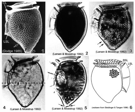
Plate 16. Dinophysis norvegica. Fig. 1. SEM: lateral view. Cell heavily areolated with pointed antapex and posterior protrusions (arrowheads). Ventral margin concave below left sulcal list (LSL)(arrow). Well developed cingular lists (CL) and LSL. Figs. 2-5. LM: lateral view. Fig. 2. Cell less robust than in Fig. 1; pointed antapex. Fig. 3. Robust cell with rounded antapex. Heavily areolated. Ventral margin straight below LSL (arrows). Fig. 4. Deepest point of cell through mid-point (dashed line), just above third rib of LSL. Fig. 5. Large posterior nucleus (n). Pointed antapex with posterior projections (arrows). Fig. 6. Line drawing. Right sulcal list depicted (RSL).
Included On The Following Pages:
- Life (creatures)
- Cellular (cellular organisms)
- Eukaryota (eukaryotes)
- SAR (Stramenopiles, Alveolates, Rhizaria)
- Alveolata (alveolates)
- Dinophyceae
- Dinophysiales
- Dinophysiaceae
- Dinophysis
- Dinophysis norvegica
- Dinoflagellata (dinoflagellates)
This image is not featured in any collections.
Source Information
- license
- cc-publicdomain
- bibliographic citation
- Faust, Maria A. and Rose A. Gulledge. Identifying Harmful Marine Dinoflagellates. Smithsonian Contributions from the United States National Herbarium, volume 42: 1-144 (including 48 plates, 1 figure and 1 table).
- original
- original media file
- visit source
- partner site
- NMNH Marine Dinoflagellates
- ID