Image of Exophiala salmonis J. W. Carmich. 1966
Description:
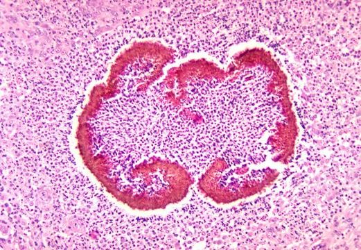
This micrograph depicts the histopathologic changes seen in black grain mycetoma due to Exophiala salmonis.
Created: 1971
Included On The Following Pages:
- Life (biota)
- Cellular
- Eukaryota (eukaryotes)
- Opisthokonta (opisthokonts)
- Nucletmycea
- Fungi (mushrooms, lichens, molds, yeasts and relatives)
- Dikarya
- Ascomycota (sac fungi)
- Eurotiomycetes
- Chaetothyriales
- Herpotrichiellaceae
- Exophiala
- Exophiala salmonis
- Pezizomycotina (Ascolichens)
- Chaetothyriomycetidae
This image is not featured in any collections.
Source Information
- license
- cc-publicdomain
- provider
- Public Health Image Library
- original
- original media file
- visit source
- partner site
- Public Health Image Library
- ID