Image of Eastern Carpenter Bee
Description:
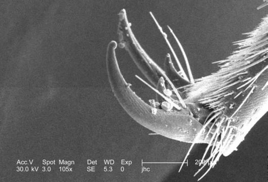
At a low magnification of 105x this scanning electron micrograph (SEM) depicted some of the morphologic details found at the distal end of a leg of a carpenter bee, Xylocopa virginica. Note the configuration of what is known as the "tarsal claw", used by the bee to grasp objects in its environment while obtaining food, or tunneling a nest. The "hairs" on the bee's leg, are not hairs in the mammalian sense, but rather than being composed of keratin, these hair-like structures are composed of "chitin", a polysaccharide, i.e., complex carbohydrate molecule, composed of monosaccharides joined together by glycosidic bonding. These hairs are sensorial, as well as protective, both insulating the bee from thermal changes in its environment, and possible physical assaults.
Created: 2006
Included On The Following Pages:
- Life (creatures)
- Cellular (cellular organisms)
- Eukaryota (eukaryotes)
- Opisthokonta (opisthokonts)
- Metazoa (Animal)
- Bilateria
- Protostomia (protostomes)
- Ecdysozoa (ecdysozoans)
- Arthropoda (arthropods)
- Pancrustacea
- Hexapoda (hexapods)
- Insecta (insects)
- Pterygota (winged insects)
- Neoptera (neopteran)
- Endopterygota (endopterygotes)
- Hymenoptera (wasps, bees, and ants)
- Apocrita (wasp)
- Aculeata
- Apoidea (bees & apoid Wasps)
- Apidae (honeybees, bumblebees, and relatives)
- Xylocopa (Carpenter Bees)
- Xylocopa virginica (Eastern Carpenter Bee)
- Panarthropoda
This image is not featured in any collections.
Source Information
- license
- cc-publicdomain
- photographer
- Janice Carr
- provider
- Public Health Image Library
- original
- original media file
- visit source
- partner site
- Public Health Image Library
- ID