Egranulosus cysts HB
Description:
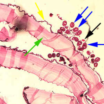
Description: English: Cross-section of an Echinococcus granulosus cyst, stained with H&E. The cyst wall is composed of an acellular laminated external layer (green arrow) and a thin, germinal (nucleated) inner layer (yellow arrow). Note the brood capsule (black arrow) with protoscoleces (blue arrows) inside. Image taken at 40x magnification. Čeština: Příčný řez stěnou cysty Echinococcus granulosus, barveno hematoxylin-eosinem. Zelená šipka - laminární, vnější, nebuněčná vrstva Žlutá šipka - zárodečná vrstva Černá šipka - dceřiná cysta Modré šipky - protoskolexy Zvětšení 40x. Date: 4 August 2016. Source: CDC-DPDx https://www.cdc.gov/dpdx/echinococcosis/gallery.html#intissue. Author: CDC.
Included On The Following Pages:
- Biota
- Eukaryota (eukaryotes)
- Unikonta
- Opisthokonta (opisthokonts)
- Distaplia
- Filozoa
- Apoikozoa
- Animalia
- Eumetazoa
- Bilateria
- Protostomia (protostomes)
- Platyzoa
- Platyhelminthes (flatworms)
- Cestoda (cestodes)
- Eucestoda
- Cyclophyllidea
- Taeniidae
- Echinococcus
- Echinococcus granulosus
This image is not featured in any collections.
Source Information
- license
- cc-publicdomain
- creator
- CDC
- source
- CDC-DPDx https://www.cdc.gov/dpdx/echinococcosis/gallery.html#intissue
- original
- original media file
- visit source
- partner site
- Wikimedia Commons
- ID