Photomicrographs from a foothill yellow-legged frog (Rana boylii)
Description:
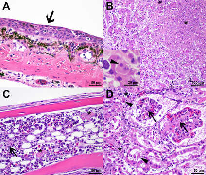
Description: English: Photomicrographs from a foothill yellow-legged frog (Rana boylii) found dead in Santa Clara, California, USA. (A) Small areas of epidermal necrosis with apoptotic keratinocytes and nuclear debris are multifocally present (arrow). (B) The liver shows randomly distributed, variably sized areas of coagulative necrosis (*). A few adjacent hepatocytes have intracytoplasmic, round, basophilic, 2 to 4-µm inclusion bodies (inset, arrowhead). (C) Within the bone marrow are necrotic hematopoietic cells (arrow) and endothelial cells with consecutive fibrin thrombi (*). (D) Different processes are present in the kidney. Glomeruli are affected due to necrosis of the endothelial cells (arrow). The interstitium, normally containing hematopoietic cells, show widespread hematolymphoid necrosis (*). Additionally, proximal tubules show epithelial necrosis with hypereosinophilic sloughed epithelial cells (arrowhead). Date: 4 February 2020, 10:55:57. Source: U.S. Geological Survey. Author: Saskia Keller, USGS National Wildlife Health Center. Public domain.
Included On The Following Pages:
- Life (creatures)
- Cellular (cellular organisms)
- Eukaryota (eukaryotes)
- Opisthokonta (opisthokonts)
- Metazoa (Animal)
- Bilateria
- Deuterostomia (deuterostomes)
- Chordata (Chordates)
- Vertebrata (vertebrates)
- Gnathostomata (jawed fish)
- Osteichthyes
- Sarcopterygii (Lobe-finned fishes)
- Tetrapoda (terrestrial vertebrates)
- Lissamphibia (amphibians)
- Anura (frogs and toads)
- Ranidae (true frogs)
- Rana
- Rana boylii (Foothill yellow-legged frog)
This image is not featured in any collections.
Source Information
- license
- cc-publicdomain
- creator
- Saskia Keller, USGS National Wildlife Health Center. Public domain.
- source
- U.S. Geological Survey
- original
- original media file
- visit source
- partner site
- Wikimedia Commons
- ID
.jpg)
.jpg)