Conidiospore-hyaloperonospora-parasitica-appressorium
Description:
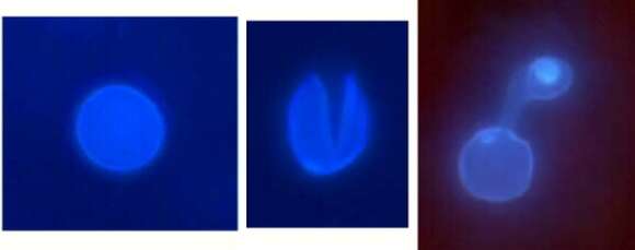
Description: English: Conidiospores of Hyaloperonospora parasitica (downy mildew) germinating on the leaf of Arabidopsis thaliana (cf.wikipedia) observed by fluorescent microscopy. Double staining: The staining with calcofluor makes the cell walls of H. parasitica fluoresceing in blue and the staining with aniline blue staining reveal callose deposition in a blue/yellow color. From left to right: fresh conidiospore intact, opening conidiospore starting to germinate (3h after inoculation), germinated conidiospore (6h after inoculation) with a short germ tube trying to penetrate between cells of A. thaliana by doing an appressorium surrounded by callose deposition. This is the first step of the short life cycle of H. parasitica. Français : Germination de conidiospores de Hyaloperonospora parasitica (Mildiou) à la surface d'une feuille d'Arabidopsis thaliana (cf.wikipedia) observée au microscope optique à fluorescence. Double coloration : Le calcofluor rend les parois cellulaires de H. parasitica fluorescentes en bleu, tandis que le bleu d'aniline permet de visualiser la déposition de callose an bleu/jaune. De gauche à droite : jeune conidiospore intact, ouverture d'un conidiospore durant sa germination (3h après inoculation), conidiospore germé (6h après inoculation) avec un court tube germinatif qui essaye de pénétrer entre les cellules d' A. thaliana avec un appressorium entouré de dépositions de callose. C'est la première étape du cycle court de H. parasitica. Date: 2002. Source: Own work. Author: Emmanuel Boutet. Permission(Reusing this file): Own work, copyleft: Multi-license with GFDL and Creative Commons CC-BY-SA-2.5 and older versions (2.0 and 1.0).
Included On The Following Pages:
- Life (creatures)
- Cellular (cellular organisms)
- Eukaryota (eukaryotes)
- SAR (Stramenopiles, Alveolates, Rhizaria)
- Stramenopiles (heterokont)
- Oomycota (oomycetes)
- Peronosporales
- Peronosporaceae (downy mildews)
- Hyaloperonospora
- Hyaloperonospora parasitica
- Hyaloperonospora parasitica
This image is not featured in any collections.
Source Information
- license
- cc-by-sa-3.0
- copyright
- Emmanuel Boutet
- creator
- Emmanuel Boutet
- original
- original media file
- visit source
- partner site
- Wikimedia Commons
- ID