Klebsiella pneumoniae on CLED agar - Detail
Description:
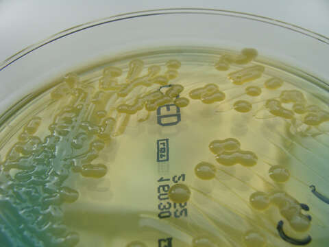
Summary.mw-parser-output table.commons-file-information-table,.mw-parser-output.fileinfotpl-type-information{border:1px solid #a2a9b1;background-color:#f8f9fa;padding:5px;font-size:95%;border-spacing:2px;box-sizing:border-box;margin:0;width:100%}.mw-parser-output table.commons-file-information-table>tbody>tr,.mw-parser-output.fileinfotpl-type-information>tbody>tr{vertical-align:top}.mw-parser-output table.commons-file-information-table>tbody>tr>td,.mw-parser-output table.commons-file-information-table>tbody>tr>th,.mw-parser-output.fileinfotpl-type-information>tbody>tr>td,.mw-parser-output.fileinfotpl-type-information>tbody>tr>th{padding:4px}.mw-parser-output.fileinfo-paramfield{background:#ccf;text-align:right;padding-right:0.4em;width:15%;font-weight:bold}.mw-parser-output.commons-file-information-table+table.commons-file-information-table,.mw-parser-output.commons-file-information-table+div.commons-file-information-table>table{border-top:0;padding-top:0;margin-top:-8px}@media only screen and (max-width:719px){.mw-parser-output table.commons-file-information-table,.mw-parser-output.commons-file-information-table.fileinfotpl-type-information{border-spacing:0;padding:0;word-break:break-word;width:100%!important}.mw-parser-output.commons-file-information-table>tbody,.mw-parser-output.fileinfotpl-type-information>tbody{display:block}.mw-parser-output.commons-file-information-table>tbody>tr>td,.mw-parser-output.commons-file-information-table>tbody>tr>th,.mw-parser-output.fileinfotpl-type-information>tbody>tr>td,.mw-parser-output.fileinfotpl-type-information>tbody>tr>th{padding:0.2em 0.4em;text-align:left;text-align:start}.mw-parser-output.commons-file-information-table>tbody>tr,.mw-parser-output.fileinfotpl-type-information>tbody>tr{display:flex;flex-direction:column}.mw-parser-output.commons-file-information-table+table.commons-file-information-table,.mw-parser-output.commons-file-information-table+div.commons-file-information-table>table{margin-top:-1px}.mw-parser-output.fileinfo-paramfield{box-sizing:border-box;flex:1 0 100%;width:100%}} Description: Klebsiella pneumoniae on Cystine Lactose Electrolyte Deficient Agar Bromothymol blue indicator in the agar changes to yellow due to acidification of the medium due to lactose fermentation by bacterial growth. Lactose fermenters appear yellow Non Lactose fermenters remain a translucent blue. Date: 13 September 2011, 05:49. Source: Klebsiella pneumoniae on C.L.E.D. Agar - detail. Author: Nathan Reading from Halesowen, UK.
Included On The Following Pages:
- Life (creatures)
- Cellular (cellular organisms)
- Bacteria
- Proteobacteria (Purple Bacteria & relatives)
- Gammaproteobacteria
- Enterobacterales
- Enterobacteriaceae
- Klebsiella
- Klebsiella pneumoniae
This image is not featured in any collections.
Source Information
- license
- cc-by-3.0
- copyright
- Nathan Reading
- creator
- Nathan Reading
- source
- Flickr user ID nathanreading
- original
- original media file
- visit source
- partner site
- Wikimedia Commons
- ID