Image of Imadateiella
Description:
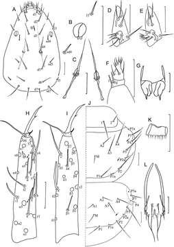
Figure 7.Imadateiella sharovi (Martynova, 1977) A Head, dorsal view B pseudoculus C canal of maxillary gland D labial palpus (specimens from samples 24 and 27) E labial palpus (specimens from samples 26, 30 and 32) F maxillary palpus G female squama genitalis H foretarsus, exterior view I foretarsus, interior view J nota, right side K comb L male squama genitalis. A–D and F–L specimen No. FE-2011062-4; E specimen No. FE-2011060-1. Arrows indicate pores. Scale bar: 20 μm.
Included On The Following Pages:
- Life (creatures)
- Cellular (cellular organisms)
- Eukaryota (eukaryotes)
- Opisthokonta (opisthokonts)
- Metazoa (Animal)
- Bilateria
- Protostomia (protostomes)
- Ecdysozoa (ecdysozoans)
- Arthropoda (arthropods)
- Pancrustacea
- Hexapoda (hexapods)
- Protura (proturans)
- Acerentomidae
- Imadateiella
- Imadateiella sharovi
- Panarthropoda
This image is not featured in any collections.
Source Information
- license
- cc-by-3.0
- copyright
- Yun Bu, Mikhail B. Potapov, Wen Ying Yin
- bibliographic citation
- Bu Y, Potapov M, Yin W (2014) Systematic and biogeographical study of Protura (Hexapoda) in Russian Far East: new data on high endemism of the group ZooKeys 424: 19–57
- original
- original media file
- visit source
- partner site
- Zookeys
- ID