Image of Xyphinus
Description:
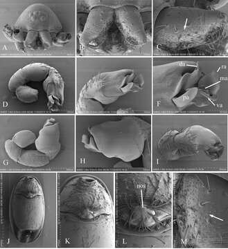
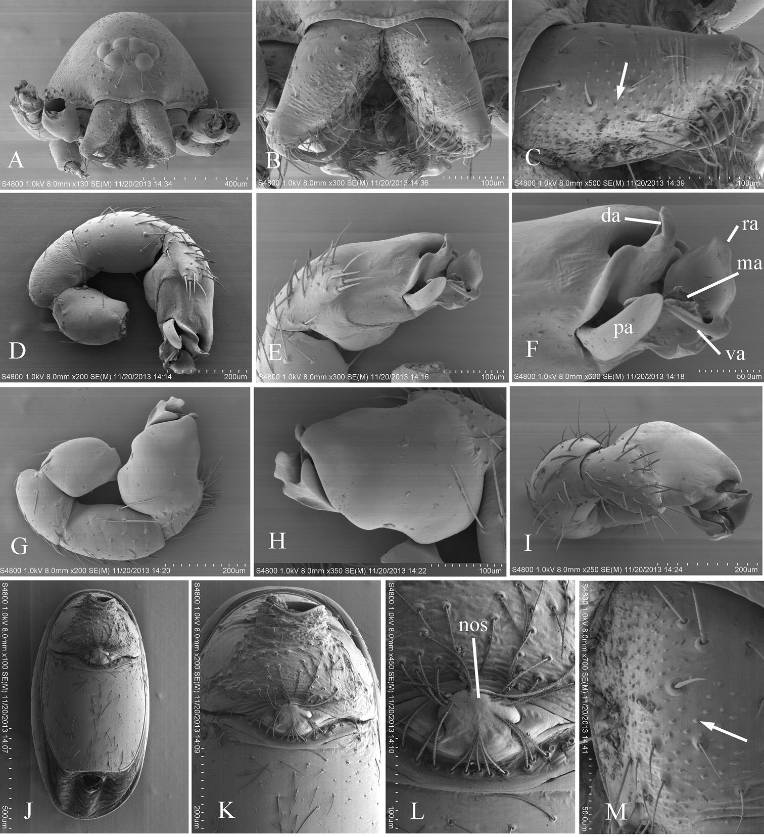
Figure 6.Xyphinus hwangi sp. n., SEM. A male prosoma, anterior view B, C, M male chelicerae, frontal view (arrow shows the small granules) D, G, I male left palp, prolateral, retrolateral and dorsal views E, H male left palpal bulb, prolateral and retrolateral views F distal part of male left palpal bulb, prolateral view J female abdomen, ventral view K, L female genital area, ventral view. Abbreviations: da = dorsal apophysis; ma = medial apophysis; nos = nose-shaped protuberance; pa = prolateral apophysis; ra = retrolateral apophysis; va = ventral apophysis.
Included On The Following Pages:
- Life (creatures)
- Cellular (cellular organisms)
- Eukaryota (eukaryotes)
- Opisthokonta (opisthokonts)
- Metazoa (Animal)
- Bilateria
- Protostomia (protostomes)
- Ecdysozoa (ecdysozoans)
- Arthropoda (arthropods)
- Chelicerata (chelicerates)
- Arachnida (arachnids)
- Araneae (spiders)
- Opisthothelae
- Araneomorphae
- Haplogynae
- Oonopidae (goblin spiders)
- Xyphinus
- Xyphinus hwangi
- Panarthropoda
This image is not featured in any collections.
Source Information
- license
- cc-by-3.0
- copyright
- Yanfeng Tong, Shuqiang Li
- bibliographic citation
- Tong Y, Li S (2014) A survey of oonopid spiders in Taiwan with descriptions of three new species ZooKeys 396: 67–86
- original
- original media file
- visit source
- partner site
- Zookeys
- ID