Life cycle of the nematode (roundworm) Toxocara canis, the most common cause of Toxocariasis in humans
Description:
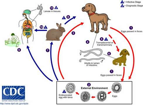
Life cycle of the nematode (roundworm) Toxocara canis, the most common cause of Toxocariasis in humans
Toxocara canis completes its life cycle in dogs, with humans acquiring the infection as accidental hosts. Unembryonated eggs are shed in the feces of the definitive host (1). Eggs embryonate and become infective in the environment (2). Following ingestion by dogs (3), the infective eggs hatch and larvae penetrate the gut wall. In younger dogs, the larvae migrate through the lungs, bronchial tree, and esophagus; adult worms develop and oviposit in the small intestine (4). In older dogs, patent infections can also occur, but larval encystment in tissues is more common (an infection is "patent" when direct evidence of the organism can be detected, e.g., in the patient’s feces or blood, regardless of whether symptoms have appeared). Encysted stages are reactivated in female dogs during late pregnancy and infect by the transplacental and transmammary routes the puppies (5), in whose small intestine adult worms become established (6). Puppies are a major source of environmental egg contamination. Toxocara canis can also be transmitted through ingestion of paratenic hosts ("transport hosts"): eggs ingested by small mammals (e.g. rabbits) hatch and larvae penetrate the gut wall and migrate into various tissues where they encyst (7). The life cycle is completed when dogs eat these hosts (8) and the larvae develop into egg-laying adult worms in the small intestine. Humans are accidental hosts who become infected by ingesting infective eggs in contaminated soil (9) or infected paratenic hosts (10). After ingestion, the eggs hatch and larvae penetrate the intestinal wall and are carried by the circulation to a wide variety of tissues (liver, heart, lungs, brain, muscle, eyes) (11). While the larvae do not undergo any further development in these sites, they can cause severe local reactions that are the basis of toxocariasis. The two main clinical presentations of toxocariasis are visceral larva migrans and ocular larva migrans. Diagnosis is usually made by serology or the finding of larvae in biopsy or autopsy specimens.
From Centers for Disease Control Parasites and Health website.
Included On The Following Pages:
- Life (creatures)
- Cellular (cellular organisms)
- Eukaryota (eukaryotes)
- Opisthokonta (opisthokonts)
- Metazoa (Animal)
- Bilateria
- Protostomia (protostomes)
- Ecdysozoa (ecdysozoans)
- Nematoda (nematodes)
- Ascarididae
- Toxocara
- Secernentea
- Ascaridida
- Toxocara canis (dog roundworm)
This image is not featured in any collections.
Source Information
- license
- cc-by-nc
- copyright
- Centers for Disease Control/Division of Parasitic Diseases and Malaria
- publisher
- Shapiro, Leo
- photographer
- Centers for Disease Control/Division of Parasitic Diseases and Malaria
- provider
- EOL Rapid Response Team
- original
- original media file
- visit source
- partner site
- EOL staff
- ID