portrait
Description:
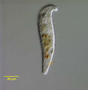
Ventrolateral surface view of the nassophorean marine ciliate, Orthodonella hamatus (Bhatia, 1936). The elongate cell is dorsoventrally flattened with the anterior end bent to the left in a blunt hook shape. The posterior is bluntly tapered. The oral aperture is right anterior. There is a cytopharyngeal apparatus composed of stout nematodesmata (not seen in this image). The longitudinal somatic kineties are uniform. There is a sigmoid post-oral frange extending obliquely from the anterior end just posterior to the cytostome almost to the right edge (seen well in this image). The ovoid macronucleus and adjacent micronucleus are located in the mid-cell. A posterior peripheral contractile vacuole is seen in this image. O. hamatus is also found in freshwater habitats. Orthodonella is a monotypic genus. Collected from a commercial saltwater aquarium in Boise, Idaho February 2004. DIC optics.
Included On The Following Pages:
- Life (creatures)
- Cellular (cellular organisms)
- Eukaryota (eukaryotes)
- SAR (Stramenopiles, Alveolates, Rhizaria)
- Alveolata (alveolates)
- Ciliophora (ciliates)
- Intramacronucleata
- Nassophorea
- Synhymeniida
- Orthodonellidae
- Orthodonella
- Orthodonella hamata
This image is not featured in any collections.
Source Information
- license
- cc-by-nc
- author
- Bill Bourland
- provider
- micro*scope
- original
- original media file
- visit source
- partner site
- micro*scope
- ID