Drawing
Description:
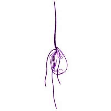
Trimastix marina Kent, 1880. Cells are 9 to 30 microns long, are broadly pyriform with an apical point. The ventral side consists of a wide groove which terminates near the posterior end in a discrete ingestion apparatus. Four flagella insert at the head of a groove in a cruciate pattern: one directed anteriorly, one posteriorly, one leftward and one rightward. The anterior and posterior flagella are one and a half to three times the length of the cell. The anterior flagellum inserts almost apically. It is substantially thickened at its base but gradually tapers to a conventional thickness. The proximal portion of the posterior flagellum usually beats within the groove with wide amplitude, short wavelength waves and two broad vanes may sometimes be discerned. The flagellum may appear attached to the posterior margin of the groove. The subequal lateral flagella are one to two times the length of the cell and are of a normal thickness. The nucleus is located anteriorly, is large, ovoid to pyriform, and contains a conspicuous, irregularly shaped nucleolus. The cell surface sometimes appears dimpled with the light microscope. Captured bacteria collect in posteriorly located food vacuoles. Remains were occasionally observed being expelled from the posterior end. Irregular threads or processes may also form posteriorly. A posterior contractile vacuole is present in cells from freshwater sites. The cells usually swim in relatively straight lines with a slow rotation and with the anterior flagellum directed anteriorly. This flagellum may also be directed ventro-posteriorly, causing motion in a more curved path. The cells may attach to the substrate by the posterior flagellum or by posterior cytoplasmic strands. Ultrastructural studies of the freshwater isolate demonstrate that the cells lack mitochondria, and confirm the presence of two opposed, very broad vanes on the posterior flagellum.
Included On The Following Pages:
- Life (creatures)
- Cellular (cellular organisms)
- Eukaryota (eukaryotes)
- Excavates (excavates)
- Metamonada (metamonad)
- Preaxostyla
- Trimastigidae
- Trimastix
- Trimastix marina
This image is not featured in any collections.
Source Information
- license
- cc-by-nc
- author
- Won Je Lee
- provider
- micro*scope
- original
- original media file
- visit source
- partner site
- micro*scope
- ID