portrait
Description:
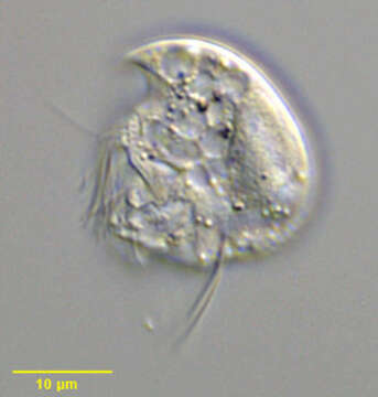
Portrait (left side) of the small Odontostome ciliate, Mylestoma anatinum. The cell is colorless with a laterally compressed rigid transparent pellicle typical of Odontostomes. The outline is roughly discoid with the dorsal convexity ending in a sharp anterior beak. The posterior is broadly rounded without spinous projections. The cytostome is located in a posterior depression (viewer's left). Somatic ciliature is strongly reduced to two long posterior cirri, an adoral zone of membranelles and a small perizonal complex anterior to the cytostome. There is a single posterior contractile vacuole and a single macronucleus. A prominent micronucleus is seen in this image at the posterior edge of the macronucleus. Refractile brownish cytoplasmic granules are seen posteriorly. Collected from bottom sediments of a freshwater aquaculture tank near Boise, Idaho December 2003. DIC optics.
Included On The Following Pages:
- Life (creatures)
- Cellular (cellular organisms)
- Eukaryota (eukaryotes)
- SAR (Stramenopiles, Alveolates, Rhizaria)
- Alveolata (alveolates)
- Ciliophora (ciliates)
- Intramacronucleata
- Plagiopylea
- Odontostomatida
- Mylestomatidae
- Mylestoma
- Mylestoma anatinum
This image is not featured in any collections.
Source Information
- license
- cc-by-nc
- author
- William Bourland
- provider
- micro*scope
- original
- original media file
- visit source
- partner site
- micro*scope
- ID