terminal cell
Description:
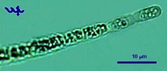
This photo shows the typical terminal cells of Aphanizomenon ovalisproum from Lake Kinneret. The filaments, 2 â 5 µm thick and 20 â 500 µm long, are terminated by an elongated hyaline cell, as seen in this picture.
Included On The Following Pages:
- Life (creatures)
- Cellular (cellular organisms)
- Bacteria
- Cyanobacteria
- Nostocales
- Aphanizomenonaceae
- Chrysosporum
- Chrysosporum ovalisporum
- Terrabacteria
- Cyanobacteria/Melainabacteria group
This image is not featured in any collections.
Source Information
- license
- cc-by-nc
- author
- Alla Alster; Tamar Zohary
- provider
- micro*scope
- original
- original media file
- visit source
- partner site
- micro*scope
- ID