Image of Mcvaughia
Description:
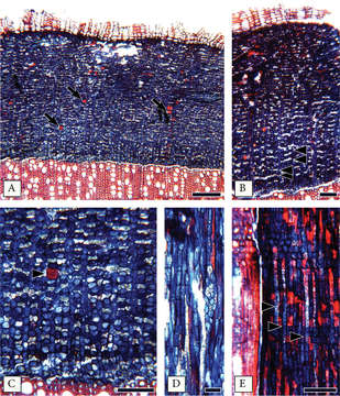
Figure 2. Secondary phloem of Mcvaughiasergipana. A–C Transverse section: A Phloem non-stratified, with scattered fiber-sclereids (arrows); Rays dilating slightly B Crystalliferous axial parenchyma arranged in diffuse-in-aggregate narrow bands (arrowhead) C Crystalliferous axial parenchyma with druse crystals, forming diffuse in aggregate bands, isolated fiber-sclereids present (arrowhead) D A ray 3 cells wide, fiber-sclereids and axial parenchyma in tangential section E Bands of crystalliferous parenchyma with druses evident also in radial section; Rays heterocellular mixed. Scale bars: 200 μm (A), 100 μm (B–C, E), 50 μm (D).
Included On The Following Pages:
- Life (creatures)
- Cellular (cellular organisms)
- Eukaryota (eukaryotes)
- Archaeplastida (plants)
- Chloroplastida (green plants)
- Spermatophytes (seed plants)
- Angiosperms (Dicotyledons)
- Eudicots
- Superrosids
- Rosids
- Malpighiales
- Malpighiaceae (Barbados cherry family )
- Mcvaughia
- NO NAME!
This image is not featured in any collections.
Source Information
- license
- cc-by-3.0
- copyright
- Rafael F. Almeida, Isabel R. Guesdon, Marcelo R. Pace, Renata M.S. Meira
- bibliographic citation
- Almeida R, Guesdon I, Pace M, Meira R (2019) Taxonomic revision of Mcvaughia W.R.Anderson (Malpighiaceae): notes on vegetative and reproductive anatomy and the description of a new species PhytoKeys (117): 45–72
- original
- original media file
- visit source
- partner site
- Phytokeys
- ID