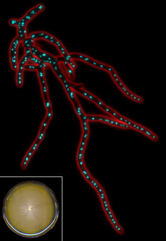
Description: English: Fluorescent micrograph of Ashbya gossypii mycelium with DAPI labeled nuclei. Blue: nuclei (DNA), red: outline of the cell (phase contrast), inlay: A. gossypii grown on a full medium petri dish. Yellowish color is the result of riboflavin production. Date: March 2007. Source: self-made (Transferred from
en.wikipedia to Commons by
Ayacop.). Author:
Spitfire ch, Philippsen Lab, Biozentrum Basel. Permission(
Reusing this file): GFDL (self made).