-
Le Calvez, Y. (1970). Contribution à l'étude des Foraminifères paléogènes du Bassin de Paris. Éditions du Centre national de la recherche scientifique. 1-326. page(s): p. 69 pl. 4 fig. 6
-
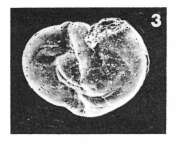
Le Calvez, Y. (1970). Contribution à l'étude des Foraminifères paléogènes du Bassin de Paris. Éditions du Centre national de la recherche scientifique. 1-326. page(s): p. 47 pl. 5 fig. 6
-
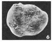
Le Calvez, Y. (1970). Contribution à l'étude des Foraminifères paléogènes du Bassin de Paris. Éditions du Centre national de la recherche scientifique. 1-326. page(s): p. 65 pl. 3 fig. 3
-
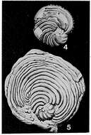
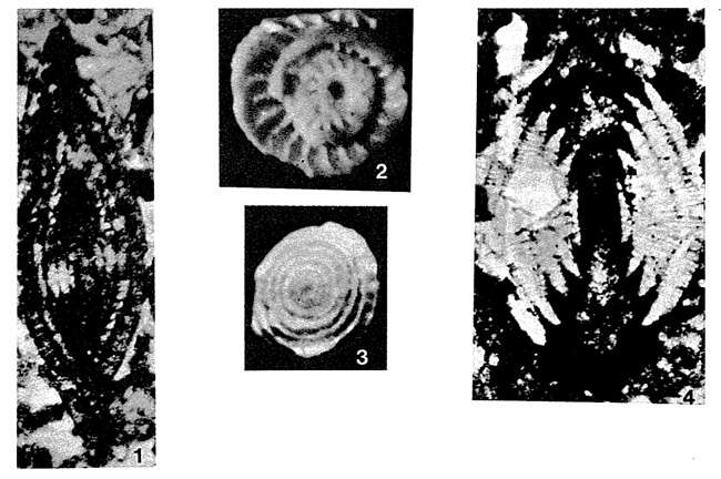
Loeblich, A. R., Tappan, H. N., 1987: Foraminiferal genera and their classification. Van Nostrand, Reinhold Co. New York 1728 pp. Plate 390, Figs. 4, 5: M. Oligocene, Bretagne, France. 3, Spiral face, x 16; 4, 5, exterior of partly decorticated topotypes, x 37; 6, edge view showing natural axial half-section, x 16 (from Loeblich and Tappan, 1964). courtesy of Michael Hesemann https://foraminifera.eu
-
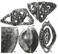
Loeblich, A. R., Tappan, H. N., 1987: Foraminiferal genera and their classification. Van Nostrand, Reinhold Co. New York 1728 pp. Plate 400, Figs. 1-4: U. Cretaceous (Senonian); 1-4, Dordogne, France. 1, Axial section showing original lenticular form, x 30; 2, equatorial half-section of free specimen, x 50; 3, detached umbilical button, possibly a result of recrystallization, x 50; 4, axial section of specimen with advanced stage of recrystallization producing umbilical buttons at each side (appearing white), x 30 (from Barrier and Neumann, 1959). courtesy of Michael Hesemann https://foraminifera.eu
-
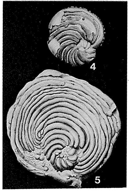
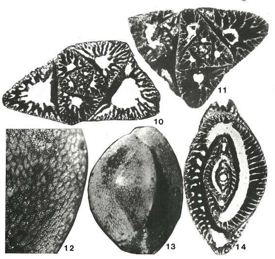
Loeblich, A. R., Tappan, H. N., 1987: Foraminiferal genera and their classification. Van Nostrand, Reinhold Co. New York 1728 pp. Plate 362, Figs. 10-14: L. Miocene (Bairnsdalian), South Australia. 10, 11, Transverse sections of microspheric tests, x 40 and x 50, respectively; 12, 13, side view of exterior, surface enlarged (magnification not given), and entire specimen, x 35; 14, longitudinal section of megalospheric test, magnification not given (from Adams, 1968).
-
Pseudohauerina occidentalis from Twin Cays, Belize
-
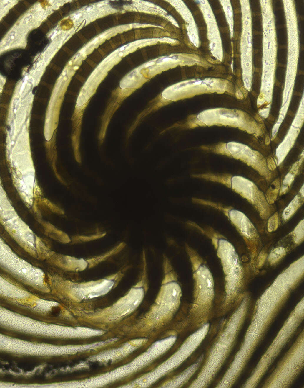
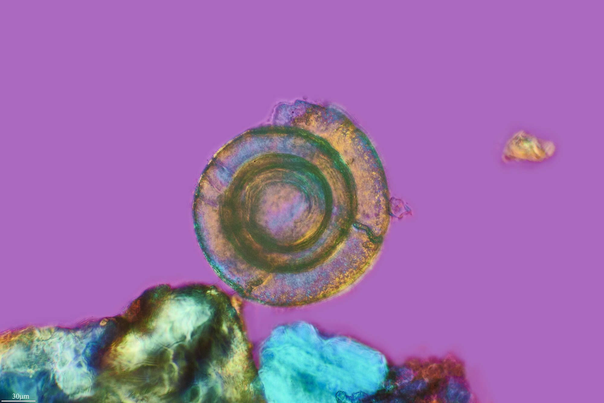
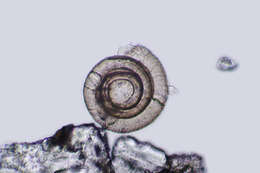
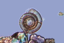
Cycloputeolina discoidea (Flint, 1899). Photomicrograph of early coil in specimen figured in Flint, 1899, Plate 49, fig. 2 (left image). Uncatalogued slide F-4446 in Cushman Collection of Foraminifera, National Museum of Natural History, Smithsonian Institution, Washington, DC.
-
Almera, Andalusia, Spain
-
Almera, Andalusia, Spain
-
Almera, Andalusia, Spain
-
Almera, Andalusia, Spain
-
Almera, Andalusia, Spain
-
Extreme macro: take a good binocular microscope, a good camera and just focus your microfossils ;)
-
Pyrgo williamsonifound on the shelf of Greenland at 192m depthrecentSend me your sand or rock ! I extract the foraminifera and shoot the images for free. Info at
www.foraminifera.eu
-
Pyrgo williamsonifound on the shore of SpitsbergenrecentSend me your sand or rock ! I extract the foraminifera and shoot the images for free. Info at
www.foraminifera.eu
-
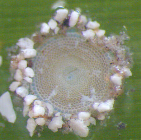
Live individual of the foraminiferan Sorites dominicensis attached to a blade of turtle grass (Thalassia testudinum) from Belize
-
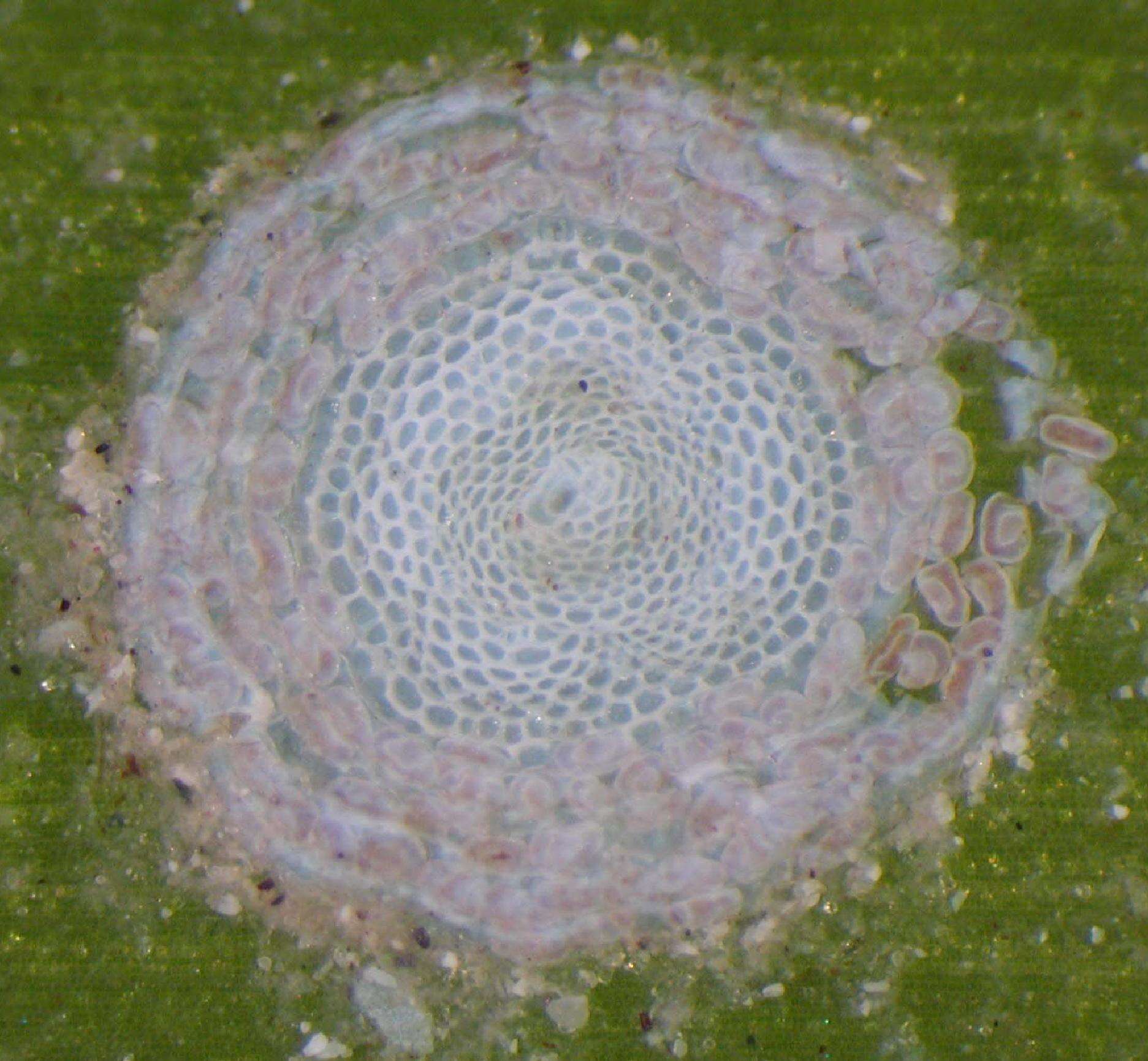
Live individual of Sorites dominicensis undergoing reproduction by multiple fission
-
-
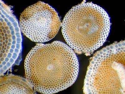
Live individuals of the foraminiferans Archaias angulatus and one specimen of Coscinospira (=Peneroplis) antillarum from Belize
-
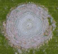
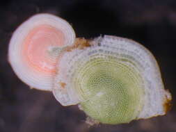
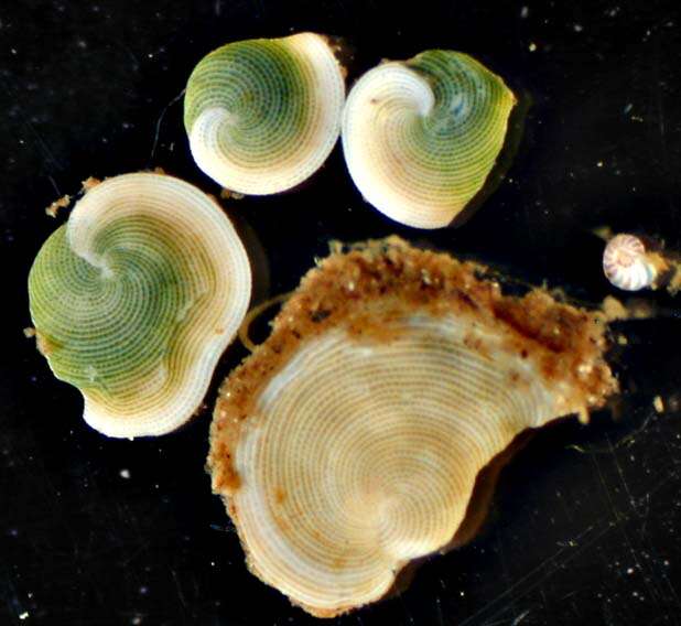
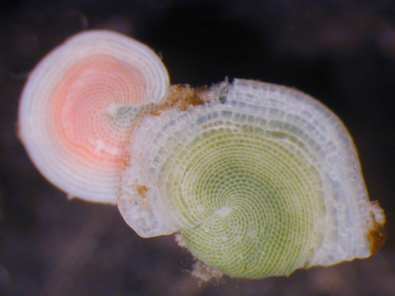
"Watermelon forams"--These foraminiferans possess chlorophyte (green algal) endosymbionts which give to the living cell a characteristic "grass green" coloration (specimen on right). Under high light (high UV?) conditions, the symbionts produce carotenoid pigments (salmon pink specimen on left).
-
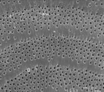
Close up of the pseudopores (pits) on the chamber surface of the foraminiferan Archaias angulatus from Belize, CA
-
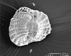
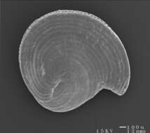
Scanning electron micrograph of a shell of the foraminiferan Archaias angulatus from Belize
-
Image courtesy of Gudmundur Gudmundsson, Icelandic Institute and Museum of Natural History. This image was originally published in J. Foram. Res. 32:308-318, and is used with permission.