-
-
2005 California Academy of Sciences
CalPhotos
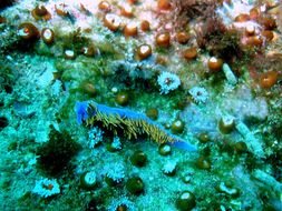
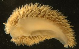
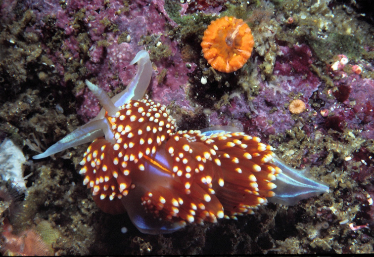
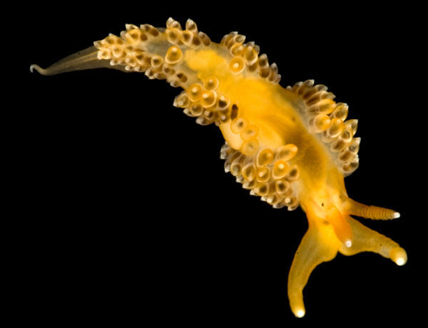
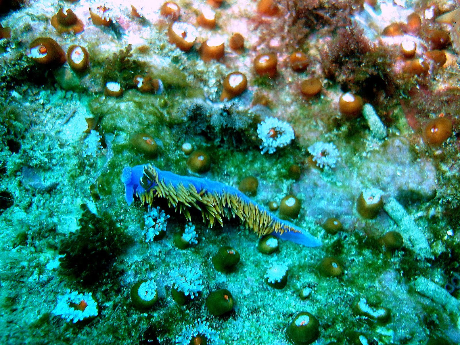
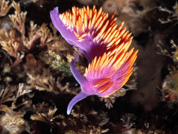
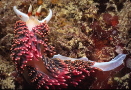
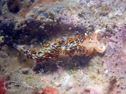
This beautiful nudibranch, about 35 mm long, feeds on the polyps of hydroids storing their stinging cells in its dorsal processes. These cells will serve in its defense. The brilliant colors may be a warning signal. When disturbed it swims by rapidly flexing its body laterally. It has a complex hermaphroditic reproductive system.
-
2010 California Academy of Sciences
CalPhotos
-
2005 California Academy of Sciences
CalPhotos
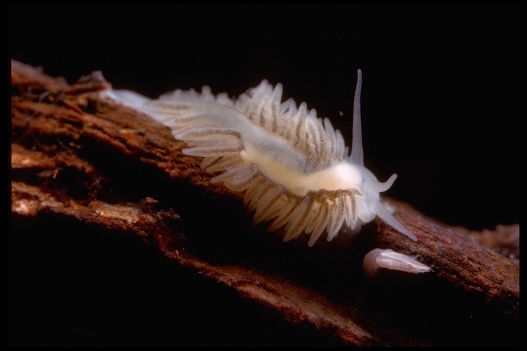
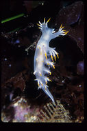
We have only found these nudibranchs on the blades of giant kelp where they feed on hydroids. Length 10 mm.
-
2005 California Academy of Sciences
CalPhotos
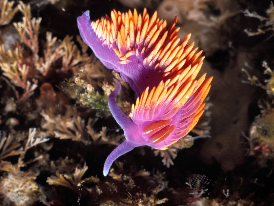
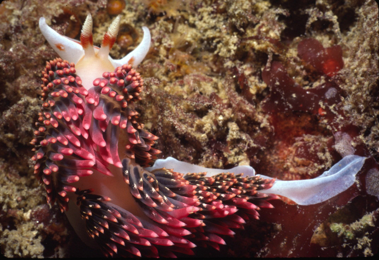
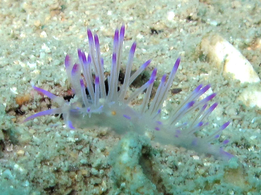
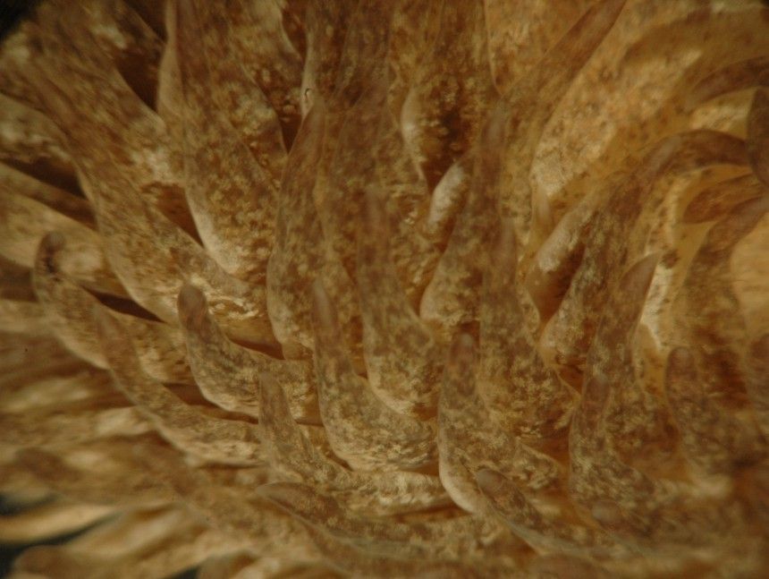
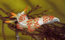
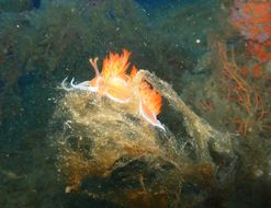
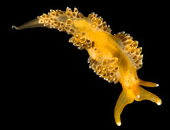
These nudibranch carnivores feed principally on hydroids, anemones and corals and store their prey's nematocysts (stinging cells) in the dorsal processes , useful for defense. Length 35 mm, depth 10 m.
-
2005 California Academy of Sciences
CalPhotos
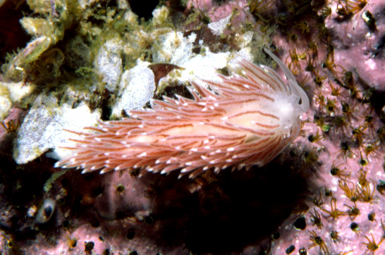
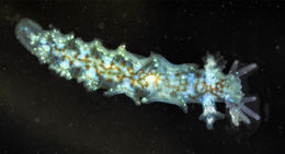
This nudibranch, found on a floating dock, was crawling on a red alga. It feeds on hydroids.
-
-
2006 California Academy of Sciences
CalPhotos
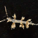
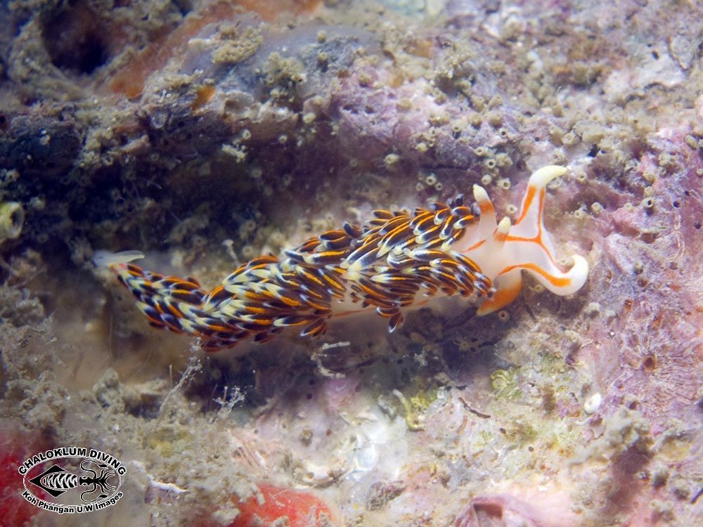
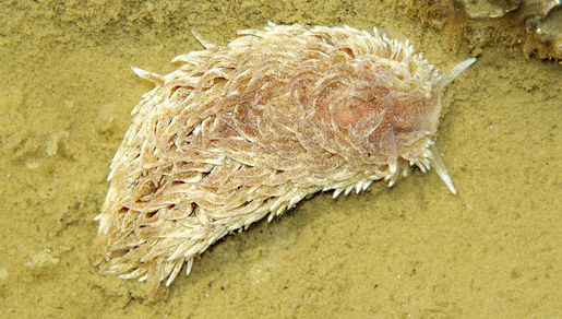
Also known with the species name of pugnax, this species is known to be very aggressive, attacking and dismembering other eolid nudibrachs. It is known to feed on cnidarian animals.
-
2006 California Academy of Sciences
CalPhotos
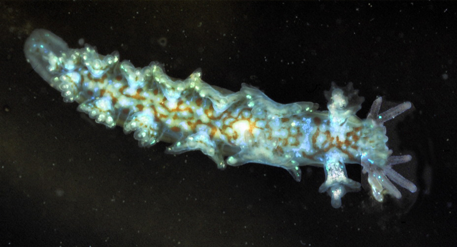
Depth 24 m. This nudibranch is known to feed on the hydroid Hydractinia.
-
2005 California Academy of Sciences
CalPhotos
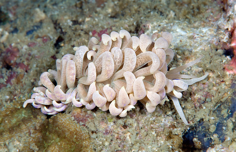
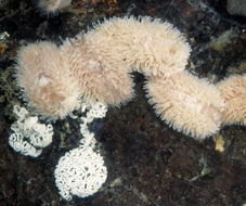
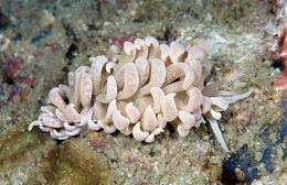
This cluster of eolid nudibranchs, mating and laying eggs, is on a wharf piling in Monterey Bay.
-
1999 California Academy of Sciences
CalPhotos
fiona
-
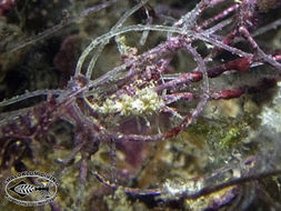
Phidiana bourailli framed in fishing line
-
Flabellina rubrolineata
-
Phidiana militaris
-
Phyllodesmium magnum
-
Melibe leonina swimming near surface, about 3 m from the bottom, in a harbor. Length about 8 cm Click the photo for a short video of this individual swimming
-
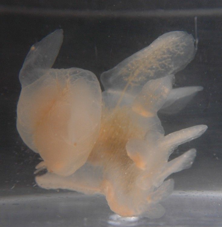
The cerata are flattened, widest above the base, and taper to a point. The hepatic diverticula cannot be readily seen within them if there is pigment present. The tips often take on the coloration of their anemone food. The mid-dorsal band, which is cerata-free but has light cororation on it, can be seen to the right.
-
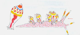
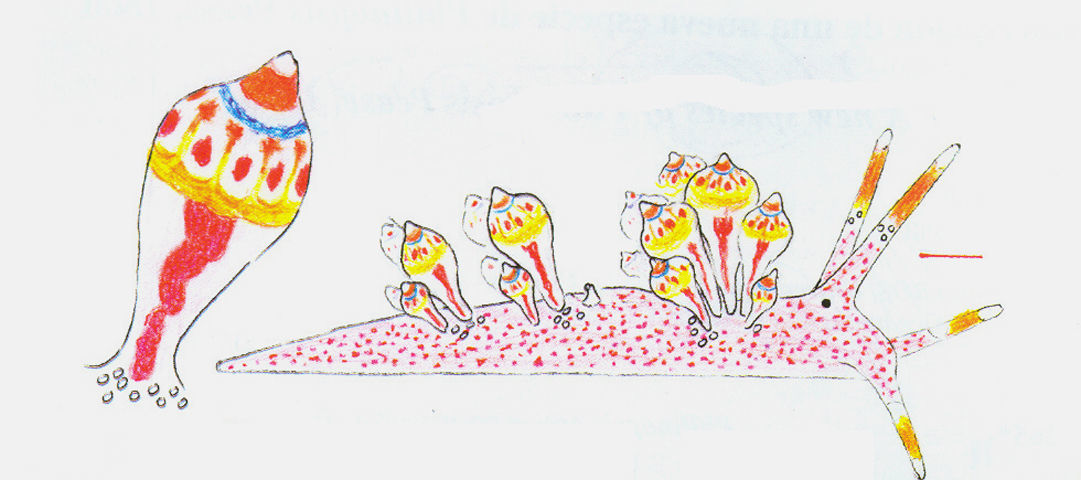
Figura 1: E. leopoldoi, dibujo esquemático del animal vivo (escala= 0,5 mm) (Caballer, Ortea y Espinosa, 2001).
-
Gray sea slug; vlokkige zeenaaktslak.
-
Phidiana sp.Mar de Cortés
-
Spanish Shawl Nudibranch
-
This is a top left view of a swimming individual, who has been swimming away but is making a strong turn to the left. The head and oral hood are visible to the right. The flaplike extensions of the oral hood are the rhinophores. The large dorsal cerata with an internal network of vessels (hepatic diverticula?) are visible at the top and right. The foot is facing down and away from view. The oral hood is closed in this view.
-
The foot tapers but is not drawn out into a long, sharp point.
-
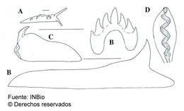
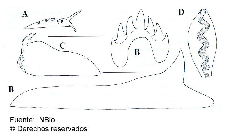
Figura 2: E. leopoldoi, A, vista lateral (escala = 1 mm). B, dientes central y lateral de la rádula (escala= 25 µm). C, mandíbula (escala= 0,5 mm). D, cerata preservado (Caballer, Ortea y Moro, 2001).