Rafael F. Almeida, Isabel R. Guesdon, Marcelo R. Pace, Renata M.S. Meira
Phytokeys
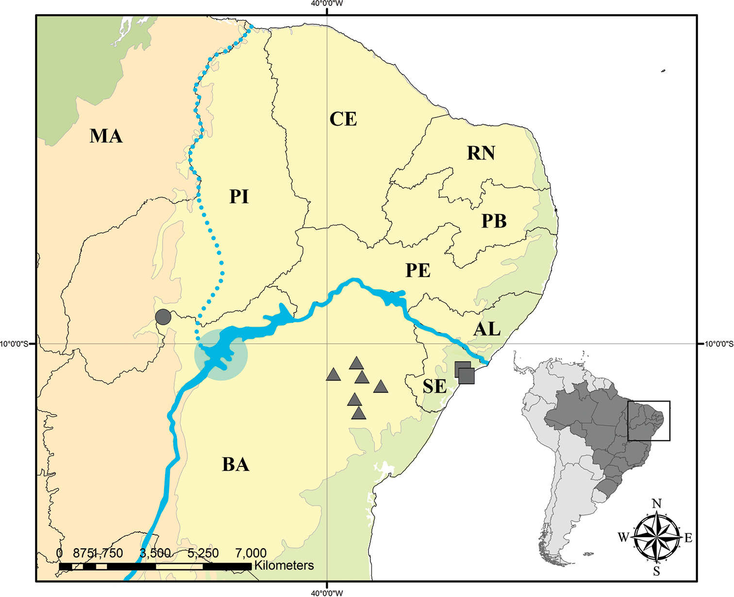
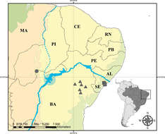
Figure 11.
Distribution map of Mcvaughia: triangle – M.bahiana, circle – M.piauhiensis, and square – M.sergipana. Solid blue line in the center represents the São Francisco River today. Dotted blue line represents the past course of São Francisco River. Blue circle represents the São Francisco paleo lake. Light green – Atlantic Forest domain, dark green – Amazon Forest domain, orange – Cerrado domain, and yellow – Caatinga domain. AL – state of Alagoas, BA – state of Bahia, CE – state of Ceará, MA – state of Maranhão, PB – state of Paraíba, PE – state of Pernambuco, PI – state of Piauí, RN – state of Rio Grande do Norte, and SE – state of Sergipe.
Rafael F. Almeida, Isabel R. Guesdon, Marcelo R. Pace, Renata M.S. Meira
Phytokeys
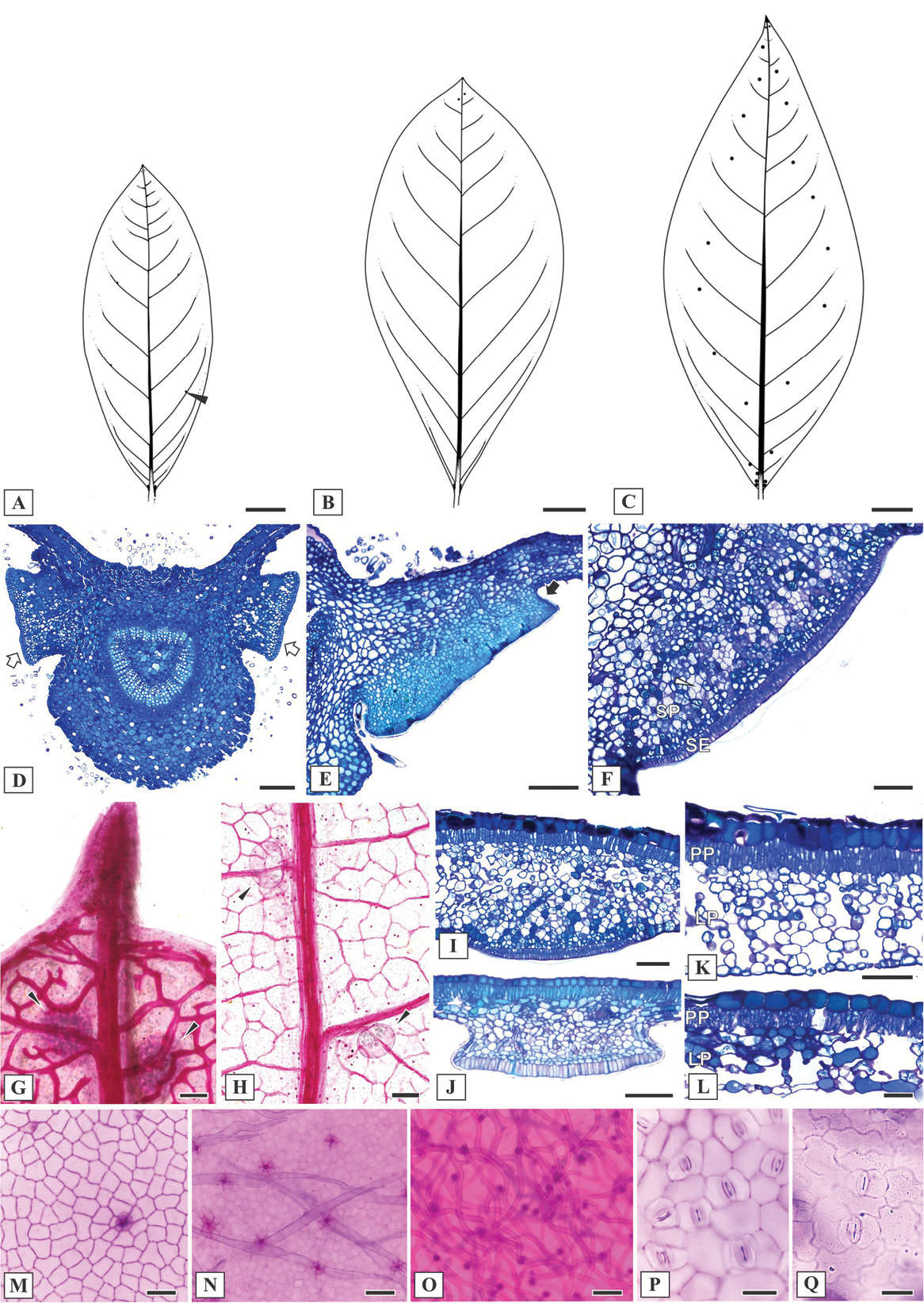
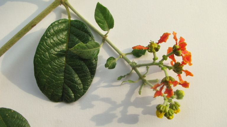
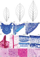
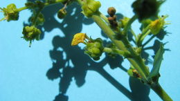
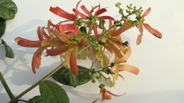
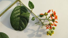
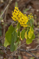
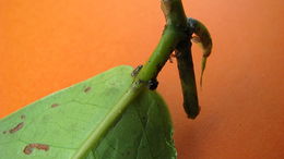
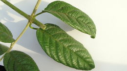
Figure 3.
Leaf morphoanatomy of Mcvaughia species. A patterns of leaf glands distribution on the abaxial leaf surface of M.bahianaB patterns of leaf glands distribution on the abaxial leaf surface of M.piauhiensisC patterns of leaf glands distribution on the abaxial leaf surface of M.sergipanaD transverse section of leaf base showing the basilaminar pair of stalked glands (white arrows) E basilaminar leaf gland with a stalk (black arrow) in M.piauhiensisF basilaminar gland in M.sergipana showing a sessile position (SE= anatomical arrangement with secretory epidermis, SP= vascularized secretory parenchyma) G–H laminar glands on the apex of cleared leaves of M.sergipana and M.bahiana respectively, note the apical tooth (G) I sessile laminar glands in M.sergipanaJ stalked laminar gland in M.piauhiensisK–L transverse sections of the leaf blade; mesophyll with uniserial palisade-like parenchyma and spongy parenchyma composed by several or few layers in M.sergipana and M.bahiana, respectively; note the idioblast with druse crystals at the mesophyll (white arrow) and the stomata distribution at the abaxial leaf surface (black arrow) M–N adaxial epidermis surface of M.piauhiensis and M.sergipana, showing scars of malpighiaceous trichomes O abaxial epidermis surface of trichomes abundance in M.bahianaP–Q outline of the anticlinal epidermal cell walls: straight in M.sergipana (P) and sinuous in M.bahiana (Q). Laminar scale bars: 1 cm (A–C), 100 μm (D, F–K, N–O), 150 μm (E), 50 μm (L–M, P–Q).
Rafael F. Almeida, Isabel R. Guesdon, Marcelo R. Pace, Renata M.S. Meira
Phytokeys
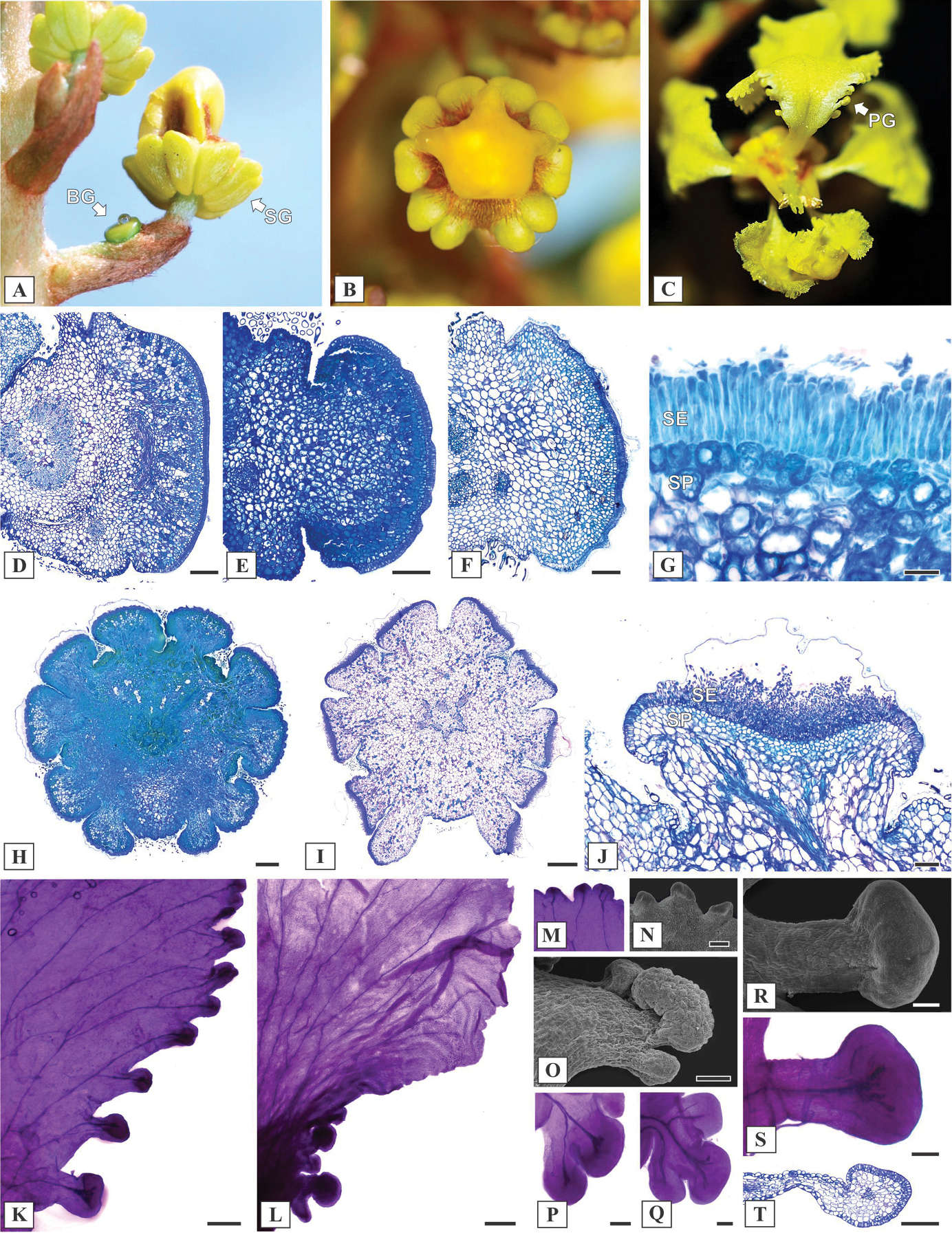
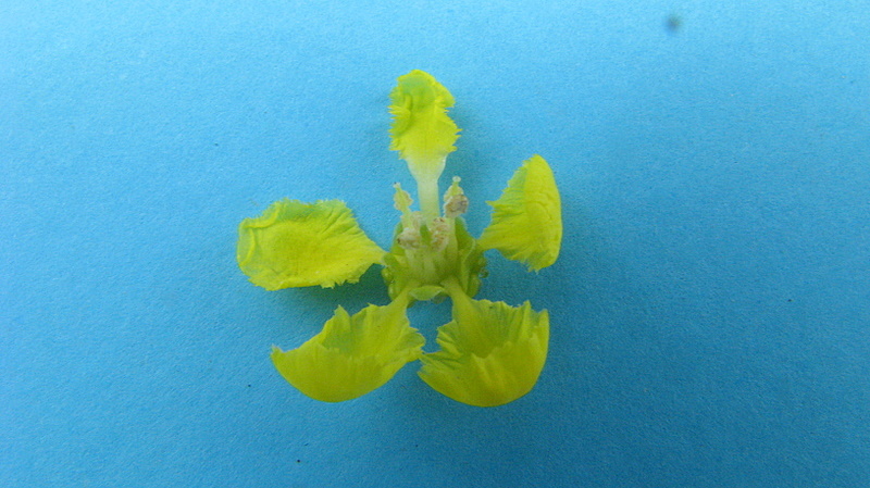
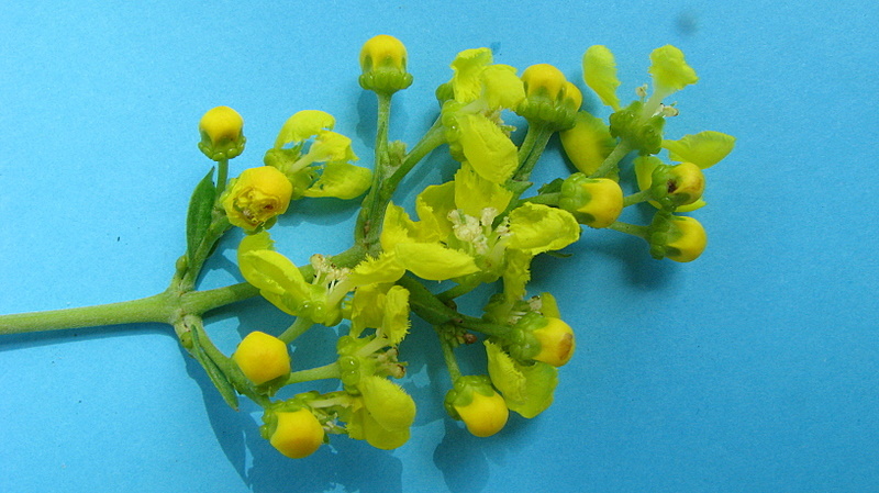
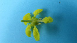
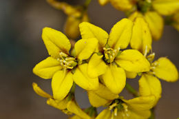
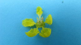
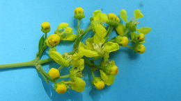
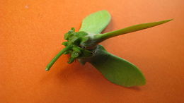
Figure 4.
Reproductive morphoanatomy of Mcvaughia species. A inflorescence during development, showing a bracteole gland (BG) and Sepal glands (SG) B ten sepal glands encircling the calyx C Petal glands (PG) along the margin of posterior petal D–F transverse section of bracteole glands in M.sergipana, M.bahiana and M.piauhiensis, respectively G anatomical arrangement of bracteole gland, with a palisade-like secretory epidermis (SE) and secretory parenchyma (SP) H–I transverse section of floral bud and anthesis flower in Mcvaughiabahiana and M.sergipana; calyx gland pair displaced at the anterior sepal J calyx gland structure, showing a secretory epidermis (SE) and vascularized secretory parenchyma (SP) K–L petal glands on the margin of petals in M.sergipana and M.bahiana respectively M–N detail of the petal glands at the apex of the petal limb in M.sergipana, cleared and in SEM image O–Q petal glands positioned at the base, M.bahiana on SEM image, M.bahiana and M.piauhiensis cleared R–T conspicuous and stalked petal glands at the base of M.sergipana, in SEM image, cleared and longitudinal section. Scale bars: 200 μm (D), 150 μm (E–F), 50 μm (G), 500 μm (H–I), 100 μm (J, P–S), 300 μm (L–M), 200 μm (N, T).
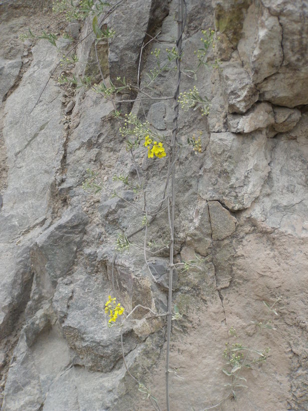
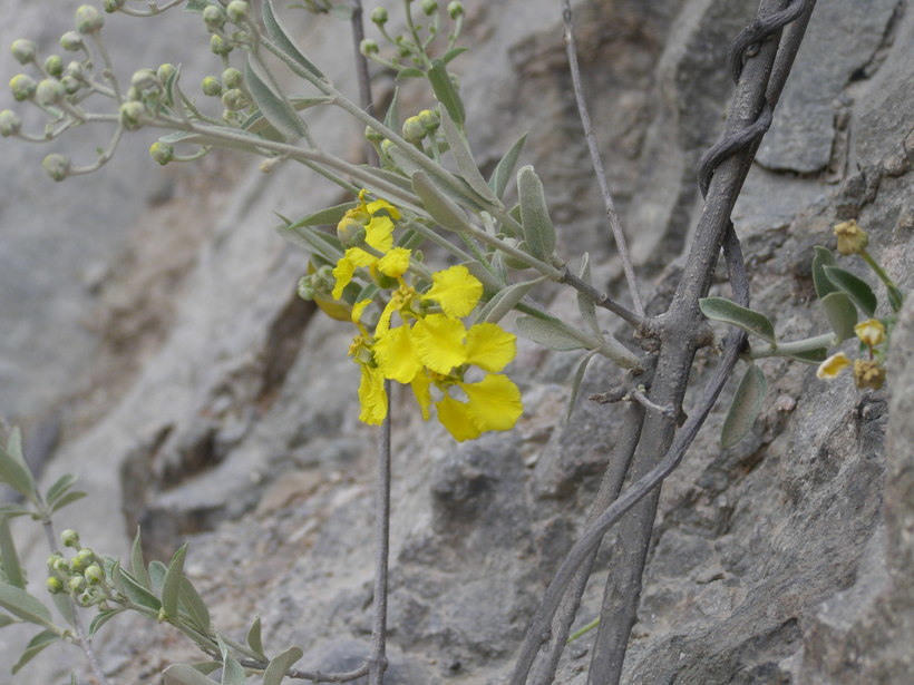
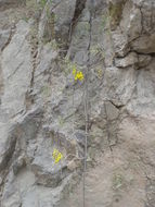
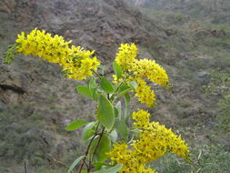
long vine draping volcanic rhyolitic tuff cliff face in lower canyon. This site is likely destroyed or no longer easily accessible due to construction of the new paved highway to Batopilas
long vine draping volcanic rhyolitic tuff cliff face in lower canyon. This site is likely destroyed or no longer easily accessible due to construction of the new paved highway to Batopilas


