-
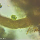
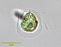
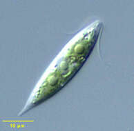
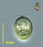
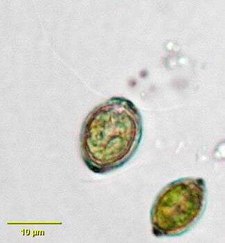
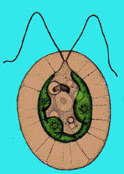
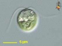
Chlamydomonas, a volvocid flagellate. The genus is very large probably with many synonymous species. Two equal length flagella emerge from a prominent anterior papilla in this species. A small contractile vacuole can be seen just posterior to the flagellar insertion site. A well-demarcated central nucleus can be seen in these images. A very small stigma is present. There is a single large cup shaped chloroplast in this species. A large pyrenoid is present in the posterior half of the cell. Some species have a gelatinous sheath although the cell wall is closely applied to the protoplast in this species. From freshwater pond near Boise, Idaho. Oblique illumination.
-
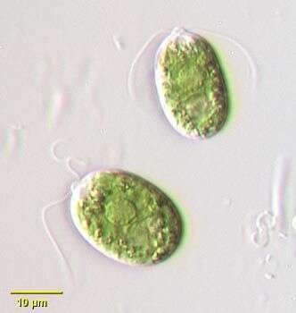
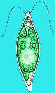
Chlamydomonas is a green alga, common in freshwater habitats like Swan Lake. The cells are small with two flagella used for locomotion. A single red eyespot (stigma) is used to sense light levels and control the direction of 'swimming'. Animations by Rosemary Arbur of flagellar beat patterns are available
here.
-
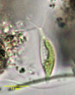
Differential interference contrast image of a cluster of cells attached to the coversl;ip by their flagella.
-
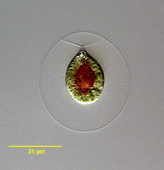
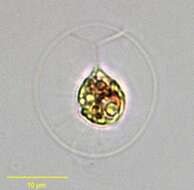
Portrait of Phacotus lenticularis, a volvocid flagellate. Lorica is composed of two shallow cup-shaped halves cemented together around the protoplast. Lorica surface is slightly rough and colored by minerals Two equal-length flagella protrude through an anterior pore in the lorica. Cup-shaped chloroplast. A stigma is present but not seen in these images. From a freshwater pond near Boise, Idaho. Brightfield.
-
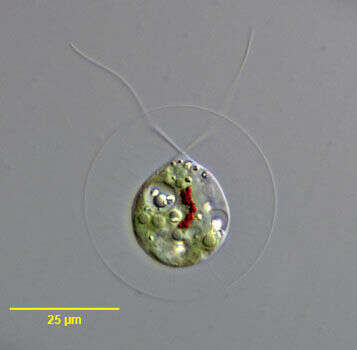
Portrait of Phacotus lenticularis, a volvocid flagellate. Lorica is composed of two shallow cup-shaped halves cemented together around the protoplast. Lorica surface is slightly rough and colored by minerals Two equal-length flagella protrude through an anterior pore in the lorica. Cup-shaped chloroplast. A stigma is present but not seen in these images. From a freshwater pond near Boise, Idaho. Phase contrast.
-
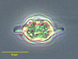
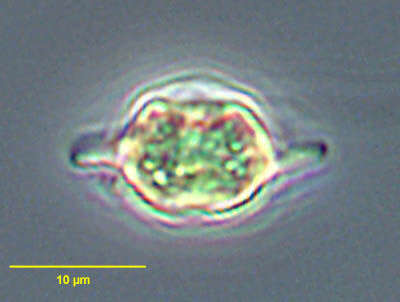
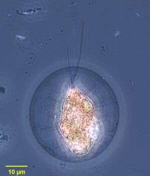
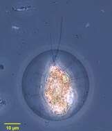
Pteromonas, a volvocid flagellate. Ovoid protoplast occupies a hyaline lorica composed of two parts that meet peripherally appearing as flattened wings in optical cross-section. Two equal flagella exit through small canals. This species has a large cup-shape chloroplast and one posterior pyrenoid. The eyespot is not well seen. An anterior contractile vacuole is seen in this image. Asexual reproduction occurs within the lorica the two halves of which are forced apart by the accumulation of gelatinous material releasing the daughter cells. From rainwater pool near Boise, Idaho. Brightfield illumination.
-
Pteromonas, a volvocid flagellate. Ovoid protoplast occupies a hyaline lorica composed of two parts that meet peripherally appearing as flattened wings in optical cross-section. Two equal flagella exit through small canals. This species has a large cup-shape chloroplast and one posterior pyrenoid. The eyespot is not well seen. An anterior contractile vacuole is seen in this image. Asexual reproduction occurs within the lorica the two halves of which are forced apart by the accumulation of gelatinous material releasing the daughter cells. From rainwater pool near Boise, Idaho. Phase contrast illumination.
-
-
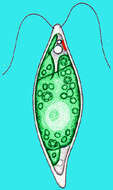
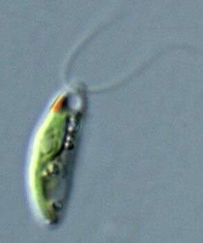
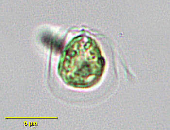
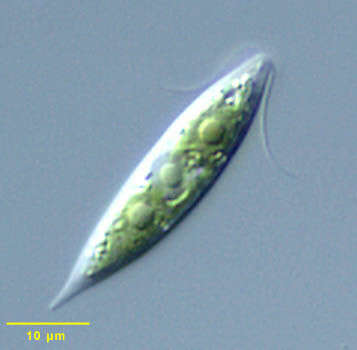
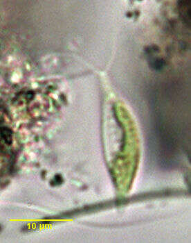
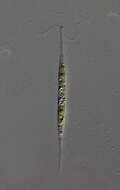
Chlorogonium, a small volvocid flagellate, with two equal anterior flagella. The cell body is fusiform. Chloroplasts are irregular. A stigma may be present. From standing rainwater pool near Boise, Idaho. Nomarski illumination.
-
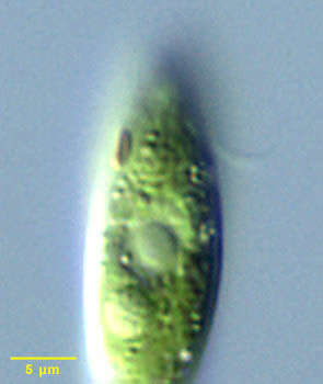
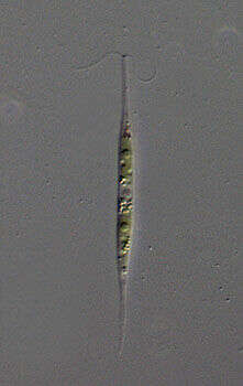
Chlorogonium, a volvocid green alga (Chlorophyta), the cell is spindle shaped with two flagella inserting at the anterior apex of the cell. The flagella beat with a breast-stroke like motion. This detail shows the anterior stigma or eyespot. Differential interference contrast optics.
-
Chlorogonium, a volvocid green alga (Chlorophyta), the cell is spindle shaped with two flagella inserting at the anterior apex of the cell. The flagella beat with a breast-stroke like motion.
-
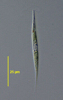
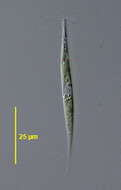
Portrait of the volvocid flagellate, Chlorogonium elongatum (Dangeard 1899). Collected from a freshwater pond near Boise, Idaho March 2005. DIC.
-
Portrait of the volvocid flagellate Chlorogonium elongatum (Dangeard 1899). Collected from a freshwater pond near Boise, Idaho March 2005. DIC
-
-
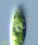
Phase contrast micrograph of living cell.
-
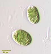
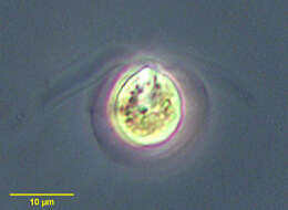
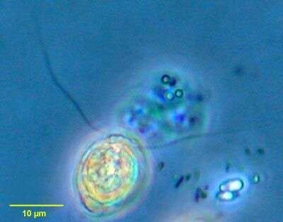
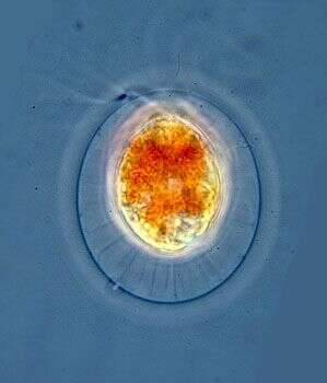
Portrait of Haematococcus pluvialis (Flotow, 1844), a widely distributed volvocid flagellate. Thin cytoplasmic strands traverse the clear mucilaginous layer to connect the protoplast to the spherical cell wall. Single large cup-shaped chloroplast. Two equal-length flagella are also seen traversing the mucilaginous layer. Reddish carotenoid pigments may obscure the stigma and chloroplast. From fish farm aquaculture pond near Boise, Idaho. Phase contrast.
-
Portrait of Haematococcus pluvialis (Flotow, 1844), a widely distributed volvocid flagellate. Thin cytoplasmic strands traverse the clear mucilaginous layer to connect the protoplast to the spherical cell wall. Single large cup-shaped chloroplast. Two equal-length flagella are also seen traversing the mucilaginous layer. Reddish carotenoid pigments may obscure the stigma and chloroplast. From fish farm aquaculture pond near Boise, Idaho. Brightfield.
-
Portrait of Haematococcus pluvialis (Flotow, 1844), a widely distributed volvocid flagellate. Thin cytoplasmic strands traverse the clear mucilaginous layer to connect the protoplast to the spherical cell wall. Single large cup-shaped chloroplast. Two equal-length flagella are also seen traversing the mucilaginous layer. Reddish carotenoid pigments (concentrated in cell center here) may obscure the stigma and chloroplast. From ephemeral freshwater pool near Boise, Idaho, March, 2005. DIC.
-
Portrait of Haematococcus pluvialis (Flotow, 1844), a widely distributed volvocid flagellate. Thin cytoplasmic strands traverse the clear mucilaginous layer to connect the protoplast to the spherical cell wall. Single large cup-shaped chloroplast. Two equal-length flagella are also seen traversing the mucilaginous layer. Reddish carotenoid pigments are concentrated in the cell center here. The inconspicuous stigma is seen at 1 o'clock near the proroplast surface. Several pyrenoids are visible here. There are multiple small contractile vacuoles. refractile cytoplasmic crystals are present in this individual. From ephemeral freshwater pool near Boise, Idaho, March, 2005. DIC.
-
Hyalogonium, a colorless volvocid flagellate with two equal anterior flagella. The body is elongate and fusiform. A stigma is present on the left. Many small starch grains are seen in the cytoplasm. This genus may be confused with the euglenid Cyclidiopsis but Hyalogonium is smaller, biflagellate with thinner flagella, and lacks large paramylon bodies and the anterior canal opening. From standing rainwater pool near Boise, Idaho. Brightfield illumination.
-
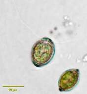
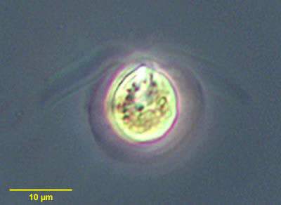
Dunaliella (done-al-ee-ella), a solitary volvocid (flagellated green algal cell). Cell surrounded by a cellulosic wall, with two similar flagella emerging from near the apex. The photosynthetic pigments are located within a cup-shaped chloroplast which has a large pyrenoid with associated polysaccharide materials located posteriorly. The nucleus is located within the cup. From marine and usually hypersaline habitats, grown commercially because of the tendency to produce large quantities of beta carotene when intensely illuminated. Differential interference contrast.
-
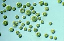
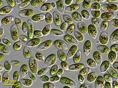
Dunaliella is a green alga and is a common member of the phytoplankton in salty water bodies. These cells were abundant in a collection taken at the margins of Mono lake (at Navy Beach). Each cell has a cup-shaped or bowl-shpaed chloroplast at the posterior end of the cell, aneriorly they have two equally long flagella. Differential interference contrast optics.