-
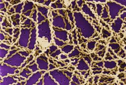
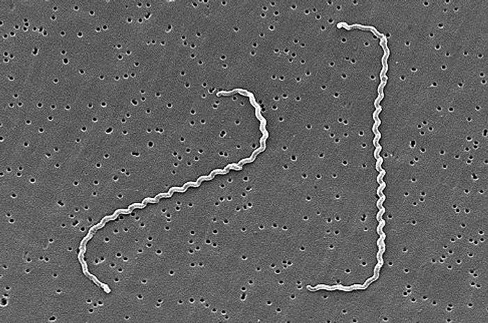
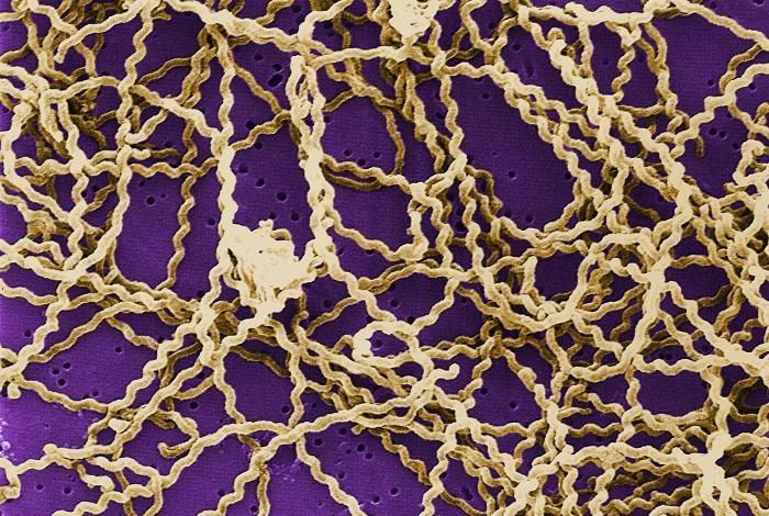
Scanning electron micrograph of Leptospira interrogans strain RGA.Created:
-
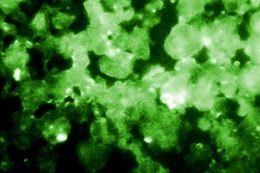
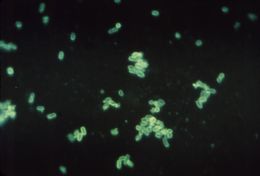
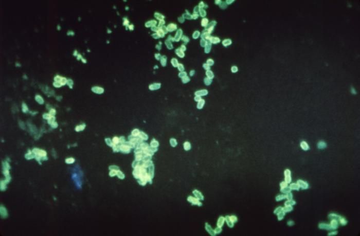
This photomicrograph depicted the results of using the Indirect Fluorescent Antibody (IFA) technique to confirm the presence of Legionella pneumophila bacteria in this human lung secretion sample from a suspected victim of Legionnaires disease.Created: 1978
-
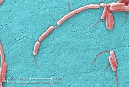
This scanning electron micrograph (SEM) depicts a number of Leptospira sp. bacteria atop a 0.1. µm polycarbonate filter.Created: 1998
-
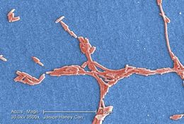
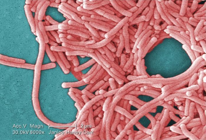
Under a moderately-high magnification of 8000X, this colorized scanning electron micrograph (SEM) depicted a large grouping of Gram-negative Legionella pneumophila bacteria. Please see PHIL 11092 through 11152 for additional SEMs of these organisms, specifically PHIL 11151 for a black and white version of this image. Of particular importance, is the presence of polar flagella, and pili, or long streamers, which due to their fragile nature, in some of these views seem to be dissociated from any of the bacteria.Created: 2009
-
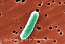
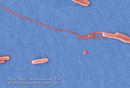
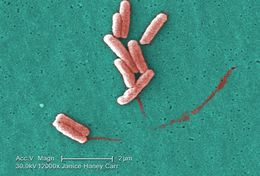
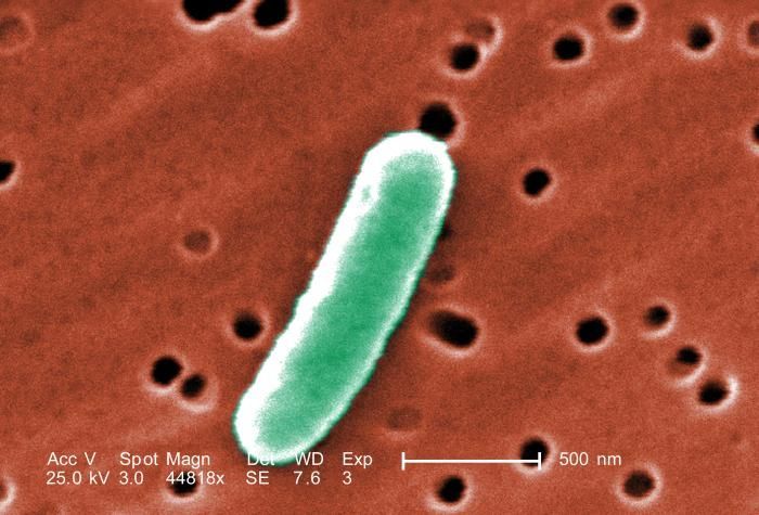
At an extremely high magnification of 44, 818X, twice that of PHIL 10574 and 10575, this colorized scanning electron micrograph (SEM) revealed some of the morphologic details displayed by a single Gram-negative Escherichia coli bacterium. This bacterium was a member of the strain, 0:169 H41 ETEC (Enterotoxigenic E. coli). See PHIL 10576 for a black and white version of this image.Enterotoxigenic E. coli, a common cause of bacterial diarrhea
Enterotoxigenic Escherichia coli, or ETEC, is an important cause of bacterial diarrheal illness. Infection with ETEC is the leading cause of travelers' diarrhea and a major cause of diarrheal disease in underdeveloped nations, especially among children. ETEC is transmitted by food or water contaminated with animal or human feces. Although ETEC causes a significant amount of illness worldwide, the infection will end on its own and is rarely life-threatening.Created: 2008
-
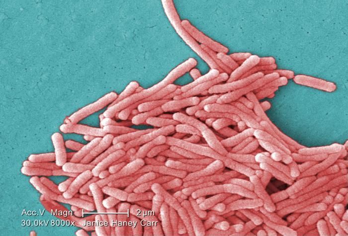
Under a moderately-high magnification of 8000X, this colorized scanning electron micrograph (SEM) depicted a large grouping of Gram-negative Legionella pneumophila bacteria. Please see PHIL 11092 through 11152 for additional SEMs of these organisms, specifically PHIL 11149 for a black and white version of this image. Of particular importance, is the presence of polar flagella, and pili, or long streamers, which due to their fragile nature, in some of these views seem to be dissociated from any of the bacteria.Created: 2009
-
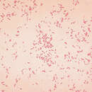
Magnified 600X, this fluorescent antibody stained photomicrograph revealed the presence of enteropathogenic Escherichia coli bacteria, which were found in a fecal smear from an infant with diarrhea. E. coli is a member of the family of bacterial organisms within the family Enteribacteriaceae, and contains the highly pathogenic strain, which has been given the label O157:H7.Created: 1960
-
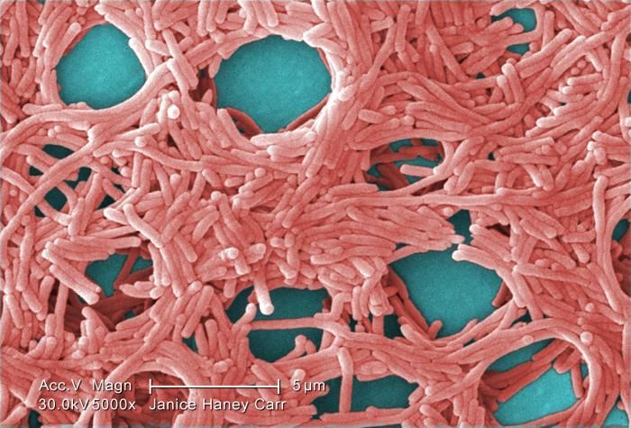
Under a moderately-high magnification of 5000X, this colorized scanning electron micrograph (SEM) depicted a large grouping of Gram-negative Legionella pneumophila bacteria. Please see PHIL 11092 through 11154 for additional SEMs of these organisms, specifically PHIL 11147 for a black and white version of this image. Of particular importance, is the presence of polar flagella, and pili, or long streamers, which due to their fragile nature, in some of these views seem to be dissociated from any of the bacteria.Created: 2009
-
Magnified 1250X, this fluorescent antibody stained photomicrograph revealed the presence of enteropathogenic Escherichia coli bacteria, which were found in a fecal smear from an infant with diarrhea. E. coli is a member of the family of bacterial organisms within the family Enteribacteriaceae, and contains the highly pathogenic strain, which has been given the label O157:H7.Created: 1960
-
Under a very very high magnification of 10000X, this colorized scanning electron micrograph (SEM) depicted a number of Gram-negative Legionella pneumophila bacteria. Please see PHIL 11092 through 11152 for additional SEMs of these organisms, specifically PHIL 11145 for a black and white version of this image. Of particular importance, is the presence of polar flagella, and pili, or long streamers, which due to their fragile nature, in some of these views seem to be dissociated from any of the bacteria.Created: 2009
-
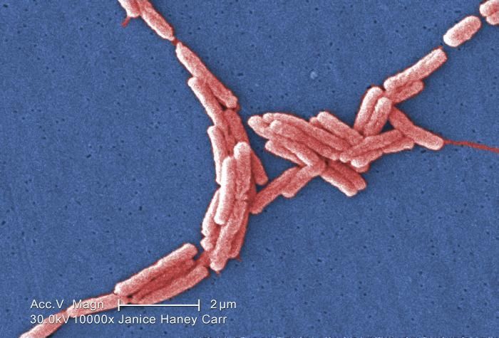
Under a high magnification of 10961x, this colorized scanning electron micrograph (SEM) depicted a number of Gram-negative Escherichia coli bacteria of the strain O157:H7. E. coli O157:H7 is one of hundreds of strains of this bacterium. Although most strains are harmless, and live in the intestines of healthy humans and animals, this strain produces a powerful toxin, which can cause severe illness.E. coli O157:H7 was first recognized as a cause of illness in 1982 during an outbreak of severe bloody diarrhea; the outbreak was traced to contaminated hamburgers. Since then, most infections have come from eating undercooked ground beef.The combination of letters and numbers in the name of the bacterium refers to the specific markers found on its surface, which distinguishes it from other types of E. coli. See PHIL 8797 for a black and white version of this image.Created: 2006
-
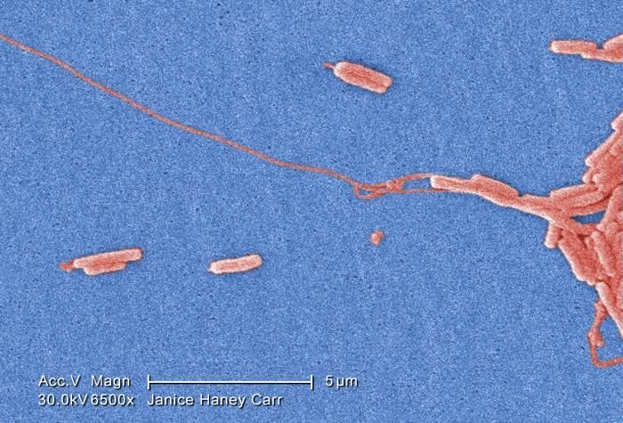
Under a very moderately-high magnification of 6500X, this colorized scanning electron micrograph (SEM) depicted a number of Gram-negative Legionella pneumophila bacteria. Please see PHIL 11092 through 11152 for additional SEMs of these organisms, specifically PHIL 11143 for a black and white version of this image. Of particular importance, is the presence of polar flagella, and pili, or long streamers, which due to their fragile nature, in some of these views seem to be dissociated from any of the bacteria.Created: 2009
-
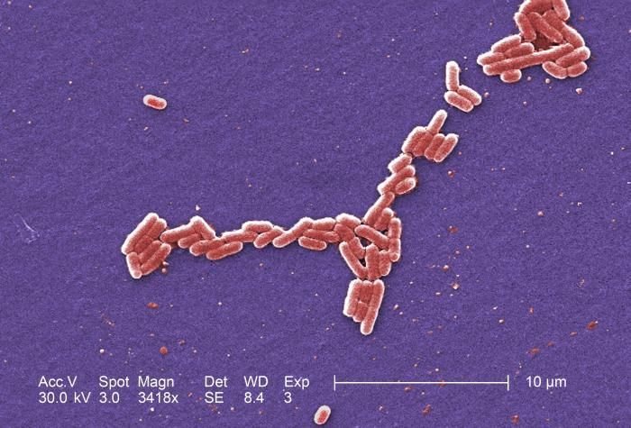
Under a magnification of 3418x, this colorized scanning electron micrograph (SEM) depicted a number of Gram-negative Escherichia coli bacteria of the strain O157:H7. E. coli O157:H7 is one of hundreds of strains of this bacterium. Although most strains are harmless, and live in the intestines of healthy humans and animals, this strain produces a powerful toxin, which can cause severe illness.E. coli O157:H7 was first recognized as a cause of illness in 1982 during an outbreak of severe bloody diarrhea; the outbreak was traced to contaminated hamburgers. Since then, most infections have come from eating undercooked ground beef.The combination of letters and numbers in the name of the bacterium refers to the specific markers found on its surface, which distinguishes it from other types of E. coli. See PHIL 8798 for a black and white version of this image.Created: 2006
-
Under a very high magnification of 15000X, this colorized scanning electron micrograph (SEM) depicted a number of Gram-negative Legionella pneumophila bacteria. Please see PHIL 11092 through 11152 for additional SEMs of these organisms, specifically PHIL 11141 for a black and white version of this image. Of particular importance, is the presence of polar flagella, and pili, or long streamers, which due to their fragile nature, in some of these views seem to be dissociated from any of the bacteria.Created: 2009
-
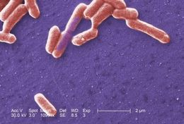
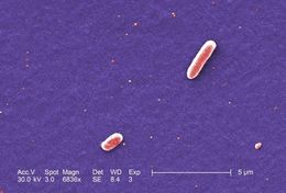
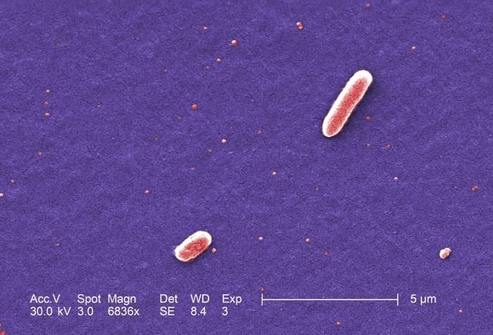
Under a magnification of 6836x, this colorized scanning electron micrograph (SEM) depicted two Gram-negative Escherichia coli bacteria of the strain O157:H7. E. coli O157:H7 is one of hundreds of strains of this bacterium. Although most strains are harmless, and live in the intestines of healthy humans and animals, this strain produces a powerful toxin, which can cause severe illness.E. coli O157:H7 was first recognized as a cause of illness in 1982 during an outbreak of severe bloody diarrhea; the outbreak was traced to contaminated hamburgers. Since then, most infections have come from eating undercooked ground beef.The combination of letters and numbers in the name of the bacterium refers to the specific markers found on its surface, which distinguishes it from other types of E. coli. See PHIL 8799 for a black and white version of this image.Created: 2006
-
Under a very high magnification of 12000X, this colorized scanning electron micrograph (SEM) depicted a number of Gram-negative Legionella pneumophila bacteria. Please see PHIL 11092 through 11152 for additional SEMs of these organisms, specifically PHIL 11139 for a black and white version of this image. Of particular importance, is the presence of polar flagella, and pili, or long streamers, which due to their fragile nature, in some of these views seem to be dissociated from any of the bacteria.Created: 2009
-
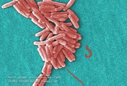
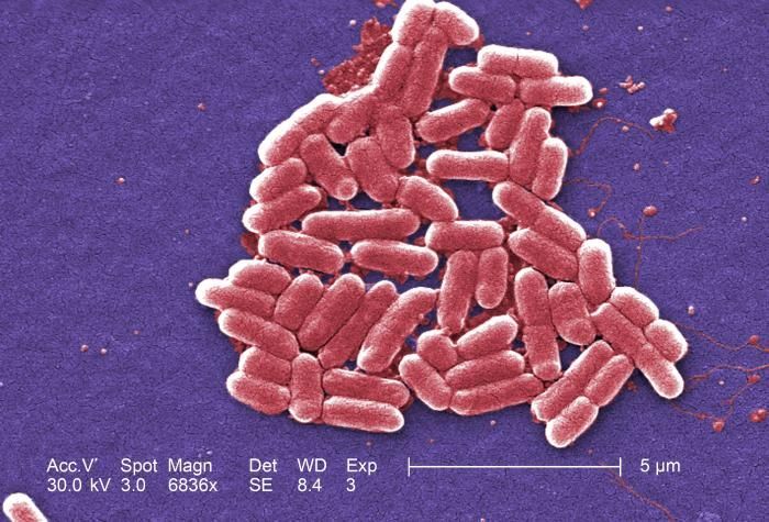
Under a magnification of 6836x, this colorized scanning electron micrograph (SEM) depicted a number of Gram-negative Escherichia coli bacteria of the strain O157:H7, which is one of hundreds of strains of this bacterium. Although most strains are harmless, and live in the intestines of healthy humans and animals, this strain produces a powerful toxin, which can cause severe illness.E. coli O157:H7 was first recognized as a cause of illness in 1982 during an outbreak of severe bloody diarrhea; the outbreak was traced to contaminated hamburgers. Since then, most infections have come from eating undercooked ground beef.The combination of letters and numbers in the name of the bacterium refers to the specific markers found on its surface, which distinguishes it from other types of E. coli. See PHIL 8800 for a black and white version of this image.Created: 2006
-
Under a moderately-high magnification of 10000X, this colorized scanning electron micrograph (SEM) depicted a number of Gram-negative Legionella pneumophila bacteria. Please see PHIL 11092 through 11152 for additional SEMs of these organisms, specifically PHIL 11138 for a colorized version of this image. Of particular importance, is the presence of polar flagella, and pili, or long streamers, which due to their fragile nature, in some of these views seem to be dissociated from any of the bacteria.Youll note that a number of these bacteria seem to display an elongated-rod morphology. L. pneumophila are known to most frequently exhibit this configuration when grown in broth, however, they can also elongate when plate-grown cells age, as it was in this case, especially when theyve been refrigerated.Created: 2009
-
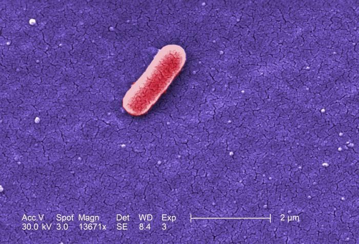
Under a high magnification of 13671x, this colorized scanning electron micrograph (SEM) depicted a single Gram-negative Escherichia coli bacterium of the strain O157:H7. E. coli O157:H7 is one of hundreds of strains of this bacterium. Although most strains are harmless, and live in the intestines of healthy humans and animals, this strain produces a powerful toxin, which can cause severe illness.E. coli O157:H7 was first recognized as a cause of illness in 1982 during an outbreak of severe bloody diarrhea; the outbreak was traced to contaminated hamburgers. Since then, most infections have come from eating undercooked ground beef.The combination of letters and numbers in the name of the bacterium refers to the specific markers found on its surface, which distinguishes it from other types of E. coli. See PHIL 8801 for a black and white version of this image.Created: 2006
-
Under a moderately-high magnification of 10000X, this scanning electron micrograph (SEM) depicted a number of Gram-negative Legionella pneumophila bacteria. Please see PHIL 11092 through 11152 for additional SEMs of these organisms, specifically PHIL 11136 for a colorized version of this image. Of particular importance, is the presence of polar flagella, and pili, or long streamers, which due to their fragile nature, in some of these views seem to be dissociated from any of the bacteria. For a better view of these cellular appendages, see the colorized version.Created: 2009
-
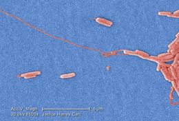
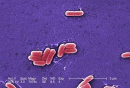
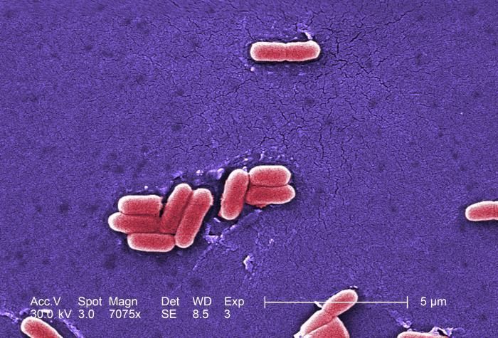
Under a magnification of 7075x, this colorized scanning electron micrograph (SEM) depicted a number of Gram-negative Escherichia coli bacteria of the strain O157:H7. E. coli O157:H7 is one of hundreds of strains of this bacterium. Although most strains are harmless, and live in the intestines of healthy humans and animals, this strain produces a powerful toxin, which can cause severe illness.E. coli O157:H7 was first recognized as a cause of illness in 1982 during an outbreak of severe bloody diarrhea; the outbreak was traced to contaminated hamburgers. Since then, most infections have come from eating undercooked ground beef.The combination of letters and numbers in the name of the bacterium refers to the specific markers found on its surface, which distinguishes it from other types of E. coli. See PHIL 8802 for a black and white version of this image.Created: 2006
-
Under a moderate magnification of 3500X, this colorized scanning electron micrograph (SEM) depicted a number of Gram-negative Legionella pneumophila bacteria. Please see PHIL 11092 through 11152 for additional SEMs of these organisms, specifically PHIL 11133 for a black and white version of this image. Of particular importance, is the presence of polar flagella, and pili, or long streamers, which due to their fragile nature, in some of these views seem to be dissociated from any of the bacteria.Created: 2009
-
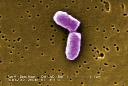
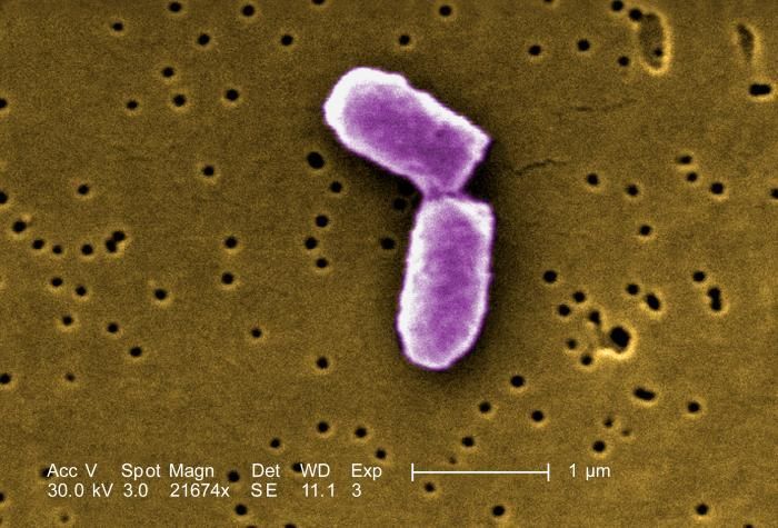
This colorized version of PHIL 7137 depicts a highly magnified scanning electron micrographic (SEM) view of a dividing Escherichia coli bacteria, clearly displaying the point at which the bacterias cell wall was dividing; Magnification 21674x.Created: 2005
-
Under a high magnification of 10000X, this colorized scanning electron micrograph (SEM) depicted a number of Gram-negative Legionella pneumophila bacteria. Please see PHIL 11092 through 11152 for additional SEMs of these organisms, specifically PHIL 11129 for a black and white version of this image. Of particular importance, is the presence of polar flagella, and pili, or long streamers, which due to their fragile nature, in some of these views seem to be dissociated from any of the bacteria.Created: 2009