-
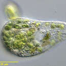
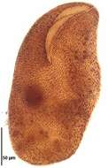
Kuklikophrya ougandae (DRAGESCO, 1972) FOISSNER, 1993. The organisms appear dark green in vivo due to large numbers of ingested cyanobacterial fragments.Collected from an organically enriched temporary freshwater pool near Boise, Idaho. May 2006.Phase contrast.
-
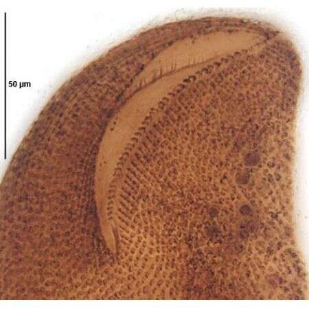
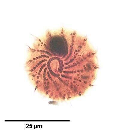
Oral infraciliature of Kuklikophrya ougandae (DRAGESCO, 1972) FOISSNER, 1993 The adoral membranelles are arrayed in an oblique line from the opening in the paraoral membrane to the left anterior apex of the cell.Stained by the silver carbonate technique (see Foissner, W. Europ. J. Protistol., 27:313-330;1991). Collected from an organically enriched temporary freshwater pool near Boise, Idaho. May 2006.Brightfield.
-
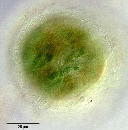
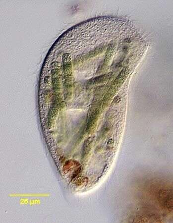
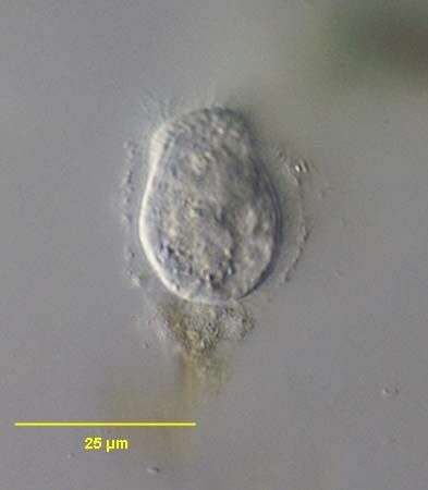
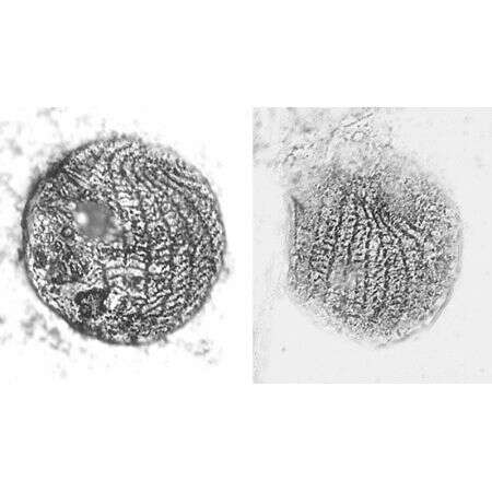
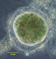
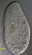
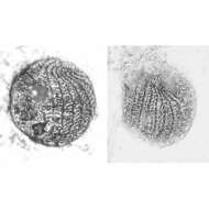
Cyst of Kuklikophrya ougandae (Dragesco,1972) Foissner 1993.Like other colpodids, K. ougandae undergoes division within cysts. The oral apparatus is visible in the center of the cell.This individual has not begun division. Cysts are tranparent with a thin but firm yellowish membrane and a thicker outer coat of mucus to which debris adhere. Ingested cyanobacteria are visible in the cytoplasm.Somatic cilia persist at this stage of encystment allowing the organism to rotate slowly within the cyst wall. Collected from an organically enriched temporary freshwater pool near Boise, Idaho. May 2006. DIC.
-
Cyst of Kuklikophrya ougandae (Dragesco,1972) Foissner 1993.Like other colpodids, K. ougandae undergoes division within cysts. This individual has not begun division. Cysts are tranparent with a thin but firm yellowish membrane and a thicker outer coat of mucus to which debris adhere. Ingested cyanobacteria are visible in the cytoplasm.Somatic cilia persist at this stage of encystment allowing the organism to rotate slowly within the cyst wall. Collected from an organically enriched temporary freshwater pool near Boise, Idaho. May 2006. Phase contrast.
-
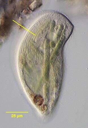
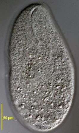
Kuklikophrya ougandae (DRAGESCO, 1972 ) FOISSNER, 1993. The organisms appear dark green in vivo due to large numbers of ingested cyanobacterial fragments (Oscillatoria in this case).The oral aperture is indicated by the yellow line.Collected from an organically enriched temporary freshwater pool near Boise, Idaho. May 2006.DIC.
-
Kuklikophrya ougandae (DRAGESCO, 1972 ) FOISSNER, 1993. The organisms appear dark green in vivo due to large numbers of ingested cyanobacterial fragments (Oscillatoria in this case).Collected from an organically enriched temporary freshwater pool near Boise, Idaho. May 2006.DIC.
-
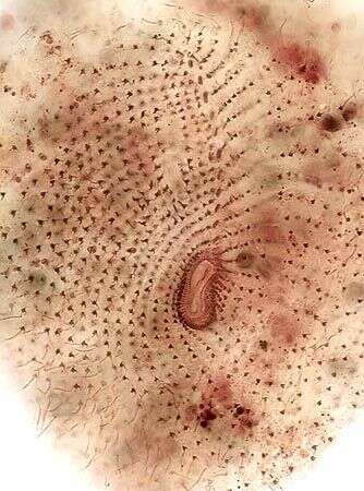
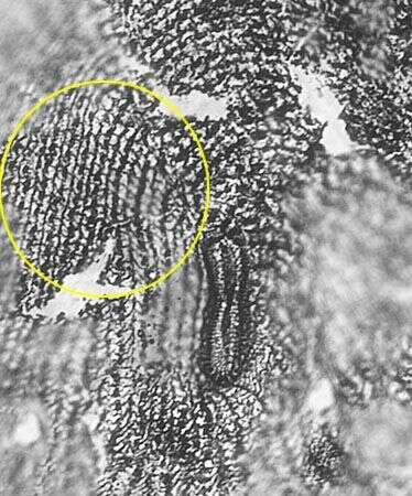
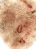
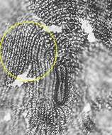
Platyophryid silverline system of Kuklikophrya ougandae (DRAGESCO, 1972) FOISSNER, 1993. Stained by the Klein-Foissner silver nitrate technique (see Foissner, W. Europ. J. Protistol., 27:313-330;1991).Brightfield.
-
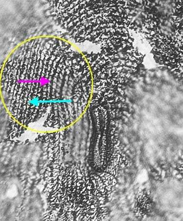
Platyophryid silverline system of Kuklikophrya ougandae (DRAGESCO, 1972) FOISSNER, 1993. The primary (kinetidal) meridians (pink arrow) alternate with thinner secondary meridians (light blue arrow) short transverse elements between the two meridians create a reticulate pattern. Stained by the Klein-Foissner silver nitrate technique (see Foissner, W. Europ. J. Protistol., 27:313-330;1991).Brightfield.
-
Woodruffides metabolicus (JOHNSON & LARSON, 1938) FOISSNER, 1987.Collected from a non-flooded Petri dish culture of soil from a grass lawn in Boise, Idaho.Febuary 2008.Stained by the protargol technique [Wilbert modification](see Foissner, W. Europ. J. Protistol., 27:313-330;1991).Brightfield.
-
Woodruffides metabolicus (JOHNSON & LARSON, 1938) FOISSNER, 1987.Stained by the protargol technique [Wilbert modification](see Foissner, W. Europ. J. Protistol., 27:313-330;1991).Brightfield.
-
Woodruffides metabolicus (JOHNSON & LARSON, 1938) FOISSNER, 1987.Collected from a non-flooded Petri dish culture of soil from a grass lawn in Boise, Idaho.Febuary 2008.Stained by the protargol technique [Wilbert modification](see Foissner, W. Europ. J. Protistol., 27:313-330;1991).DIC.
-
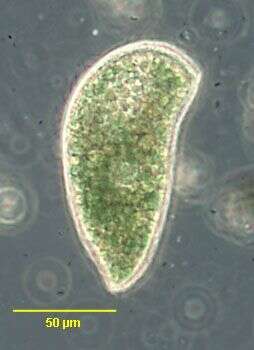
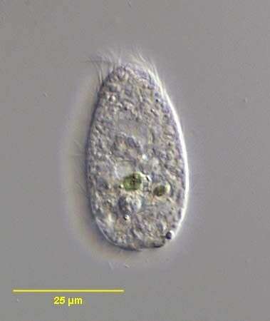
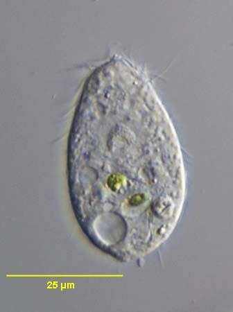
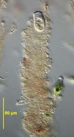
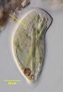
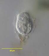
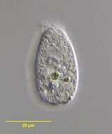
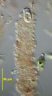
Sagittaria hyalina (FOISSNER, CZAPIK & WIACKOWSKI, 1981) just beginning its mucus case. Unlike the tube-dwelling colpodid, Maryna, Sagittaria usually has its anterior end protruding from the mucus tube.Collected from an ephemeral puddle on the lawn of a public park in Boise, Idaho.September 2006.DIC.
-
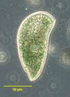
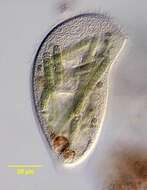
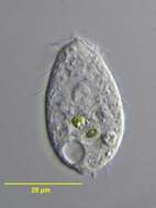
Sagittaria hyalina (FOISSNER, CZAPIK & WIACKOWSKI, 1981) which has fled its mucus case. Collected from an ephemeral puddle on the lawn of a public park in Boise, Idaho.September 2006.DIC.
-
Sagittaria hyalina (FOISSNER, CZAPIK & WIACKOWSKI, 1981) which has fled its mucus case. Collected from an ephemeral puddle on the lawn of a public park in Boise, Idaho.September 2006.DIC.
-
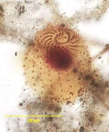
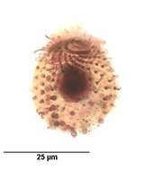
Infraciliature of the cyrtolophosidid ciliate, Sagittaria hyalina (FOISSNER, CZAPIK & WIACKOWSKI, 1981) viewed from the right apicoolateral aspect. Collected from an ephemeral puddle on the lawn of a public park in Boise, Idaho.September 2006.Stained by the silver carbonate technique (see Foissner, W. Europ. J. Protistol., 27:313-330;1991).Brightfield.
-
Infraciliature of the cyrtolophosidid ciliate, Sagittaria hyalina (FOISSNER, CZAPIK & WIACKOWSKI, 1981) viewed from the right apicoolateral aspect. Larger densely stained circular structures are mucocysts which the organism extrudes to form its mucus case.Collected from an ephemeral puddle on the lawn of a public park in Boise, Idaho.September 2006.Stained by the silver carbonate technique (see Foissner, W. Europ. J. Protistol., 27:313-330;1991).Brightfield.
-
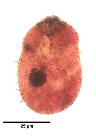
Sagittaria hyalina (FOISSNER, CZAPIK & WIACKOWSKI, 1981). The densely stained macronucleus and ellipsoid micronucleus in its perinuclear space (11 o'clock) are seen here.Collected from an ephemeral puddle on the lawn of a public park in Boise, Idaho.September 2006.Stained by the silver carbonate technique (see Foissner, W. Europ. J. Protistol., 27:313-330;1991).Brightfield.
-
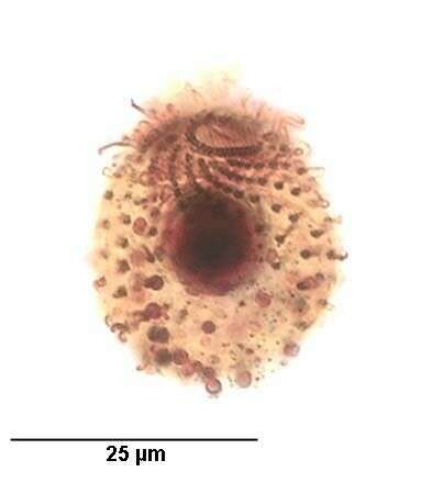
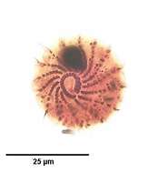
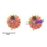
Infraciliature of the cyrtolophosidid ciliate, Sagittaria hyalina (FOISSNER, CZAPIK & WIACKOWSKI, 1981) viewed from the anterior apical aspect.The right paraoral membrane consists of ciliated dikinetids.Four adoral membranelles lie along the left border of the cytostome between the two ends of the C-shaped paraoral membrane. Collected from an ephemeral puddle on the lawn of a public park in Boise, Idaho.September 2006.Stained by the silver carbonate technique (see Foissner, W. Europ. J. Protistol., 27:313-330;1991).Brightfield.
-
Infraciliature of the cyrtolophosidid ciliate, Sagittaria hyalina (FOISSNER, CZAPIK & WIACKOWSKI, 1981) viewed from the anterior apical aspect.The right paraoral membrane consists of ciliated dikinetids (green line).Four adoral membranelles (blue lines) lie along the left border of the cytostome between the two ends of the C-shaped paraoral membrane. About 15 somatic kineties spiral around the long axis to abut the cytostome anteriorly (pink line). Collected from an ephemeral puddle on the lawn of a public park in Boise, Idaho.September 2006.Stained by the silver carbonate technique (see Foissner, W. Europ. J. Protistol., 27:313-330;1991).Brightfield.
-
Sagittaria hyalina (FOISSNER, CZAPIK & WIACKOWSKI, 1981) seen in its mucus case. Unlike the tube-dwelling colpodid, Maryna, Sagittaria usually has its anterior end protruding from the mucus tube.Collected from an ephemeral puddle on the lawn of a public park in Boise, Idaho.September 2006.Stained by the silver carbonate technique (see Foissner, W. Europ. J. Protistol., 27:313-330;1991).DIC.
-
Stained by the Klein-Foissner dry silver nitrate technique (see Foissner, W. Europ. J. Protistol., 27:313-330;1991).