-
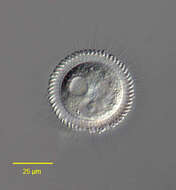
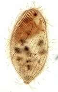
Pera, Faro, Portugal
-
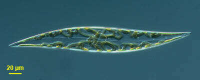
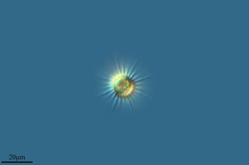
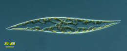
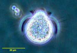
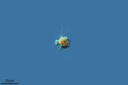
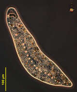
This pennate diatom was found in a plankton tow from Nantucket Sound off Martha's Vineyard - Massachusetts, USA. Image by Jeffrey Cole and Micah Dunthorn.
-
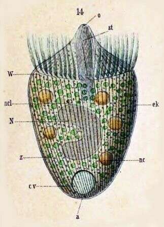
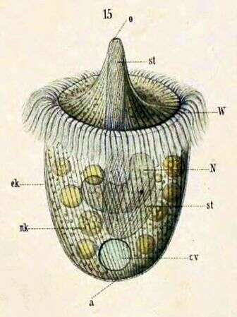
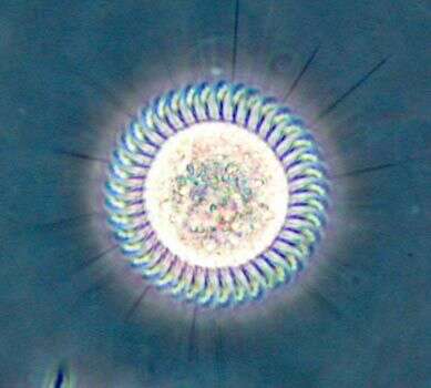
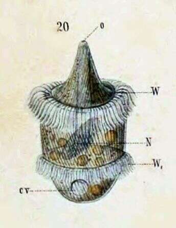
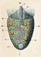
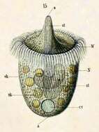
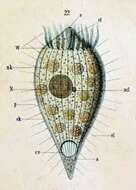
Originally described by Schewiakoff under the name Didinium balbianii. An individual with weakly projecting mouth cone, traveling backwards. a--Anus cv--Contractile vacuole ek--Ectoplasm N--Macronucleus ncl--Micronucleus o--Mouth st--Cytopharyngeal basket W--Ciliated ring z -- Zoochlorellae
-
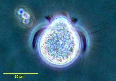
Portrait of the haptorid ciliate, Askenasia volvox (Eichwald,1852) Kahl, 1930. The cell is spherical posteriorly and the anterior is a truncate cone.The cytostome is at the anterior apex.The cytostome is surrounded by an undulating line of granules (seen only in silver impregnated specimens).Somatic cilia are arranged in three (anterior,middle and posterior)girdles.The posterior girdle consists of long stiff bristles.The anterior cilia are directed forward and the middle girdle cilia are longer , curving backwards in a "sickle" configuration.These cilia produce the saltatory locomotion typical of this genus.The posterior of the cell is unciliated.The central macronucleus is C-shaped. There is a single subequatorial contractile vacuole. From a freshwater pond near Boise, Idaho. Phase contrast.
-
-
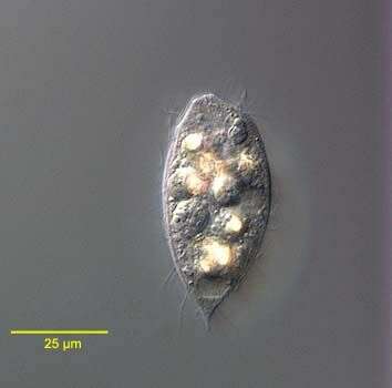
Portrait of the haptorid ciliate, Acropisthium mutabile (Perty, 1852). The cell body is ovoid. The posterior tapers to a short point. The anterior end forms a blunt snout with an apical cytostome. Short trichites support the cytopharynx. There is wreath of longer cilia just posterior to the bare anterior snout. The uniform longitudinal somatic kineties are are widely spaced. Three anterior rows of clavate cilia form a dorsal brush. The cytoplasm contains highly refractile crystaline inclusions. The spherical macronucleus is posterior. There is a single posterior terminal contractile vacuole. Collected from freshwater pond near Boise, Idaho May 2004. DIC optics.
-
Enchelyodon armatus (KAHL, 1926) KAHL, 1930.DIC.
-
Dorsolateral view of the infraciliature of Spirozona caudata (Kahl,1926) in early division. The stomatogenic field of the opisthe is seen as a patch of kinetosomes adjacent to the first somatic kinety (k1) (black arrow).Stained by the silver carbonate technique (see Foissner, W. Europ. J. Protistol., 27:313-330;1991).Brightfield.
-
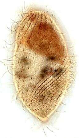
Pera, Faro, Portugal
-
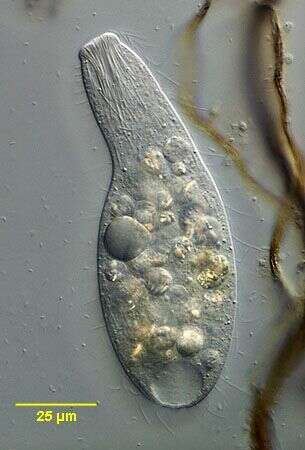
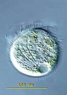
Pelagovasicola (pee-ladge-o-vee-sick-o-la) cinctum is a very fast swimming obovoid ciliate measuring 50 - 180 X 40 - 85 microns. It is common in plankton of lakes and ponds. The body is surrounded by 5-7 distinct ciliary girdles. The posterior fifth of the cell is unciliated. The contractile vacuole lies in the posterior end and has about 20 radial collecting channels. The macronucleus is kidney-shaped and lies in the mid-body. Extrusomes are arranged in the margin of the oral dome, occasionally extruded as bundles of fine filaments. This free-swimming specimen was collected in the plankton of a bog pond near Konstanz, Germany. 115 X 92 microns. Differential interference contrast.
-
Originally described by Schewiakoff under the name Didinium balbianii. Shown traveling forward, with mouth cone extended. a--Anus cv--contractile vacuole ek--Ectoplasm N--Macronucleus nk--Food particle o--Mouth st--Cytopharyngeal basket W--Ciliated ring
-
Posterior apical view of the haptorid ciliate,Askenasia volvox (Eichwald,1852) Kahl, 1930. The cell is spherical posteriorly and the anterior is a truncate cone.The cytostome is at the anterior apex.The cytostome is surrounded by an undulating line of granules (seen only in silver impregnated specimens).Somatic cilia are arranged in three (anterior,middle and posterior)girdles.The posterior girdle consists of long stiff bristles (seen here).The anterior cilia are directed forward and the middle girdle cilia are longer,curving backwards in a "sickle" configuration.These cilia produce the saltatory locomotion typical of this genus (they are seen well in this image).The posterior of the cell is unciliated.The central macronucleus is C-shaped. There is a single subequatorial contractile vacuole. From a freshwater pond near Boise, Idaho. Phase contrast.
-
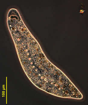
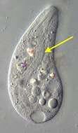
Homalozoon, a elongate ribbon-like predatory ciliate. The body is truncated (cut) off at the front end where the mouth is located, and pointed posteriorly. It has rows of cilia mostly on the ventral side, it glides over the substrate, sometimes contracting. Feeds on detritus and other protists. This image shows the line of contractile vacuoles which extends along the body. Flattened. Phase contrast micrograph.
-
Portrait of the haptorid ciliate, Acropisthium mutabile (Perty, 1852). The cell body is ovoid. The posterior tapers to a short point. The anterior end forms a blunt snout with an apical cytostome. Short trichites support the cytopharynx. There is wreath of longer cilia just posterior to the bare anterior snout. The uniform longitudinal somatic kineties are are widely spaced. Three anterior rows of clavate cilia form a dorsal brush. The cytoplasm contains highly refractile crystaline inclusions. The ellipsoid macronucleus is seen just anterior to the contractile vacuole. There is a single posterior terminal contractile vacuole. Collected from freshwater pond near Boise, Idaho February 2005. DIC optics.
-
In vivo portrait of Enchelyodon armatus (KAHL,1926),KAHL,1930 demonstrating the band-form macronucleus.
-
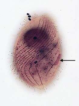
Infraciliature (dorsal view) of the trichostomatid ciliate, Spirozona caudata (Kahl,1926). The cell is elongate and rounded anteriorly and tapers posteriorly to a narrow truncate cone. The cell is ellipsoid in cross section. The cytostome is located in the anterior 1/4. Its right margin is curved and the left relatively straight. There are several oral polykinetids on the left and an undulating membrane on the right. The somatic ciliature is distinctive with a wide swath of closely spaced kineties spiraling from the right anterior to the posterior midline. A single spiral kinety of more densely packed kinetosomes bearing longer cilia originates to the right of the cytostome and spirals around the right side to the dorsum of the cell. There are 3 postoral kineties and one short paraoral kinetid on the left margin of the cytostome. More widely spaced kineties with less densely packed kinetids originate to the left of the cytostome and follow a less spiral course to the posterior end ventrally. The narrow truncate cone of the posterior end bears a circular row of kinetids.There is a small unciliated anterior apical area or "frontal plate". The spherical macronucleus and micronucleus are located in the anterior half. The single contractile vacuole is located at the posterior end. Collected from sapropelic bottom sediments from slow flowing freshwater near Boise, Idaho March,2007. Stained by the silver carbonate technique (see Foissner, W.Europ. J. Protistol.27,313-330;1991). Brightfield optics.
-
Pera, Faro, Portugal
-
Pelagovasicola (pee-ladge-o-vee-sick-o-la) cinctum is a very fast swimming obovoid ciliate measuring 50 - 180 X 40 - 85 microns. It is common in plankton of lakes and ponds. The body is surrounded by 5-7 distinct ciliary girdles. The posterior fifth of the cell is unciliated. The contractile vacuole lies in the posterior end and has about 20 radial collecting channels. The macronucleus is kidney-shaped and lies in the mid-body. Extrusomes are arranged in the margin of the oral dome, occasionally extruded as bundles of fine filaments. This slightly squashed specimen was collected in the plankton of a bog pond near Konstanz, Germany, and this images emphasizes the radial collecting channels of the contractile vacuole. Differential interference contrast.
-
Originally described by Schewiakoff under the name Didinium balbianii. Schewiakoff describes this as an individual apprehended in division. o--Mouth N--Macronucleus W--Ciliated ring W1--New ciliated ring, prepared for offspring
-
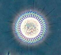
Posterior apical view of the haptorid ciliate,Askenasia volvox (Eichwald,1852) Kahl, 1930. The cell is spherical posteriorly and the anterior is a truncate cone.The cytostome is at the anterior apex.The cytostome is surrounded by an undulating line of granules (seen only in silver impregnated specimens).Somatic cilia are arranged in three (anterior,middle and posterior)girdles.The posterior girdle consists of long stiff bristles (seen here).The anterior cilia are directed forward and the middle girdle cilia are longer,curving backwards in a "sickle" configuration.These cilia produce the saltatory locomotion typical of this genus (they are seen well in this image).The posterior of the cell is unciliated.The central macronucleus is C-shaped. There is a single subequatorial contractile vacuole (seen here to viewer's left). From a freshwater pond near Boise, Idaho.DIC.
-
Homalozoon, a elongate-ribbon like predatory ciliate. The body is truncated (cut) off at the front end where the mouth is located, and pointed posteriorly. It has rows of cilia mostly on the ventral side, it glides over the substrate, sometimes contracting. Feeds on detritus and other protists. This image shows the macronucleus, which takes to form of a row of ellip[tical beads attached end-to-end. The micronuclei are small round dark structures adjacent to the macronucleus. Phase contrast micrograph.
-
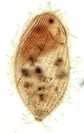
Originally described as Dinophrya liberkuhnii (Butschli) a -- Anus cl -- Cilia cv -- Contractile vacuole ek -- Ectoplasm N -- Macronucleus ncl -- Micronucleus nk -- Food particle o -- Mouth nk -- Food particle p -- Pellicle st -- Cytopharyngeal basket W -- Ciliated ring
-
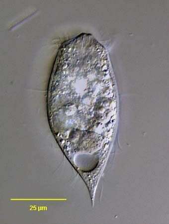
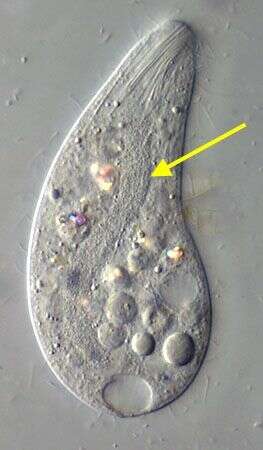
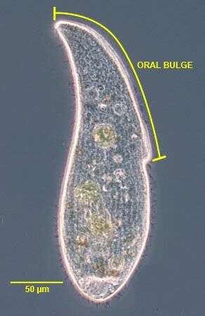
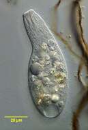
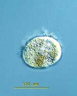
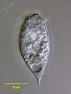
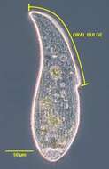
The long oral bulge (~50% of cell length) is one of the main distinguishing features of this subspecies of A. cultriforme. This specimen is somewhat stouter than the cells described by Foissner (Protistology 4 (1), 5-55 (2005) probably due to contraction after transfer from the culture dish to the slide. When observed undisturbed under the dissecting microscope the cells appear more slender.Phase contrast.
-
Infraciliature (ventral view) of the trichostomatid ciliate, Spirozona caudata (Kahl,1926).