Plate 11
Açıklama:
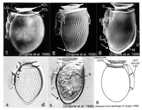
Plate 11. Dinophysis acuminata. Figs. 1-5. SEM: lateral view. Fig. 1. Cell oval and rotund; thecal surface with shallow depressions and scattered pores. Left sulcal list (LSL) extends beyond midpoint of cell. Well-developed cingular lists: anterior cingular list (ACL); posterior cingular list (PCL). C=cingulum. Fig. 2. Long and narrow cell with prominent surface areolae, each with a pore. Antapex tapered and ventrally off-center. Small posterior protrusion present (arrow). Fig. 3. Long and narrow cell. Thecal surface smooth with small scattered pores. Megacytic zone (M) void of pores. Posterior protrusions on antapex (arrow). Figs. 4-5. LM: lateral view. Fig. 4. Surface areolae and tapered antapex (from Larsen & Moestrup 1992: fig. 1d). Fig. 5. Large dorsal nucleus (N). Small, blunt projections on tapered antapex (arrow). Fig. 6. Line drawing.
Aşağıdaki Sayfalarda Bulunmaktadır:
- Life
- Cellular
- Eukaryota (Ökaryot)
- SAR (Stramenopiles, Alveolates, Rhizaria)
- Alveolata
- Dinophyceae (Ateşrengi algler)
- Dinophysiales
- Dinophysiaceae
- Dinophysis
- Dinophysis acuminata
- Dinoflagellata
Bu resim hiçbir koleksiyonda yer almıyor.
Kaynak Bilgileri
- lisans
- cc-publicdomain
- bibliyografik atıf
- Faust, Maria A. and Rose A. Gulledge. Identifying Harmful Marine Dinoflagellates. Smithsonian Contributions from the United States National Herbarium, volume 42: 1-144 (including 48 plates, 1 figure and 1 table).
- orijinal
- orijinal medya dosyası
- kaynağı ziyaret et
- ortak site
- NMNH Marine Dinoflagellates
- ID