in vivo, ventral view
Açıklama:
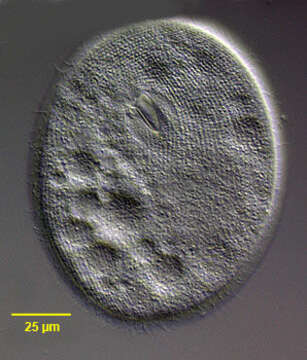
Ventral view of Epenardia myriophylli (Penard, 1922) Corliss, 1971, a large glaucomid ciliate. This cell is slighlty compressed. The body is ellipsoid with minimal dorsoventral flattening. The relatively small obliquely oriented cytostome is in the anterior 1/4. The three adoral membranelles are located in the deep buccal cavity. There is an undulating membrane on the right. E. myriophylli differs from Glaucoma species by its dense (80-110) somatic kineties and the broad M3 which is wider than M2 and a preoral suture in the long axis. There are approximately 9 postoral kineties. The spherical macronucleus and micronucleus are centrally located. The contractile vacuole empties through a single excretory pore on the right dorsal surface. The pellicle is pitted with regular square depressions. Collected from a freshwater drainage ditch near Boise, Idaho. April 2005. DIC
Aşağıdaki Sayfalarda Bulunmaktadır:
- Life
- Cellular
- Eukaryota (Ökaryot)
- SAR (Stramenopiles, Alveolates, Rhizaria)
- Alveolata
- Ciliophora (Silliler)
- Intramacronucleata
- Oligohymenophorea
- Hymenostomatida
- Tetrahymenina
- Glaucomidae
- Epenardia
- Epenardia myriophylli
Bu resim hiçbir koleksiyonda yer almıyor.
Kaynak Bilgileri
- lisans
- cc-by-nc
- yazar
- William Bourland
- sağlayıcı
- micro*scope
- orijinal
- orijinal medya dosyası
- kaynağı ziyaret et
- ortak site
- micro*scope
- ID