Dorsal infraciliature
Açıklama:
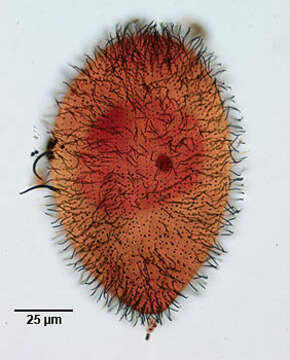
Infraciliature (dorsal aspect) of Glaucoma frontata (Stokes,1886) Kahl,1931, a hymenostome ciliate. While most Glaucoma species are ovoid, this elongate species is bluntly rounded anteriorly and tapers posteriorly to rounded tail. Dorsoventrally flattened. The oral aperture is in the anterior third and slightly oblique to the long axis of the body. There is an inconspicuous undulating membrane on the right of the oral aperture and three well developed membranelles. A small rectangular structure at the anterior end of membranelle 2 is termed the âx-bodyâ. The somatic ciliation (about 40 longitudinal kineties) is uniform with a short preoral suture and 5 postoral kineties. The right ventral kineties arch over the cytostome to abut the preoral suture. The ovoid macronucleus and single micronucleus are central. The single contractile vacuole is located laterally in the midbody. Collected from freshwater pond near Boise, Idaho August 2003. Stained by the silver carbonate technique (see Foissner, W.Europ. J. Protistol.27,313-330;1991). Brightfield.
Aşağıdaki Sayfalarda Bulunmaktadır:
- Life
- Cellular
- Eukaryota (Ökaryot)
- SAR (Stramenopiles, Alveolates, Rhizaria)
- Alveolata
- Ciliophora (Silliler)
- Intramacronucleata
- Oligohymenophorea
- Hymenostomatida
- Tetrahymenina
- Glaucomidae
- Glaucoma
- Glaucoma frontata
Bu resim hiçbir koleksiyonda yer almıyor.
Kaynak Bilgileri
- lisans
- cc-by-nc
- yazar
- William Bourland
- sağlayıcı
- micro*scope
- orijinal
- orijinal medya dosyası
- kaynağı ziyaret et
- ortak site
- micro*scope
- ID