Cortical detail, in vivo
Açıklama:
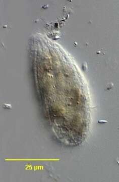
In vivo portrait of the colpodid ciliate, Platyophrya vorax (Kahl,1926). Theflask-shaped cells are very flexible. The relatively small ovoid anterior cytostome is subapical. There is a right paraoral membrane composed of dikinetids and four rectangular left adoral membranelles. There is a postoral "pseudomembrane" consisting of closely spaced anterior dikinetids of somatic kineties. The right side is more densely ciliated than the left. The slightly spiralled somatic kineties lie in shallow cortical furrows (seen here). The central spherical macronucleus has a small central nucleolus and an adjacent micronucleus. The single contractile vacuole is subterminal posteriorly. P. vorax lacks endosymbiotic algae (present in P. sphagni which also has more numerous adoral membranelles and somatic kineties).Collected from temporary puddles with heavy growth of diatoms in a meadow near Boise, Idaho. 43°41'45.09"N 116°13'55.29"W elev.3191 ft. March 2006. DIC.
Aşağıdaki Sayfalarda Bulunmaktadır:
- Life
- Cellular
- Eukaryota (Ökaryot)
- SAR (Stramenopiles, Alveolates, Rhizaria)
- Alveolata
- Ciliophora (Silliler)
- Intramacronucleata
- Colpodea
- Cyrtolophosidida
- Platyophryidae
- Platyophrya
- Platyophrya vorax
Bu resim hiçbir koleksiyonda yer almıyor.
Kaynak Bilgileri
- lisans
- cc-by-nc
- yazar
- William Bourland
- sağlayıcı
- micro*scope
- orijinal
- orijinal medya dosyası
- kaynağı ziyaret et
- ortak site
- micro*scope
- ID