Viruses-07-01700-g003

Açıklama:
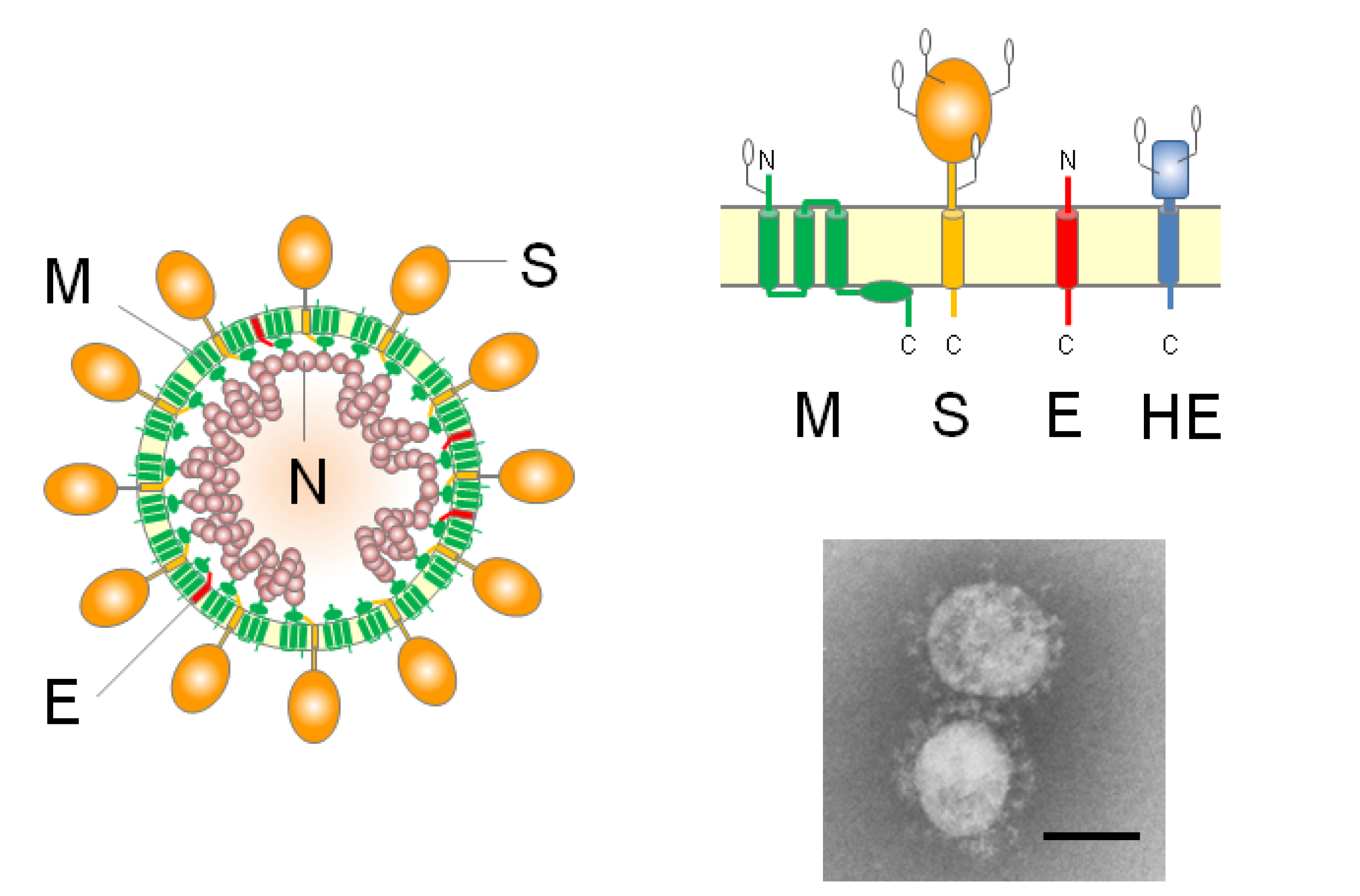
Description: English: (left) Schematic diagrams of coronavirus virions; (right top) The topology of the four structural envelope proteins. All proteins are depicted as monomers, but the S and HE proteins form homotrimers and homodimers, respectively. Oligosaccharides are shown on the M, S, and HE proteins. Although a number are omitted, the S and HE proteins contain 21 to 35 and 9 (BCoV HE) potential N-glycosylation sites, respectively; (right bottom) Electron micrograph of SCoV. Bar: 100 nm. (EM image courtesy of Dr. Nagata at National Institute of Infectious Diseases). Date: 3 April 2015. Source: https://www.mdpi.com/1999-4915/7/4/1700/htm. Author: Makoto Ujike and Fumihiro Taguchi. Other versions: : All biology images in this gallery could be re-created using vector graphics as SVG files. This has several advantages; see Commons:Media for cleanup for more information. If an SVG form of this image is available, please upload it and afterwards replace this template with {{vector version available|new image name}}..
Aşağıdaki Sayfalarda Bulunmaktadır:
Bu resim hiçbir koleksiyonda yer almıyor.
Kaynak Bilgileri
- lisans
- cc-by-3.0
- telif hakkı
- Makoto Ujike and Fumihiro Taguchi
- oluşturan
- Makoto Ujike and Fumihiro Taguchi
- kaynak
- https://www.mdpi.com/1999-4915/7/4/1700/htm
- orijinal
- orijinal medya dosyası
- kaynağı ziyaret et
- ortak site
- Wikimedia Commons
- ID