-
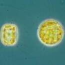
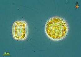
Thalassiosira (tha-lassy-owe-sire-a) eccentrica is one of the hundred or so species in the diverse and common genus of centric diatoms frequently found in marine waters. Some species can be very large. Species are distinguished primarily by the pattern of sculpting in the valve elements of the shell or frustule. The cell to the left is seen in girdle view, the one to the right in valve view. The margins of the valves give rise to a small number of fine chitinous filaments which are believed to function in flotation. These filaments are evident in arising from the cell to the right. With many small golden plastids. Phase contrast microscopy.
data on this strain.
-
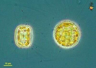
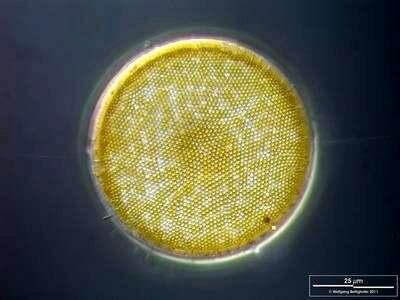
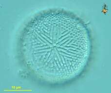
Thalassiosira (tha-lassy-owe-sire-a) eccentrica is one of the hundred or so species in the diverse and common genus of centric diatoms often found in marine waters. Some species can be very large. Species are distinguished primarily by the pattern of sculpting in the valve elements of the shell or frustule. This species has a seven-fold symmetry in the strutting which lies immediately below the surface of the valve, and can be seen in this isolated valve. Differential interference microscopy.
data on this strain.
-
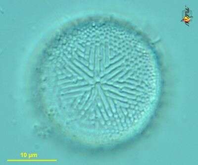
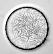
Thalassiosira (tha-lassy-owe-sire-a) eccentrica is one of the hundred or so species in the diverse and common genus of centric diatoms often found in marine waters. Some species can be very large. Species are distinguished primarily by the pattern of sculpting in the valve elements of the shell or frustule, and this shows the surface of an isolated valve. Differential interference microscopy.
data on this strain.
-
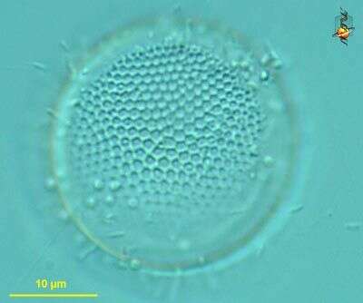
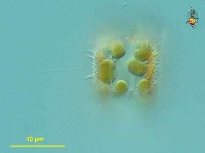
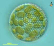
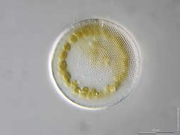
Thalassiosira (tha-lassy-owe-sire-a) eccentrica is one of the hundred or so species in the diverse and common genus of centric diatoms frequently found in marine waters. Some species can be very large. Species are distinguished primarily by the pattern of sculpting in the valve elements of the shell or frustule. The margins of the valve have a number of stout processes and these give rise to the chitinous threads. This is a girdle view and shows the processes as well as the plastids.
data on this strain.
-
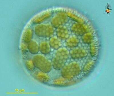
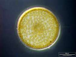
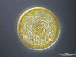
Thalassiosira (tha-lassy-owe-sire-a) eccentrica is one of the hundred or so species in the diverse and common genus of centric diatoms commonly found in marine waters. Some species can be very large. Species are distinguished primarily by the pattern of sculpting in the valve elements of the shell or frustule. This is the valve view showing the sculpting of the valve and the underlying plastids. Differential interference microscopy.
data on this strain.
-
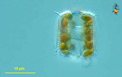
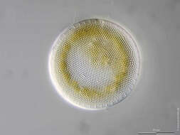
Thalassiosira (tha-lassy-owe-sire-a) eccentrica , is one of the hundred or so species in the diverse and common genus of centric diatoms commonly found in marine waters. Some species can be very large. Species are distinguished primarily by the pattern of sculpting in the valve elements of the shell or frustule. This is the girdle view showing the plastids located near the valves and a central nucleus. Differential interference microscopy.
data on this strain.
-
-
One labiate process is visible together with some chitinous spines. Scale bar indicates 25 µm. The image was built up using several photomicrographic frames with manual stacking technique. Sample from North Sea near Heligoland (spring diatom bloom). Images were taken using Zeiss Universal with Olympus C7070 CCD camera.
-
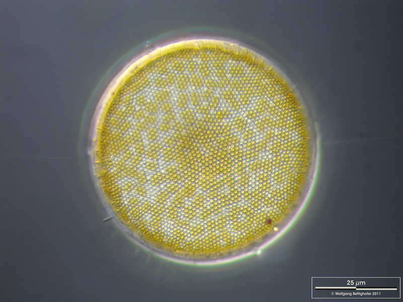
Thalassiosira eccentrica The specimen was gathered in the Kieler Förde (German Baltic Sea). Scale bar indicates 25 µm. The image was built up using several photomicrographic frames with manual stacking technique. Images were taken using Zeiss Axioplan with MFT camera Olympus OM-D E-M5 II.Image under Creative Commons License V 3.0 (CC BY-NC-SA). Place name: Baltic Sea, Kieler Förde, Kiel Fjord (Germany) Latitude: 54.3894126 Longitude: 10.1749055 Multiebenen-Abbildung, manuell gestapelt. Der Messbalken markiert eine Länge von 25 µm. Probe aus der Kieler Förde. Mikrotechnik: Zeiss Axioplan Kamera: Olympus OM-D M5 MKII.Creative Commons License V 3.0 (CC BY-NC-SA). For permission to use of (high-resolution) images please contact postmaster@protisten.de.
-
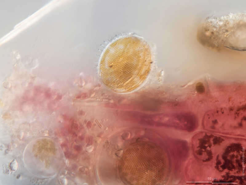
Thalassiosira eccentrica The specimen was gathered in the Kieler Förde (German Baltic Sea). Scale bar indicates 50 µm. The image was built up using several photomicrographic frames with manual stacking technique. Images were taken using Zeiss Axioplan with MFT camera Olympus OM-D E-M5 II.Image under Creative Commons License V 3.0 (CC BY-NC-SA). Place name: Baltic Sea, Kieler Förde, Kiel Fjord (Germany) Latitude: 54.3894126 Longitude: 10.1749055 Multiebenen-Abbildung, manuell gestapelt. Der Messbalken markiert eine Länge von 50 µm. Probe aus der Kieler Förde. Mikrotechnik: Zeiss Axioplan Kamera: Olympus OM-D M5 MKII.Creative Commons License V 3.0 (CC BY-NC-SA). For permission to use of (high-resolution) images please contact postmaster@protisten.de.
-
Thalassiosira eccentrica The specimen was gathered in the Kieler Förde (German Baltic Sea). Scale bar indicates 25 µm. The image was built up using several photomicrographic frames with manual stacking technique. Images were taken using Zeiss Axioplan with MFT camera Olympus OM-D E-M5 II.Image under Creative Commons License V 3.0 (CC BY-NC-SA). Place name: Baltic Sea, Kieler Förde, Kiel Fjord (Germany) Latitude: 54.3894126 Longitude: 10.1749055 Multiebenen-Abbildung, manuell gestapelt. Der Messbalken markiert eine Länge von 25 µm. Probe aus der Kieler Förde. Mikrotechnik: Zeiss Axioplan Kamera: Olympus OM-D M5 MKII.Creative Commons License V 3.0 (CC BY-NC-SA). For permission to use of (high-resolution) images please contact postmaster@protisten.de.
-
Thalassiosira eccentrica One labiate process is visible together with some chitinous spines. Scale bar indicates 25 µm. The image was built up using several photomicrographic frames with manual stacking technique. Sample from North Sea near Heligoland (spring diatom bloom). Images were taken using Zeiss Universal with Olympus C7070 CCD camera.Image under Creative Commons License V 3.0 (CC BY-NC-SA). Place name: North Sea around Heligoland Latitude: 54.186311 Longitude: 7.895034 Ein Silikatfortsatz vom Typ labiate ist sichtbar, zusammen mit chitinösen Schwebefortsätzen. Multiebenen-Abbildung, manuell gestapelt. Der Messbalken markiert eine Länge von 25 µm. Probe aus der Nordsee vor Helgoland in der Zeit der Frühjahrsblüte. Mikrotechnik: Zeiss Universal, Kamera: Olympus C7070.Creative Commons License V 3.0 (CC BY-NC-SA). For permission to use of (high-resolution) images please contact postmaster@protisten.de.