-
Sergio I. Salazar-Vallejo, Galina Buzhinskaja
Zookeys
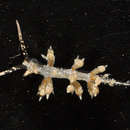
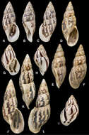
Figure 1.Caulleryaspis fauchaldi sp. n. A Holotype (LACM 5360), ventral view B Anterior end, frontal view C Ventro-caudal shield, frontal view D Paratype (LACM 5361), ventral view E Ventro-caudal shield, frontal view F Posterior end, dorsal view. Bars: A 1.8 mm B, C, E, F 0.6 mm D 2 mm.
-
María E. García-Garza, J.A. de León-González
Zookeys
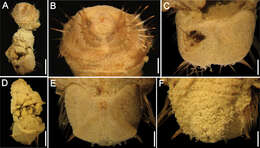
Figure 2.Amphictene helenae. Holotype. A ventral view of anterior end B dorsal view of anterior end C lateral view of anterior end, showing tentacular cirri D notochaetae from 7th chaetiger E front view of bayonet shaped neurochaetae from 7th chaetiger F lateral view of neurochaetae from 7th chaetiger G dorsal view of scaphe (G’) scaphal hooks detail H ventral view, anal papillae I tube of holotype. Bar scale = A, B, C, G, H = 1mm; D = 50mm E, F = 10mm; I = 3mm.
-
Pei Wang, Qiong Xiao, Wei-Chuan Zhou, Chung-Chi Hwang
Zookeys
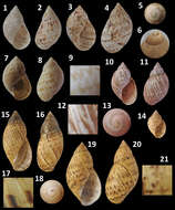
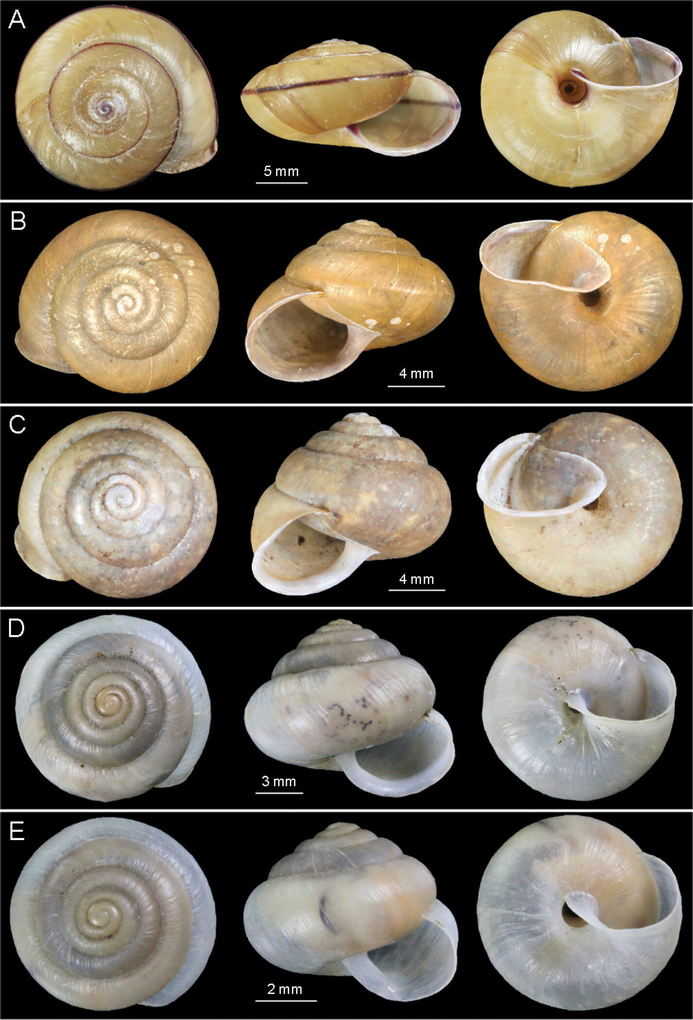
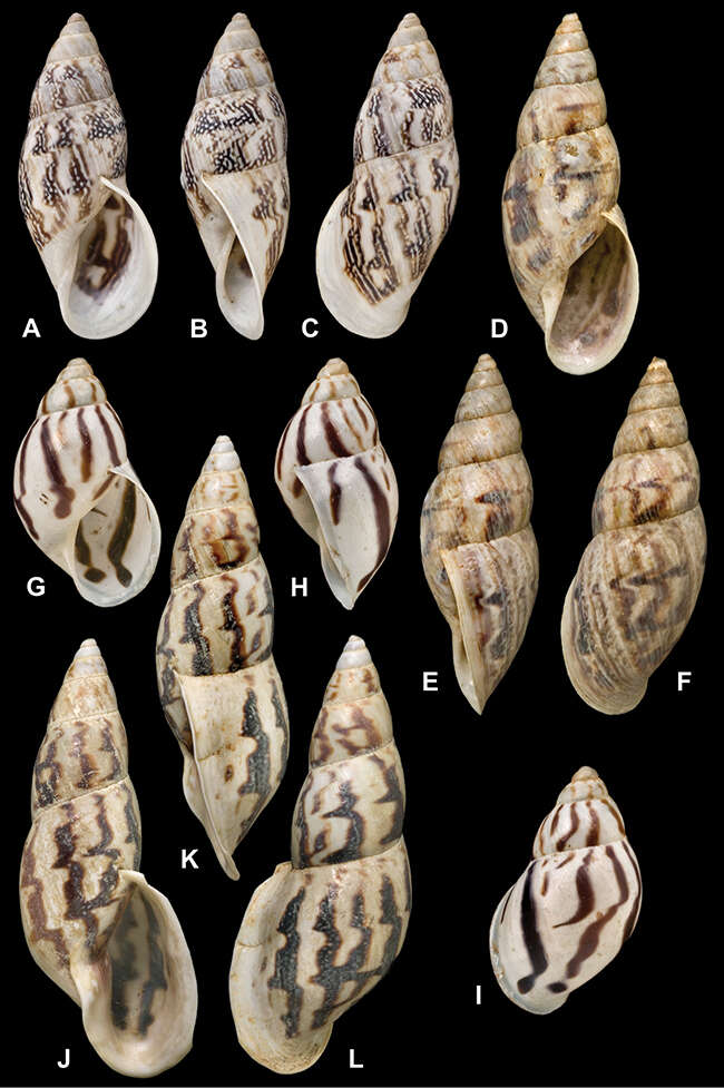
Figure 2.Photographs of shells. A Satsuma mellea stenozona (Moellendorff, 1884) (FJIQBC 18221, Fuzhou, China) B Satsuma meridionalis (Moellendorff, 1884) (FJIQBC 18415, Guangdong, China) C Satsuma uncopila (Heude, 1882) (FJIQBC 18417, Hangzhou, China) D Bradybaena virgo virgo (Pilsbry, 1927) (FJIQBC 18432, Haerbin, China) E Bradybaena virgo mongolia subsp. n. (Holotype, FJIQBC 18466, Inner Mongolia, China).
-
Abraham S.H. Breure, Jonathan D. Ablett
Zookeys
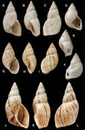
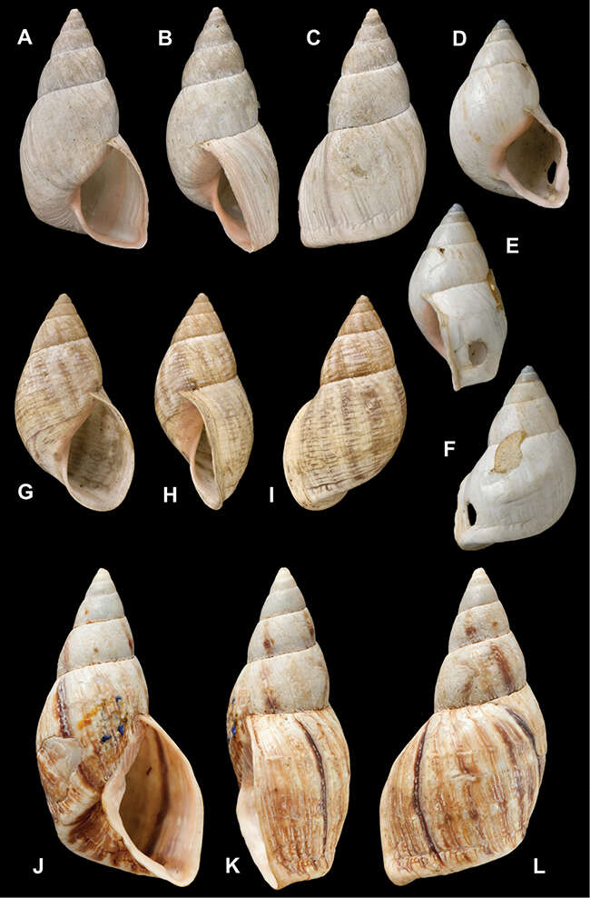
Figure 26.Drymaeus species. A–C Drymaeus (Drymaeus) abscissus (Pfeiffer, 1855), lectotype NHMUK 1975497 (H = 27.8) D–F Drymaeus (Drymaeus) fallax (Pfeiffer, 1853), lectotype NHMUK 1969142 (H = 22.4) G–I Drymaeus (Drymaeus) bourcieri (Pfeiffer, 1853), lectotype NHMUK 1975446 (H = 23.9) J–L Drymaeus (Drymaeus) abruptus (Rolle, 1904), syntype NHMUK 1947.2.10.1 (H = 42.5). All enlarged.
-
Abraham S.H. Breure, Jonathan D. Ablett
Zookeys
Figure 35.Drymaeus species. A–C Drymaeus (Drymaeus) volsus Fulton, 1907, lectotype NHMUK 1907.5.3.162 (H = 30.3) D–F Drymaeus (Drymaeus) yungasensis (d’Orbigny, 1837), lectotype NHMUK 1854.12.4.134 (H = 32.6) G–I Drymaeus (Drymaeus) lucidus (da Costa, 1898), lectotype NHMUK 1907.11.21.44 (H = 18.6) J–L Drymaeus (Drymaeus) sykesi da Costa, 1906, holotype NHMUK 1907.11.21.4 (H = 51.7). All enlarged.
-
Juan Francisco Araya, Ricardo Catalán
Zookeys
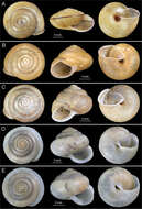
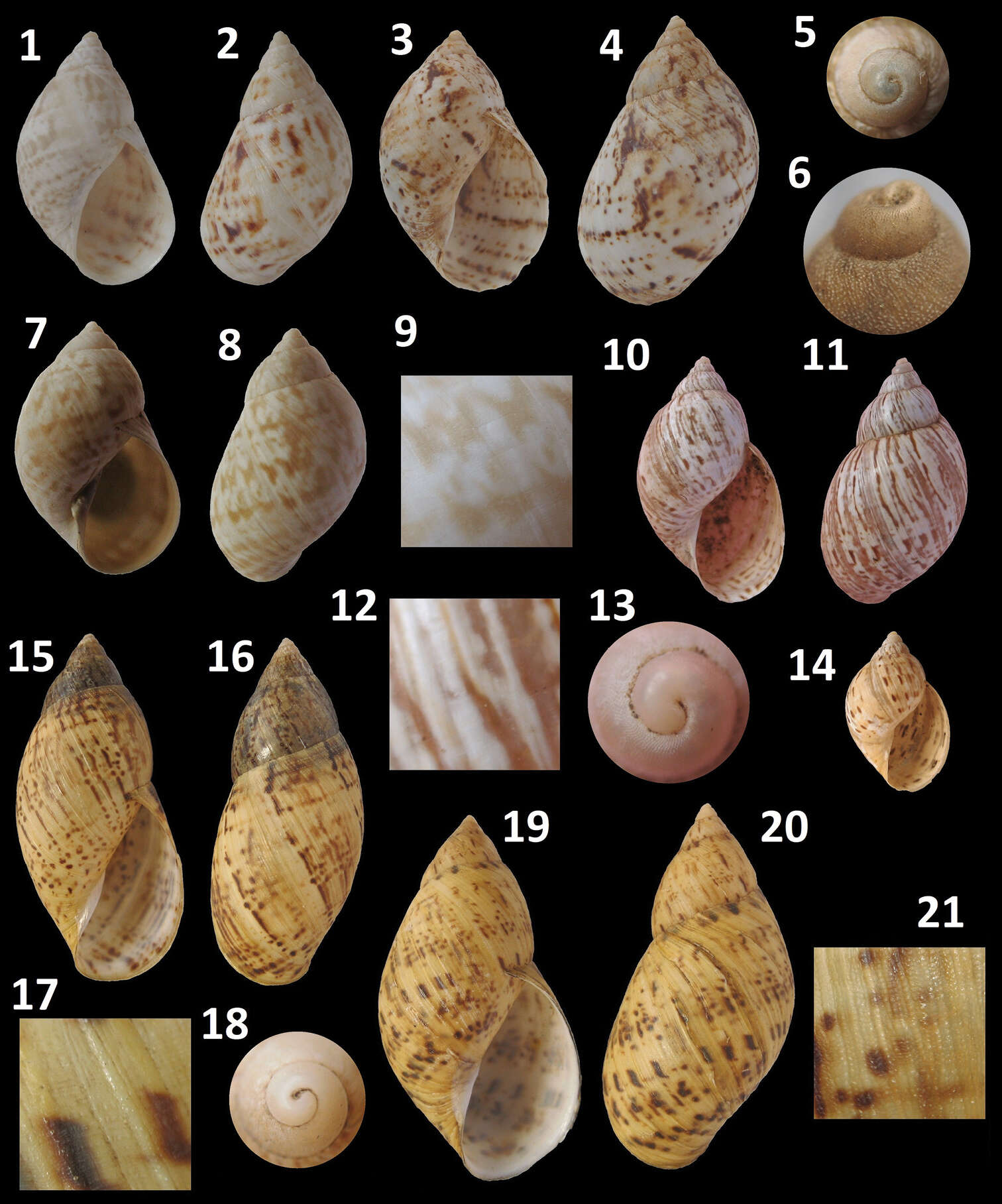
Figure 3.Plectostylus shells. Plectostylus broderipii, Aguas Verdes, Commune of Caldera, 24 mm: 1 Ventral view2 Dorsal view. El Morro hill, Commune of Caldera, 28.8 mm: 3 Ventral view4 Dorsal view 5 Detail of protoconchPlectostylus coturnix, El Morro hill, Commune of Caldera, 27.5 mm: 6 Detail of protoconch7 Ventral view8 Dorsal view 9 Detail of sculpture. Plectostylus elegans, Barranquilla, Commune of Caldera, 24 mm: 10 Ventral view11 Dorsal view 12 Detail of sculpture. 13 Detail of protoconch. 14 Juvenile shell. Plectostylus punctulifer, Fray Jorge National Park, Coquimbo Region, Chile, 20.2 mm: 15 Ventral view16 Dorsal view 17 Detail of sculpture. 18 Detail of protoconch. Plectostylus variegatus, Vallenar, Province of Huasco, 50.5 mm.: 19 Ventral view20 Dorsal view21 Detail of sculpture.
-
Daniel Fernández Marchán, Rosa Fernández, Marta Novo, Darío J. Díaz Cosín
Zookeys
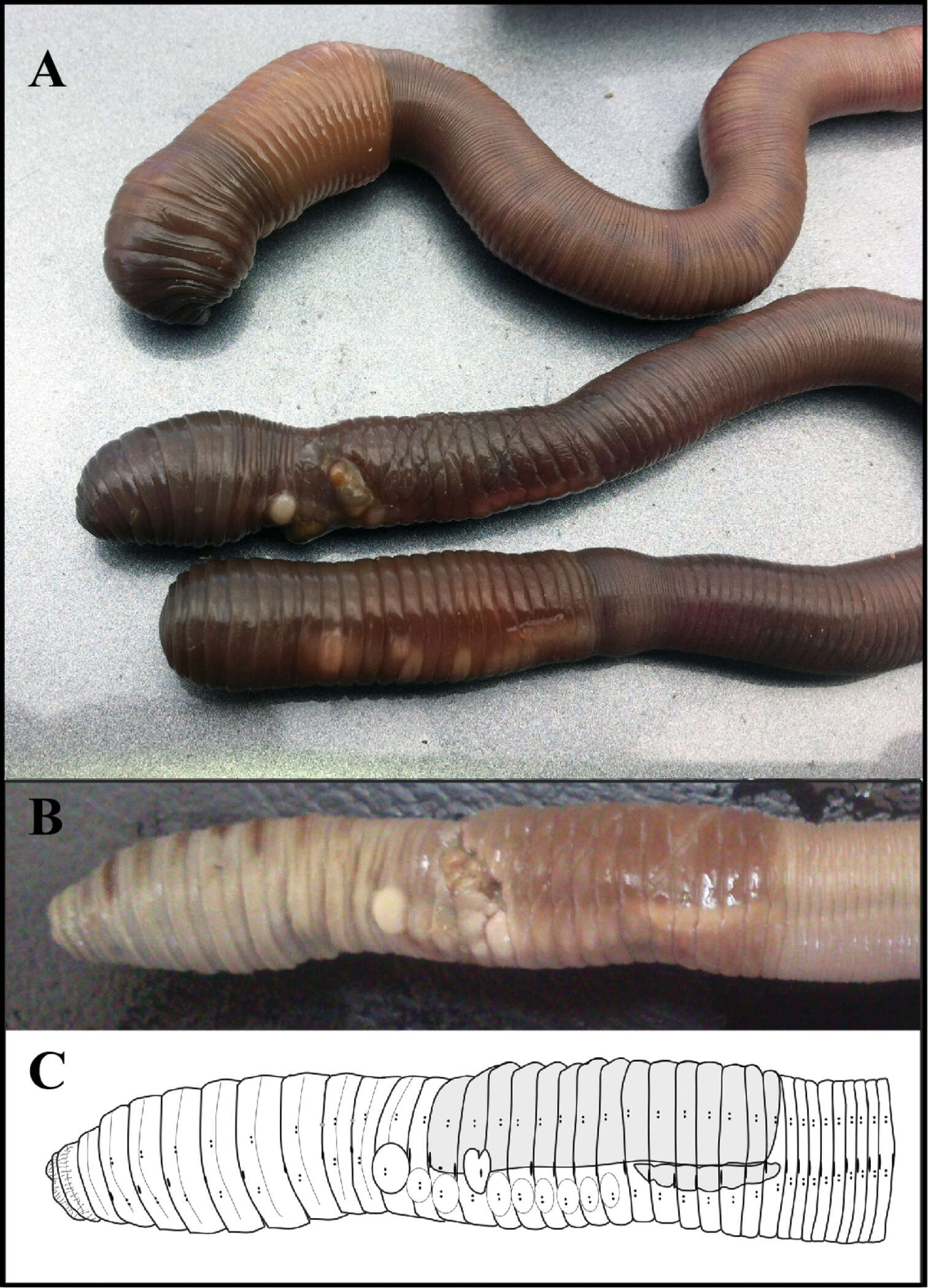
Figure 2.(A) Live specimens of Hormogaster joseantonioi sp.n. External morphology of a fixed specimen, shown in a picture (B) and diagram (C).
-
Shinri Tomioka, Eijiroh Nishi, Hiroshi Kajihara
Zookeys
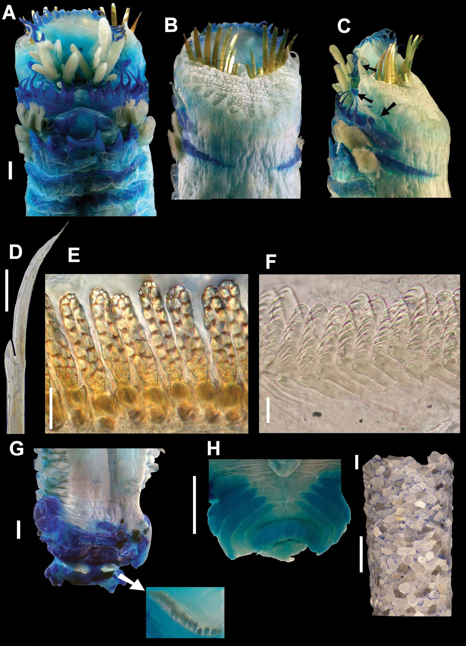
Figure 2.Mediomastus duobalteus sp. n., holotype, CBM-ZW 1088. A Anterior end of body, left lateral view B abdominal segments, left lateral view.
-
Jaime Gonzalez-Cueto, Sigmer Quiroga, Jon Norenburg
Zookeys
Figure 2.A–B Tubulanus rhabdotus: B detail of the head, mouth and lateral organ (modified from Corrêa 1954) C–E Baseodiscus delineatus: C entire specimen, the worm has expulsed the proboscis D ventral detail of the head E dorsal detail of the head F–I Dushia atra: G dorsal detail of the head H ventral detail of the head I detail of the tail showing the caudal cirrus J–L Lineus stigmatus: J entire specimen, the worm autotomized K ventral detail of the head L lateral detail of the head. b brain, c cirrus, cg cephalic grooves, e eyes, lo lateral organ, m mouth, p proboscis, pp proboscis pore.
-
-
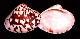
Veneto, Italy
-
-
-
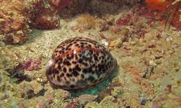
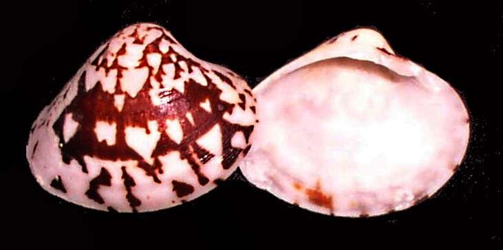
Mabul, Sabah, Malaysia
-
-
-
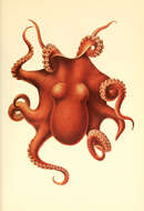
The Cephalopod. atlasJerusalem :Israel Program for Scientific Translations ; Springfield, Va. : available from the U. S. National Technical Information Service,1975.
biodiversitylibrary.org/page/32124297
-
-
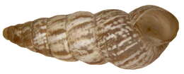
Family: HelicidaeSize: 10-20 x 4-7 mmDistribution: Mediterranean, AtlanticLocation: Tunisia, Hammametleg.det. U.Schmidt, 1993Photo: U.Schmidt, 2008
-
-
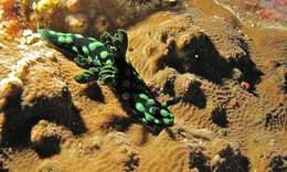
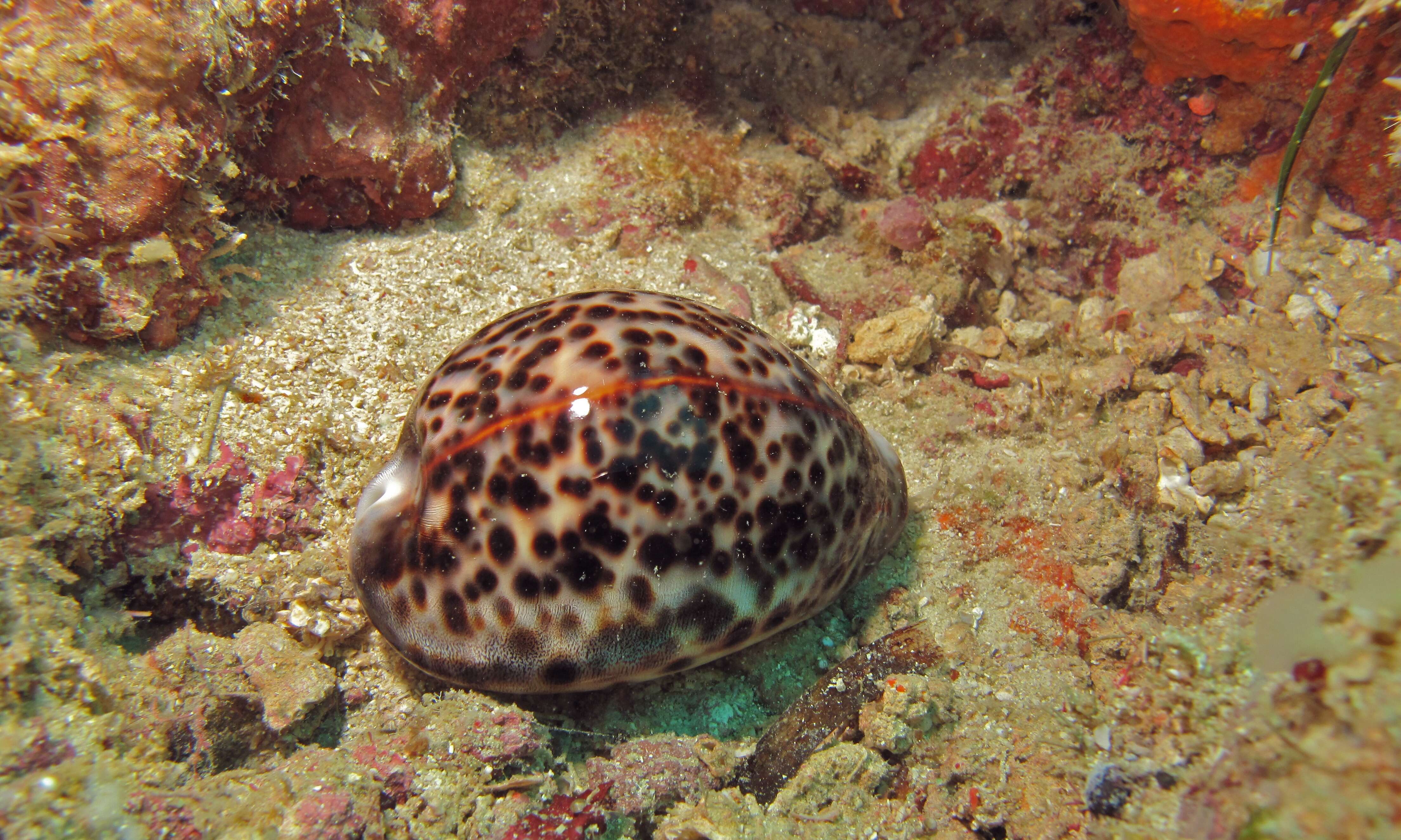
Bunaken, North Sulawesi, Indonesia
-
-
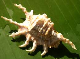
Family: BuccinidaeSize: 45 mmDistribution: Baja California bis PeruPhoto: U.Schmidt, 2007
-
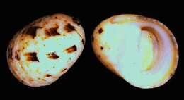
Scientific name: Diodora cayenensis