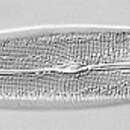
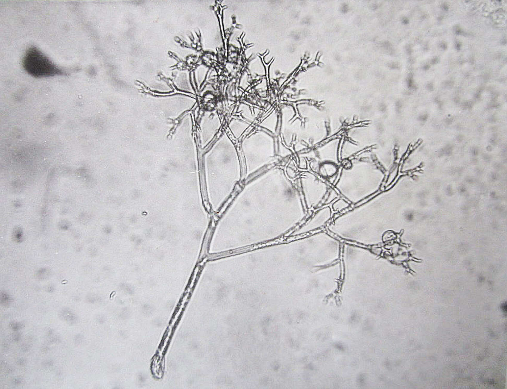
Description: Čeština: P. cambivora strain no 22-06 from Czech Culture of Phytopathogenic Oomycetes (photo K. Černý). Date: 4 June 2008. Source: Own work. Author: Karel Černý.
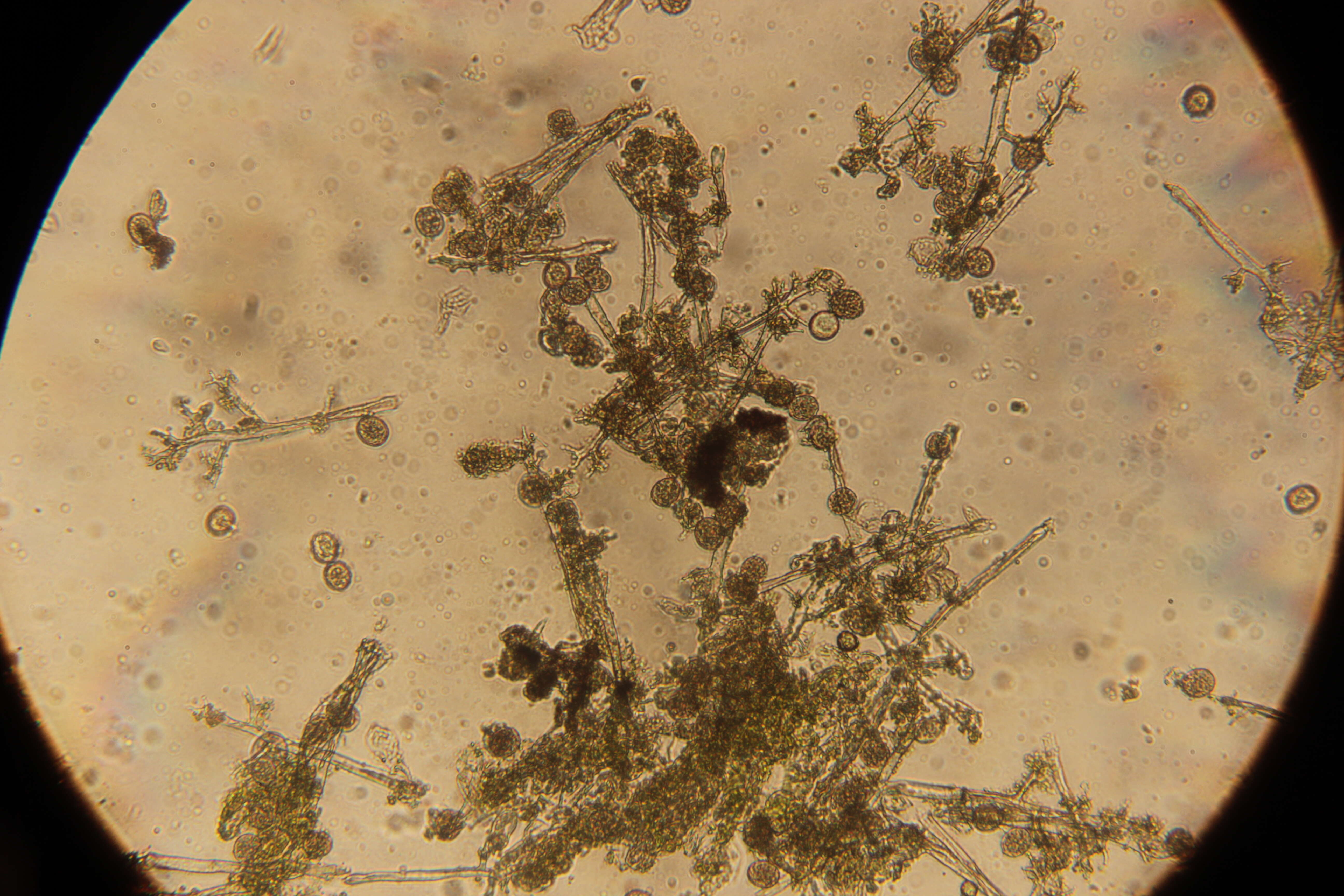
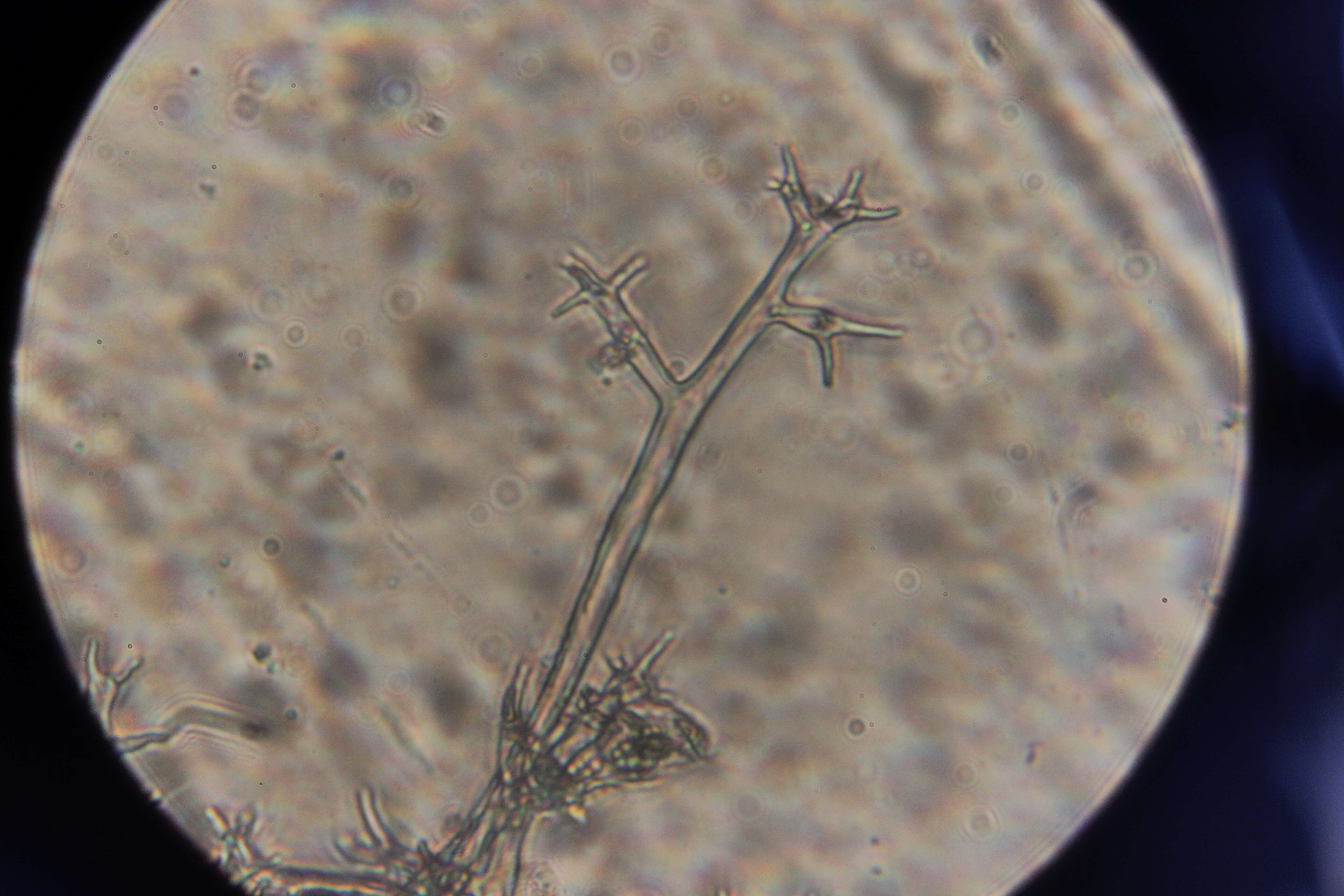
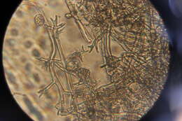
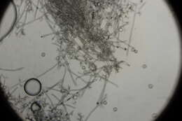
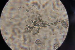
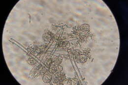
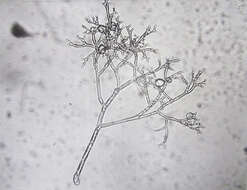
Description: Македонски: Дихотомно разгранет конидиофор од Peronospora tabacina каде јасно се гледаат двете стеригми. English: Dichotomous branched conidiophores of Peronospora tabacina where clearly we can see the both sterigmata.English: Dichotomous branched conidiophores of Peronospora tabacina. Date: 13 December 2015, 10:51:45. Source: Own work. Author: Tashkoskip.
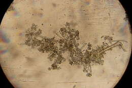
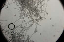
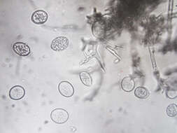
Description: Македонски: Конидии од Peronospora tabacina. Имаат лимонеста форма, безбојни или светложолти со различна големина 10-25x10-22µm.Конидиите ‘ртат во влажни услови и при оптимална температура од 15-23(oС), кога има и најмасова појава на зараза кај тутунот. English: Conidia of Peronospora tabacina. They have lemon form, discolored, or bright yellow with different size 10-25x10-22μm.The conidia germinate in damp conditions and at the optimum temperature of 15-23 (oC), when there are the most outbreak of disease among tobacco.English: Conidia of Peronospora tabacina. Date: 22 February 2013, 10:15:28. Source: Own work. Author: Tashkoskip.
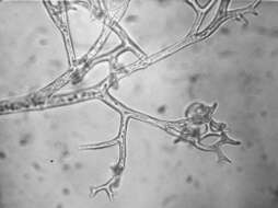
Description: Македонски: Конидиофори од Peronospora hyoscyami f.sp. tabacina — причинител на пламеницата кај тутунот. Конидиофорите кај овој патоген се дихотомно разгранети, на крајот завршуваат со по два запци или стеригми а на секоја стеригма се формира по една конидија. English: Conidophores of Peronospora hyoscyami f.sp. tabacina, a cause of tobacco disease. Conidiophores with this pathogen are dichotomous branched, eventually end with two teeth or sterigmata and on each sterigma is formed one conidia. Date: 13 December 2015, 10:50:39. Source: Own work. Author: Tashkoskip.
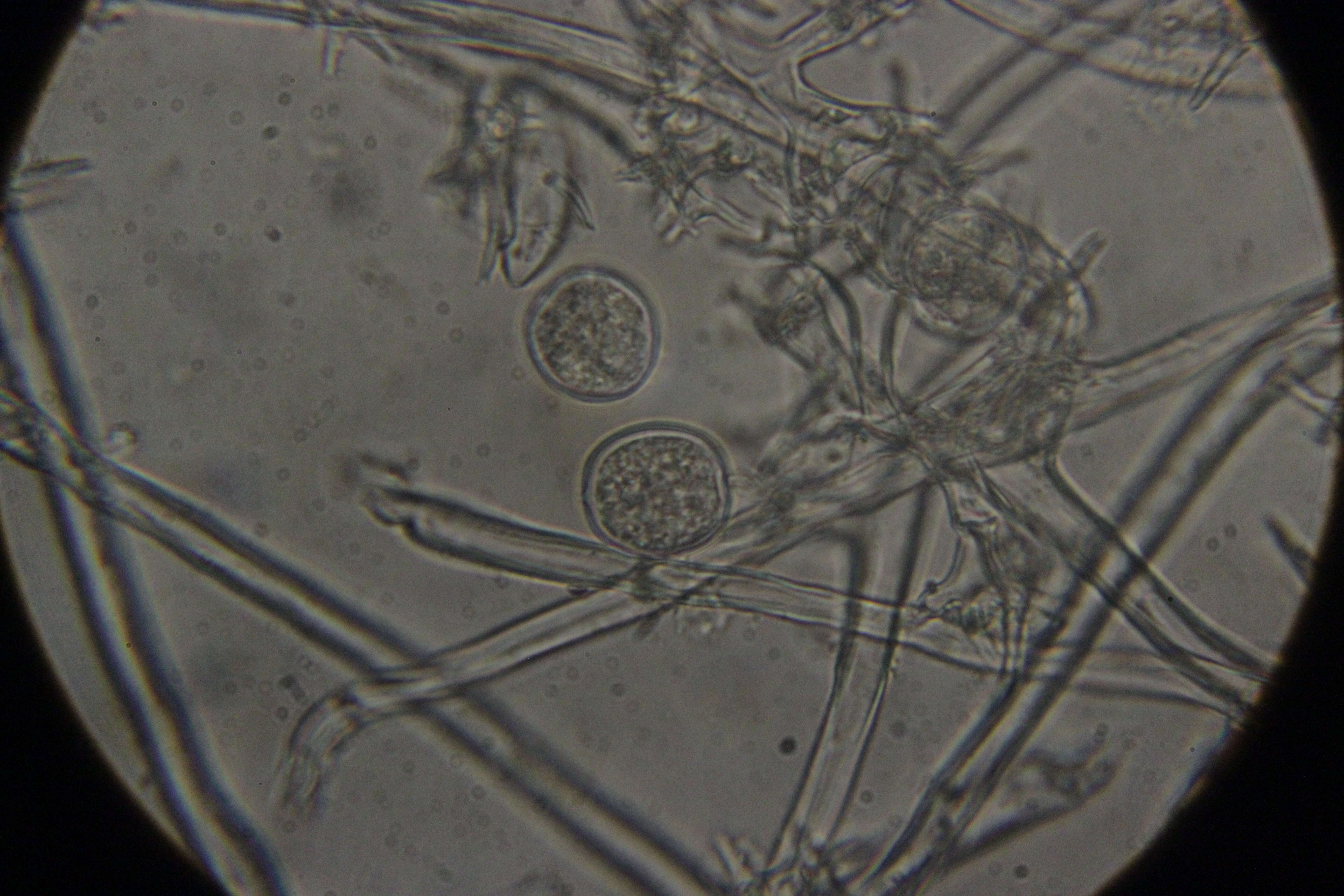
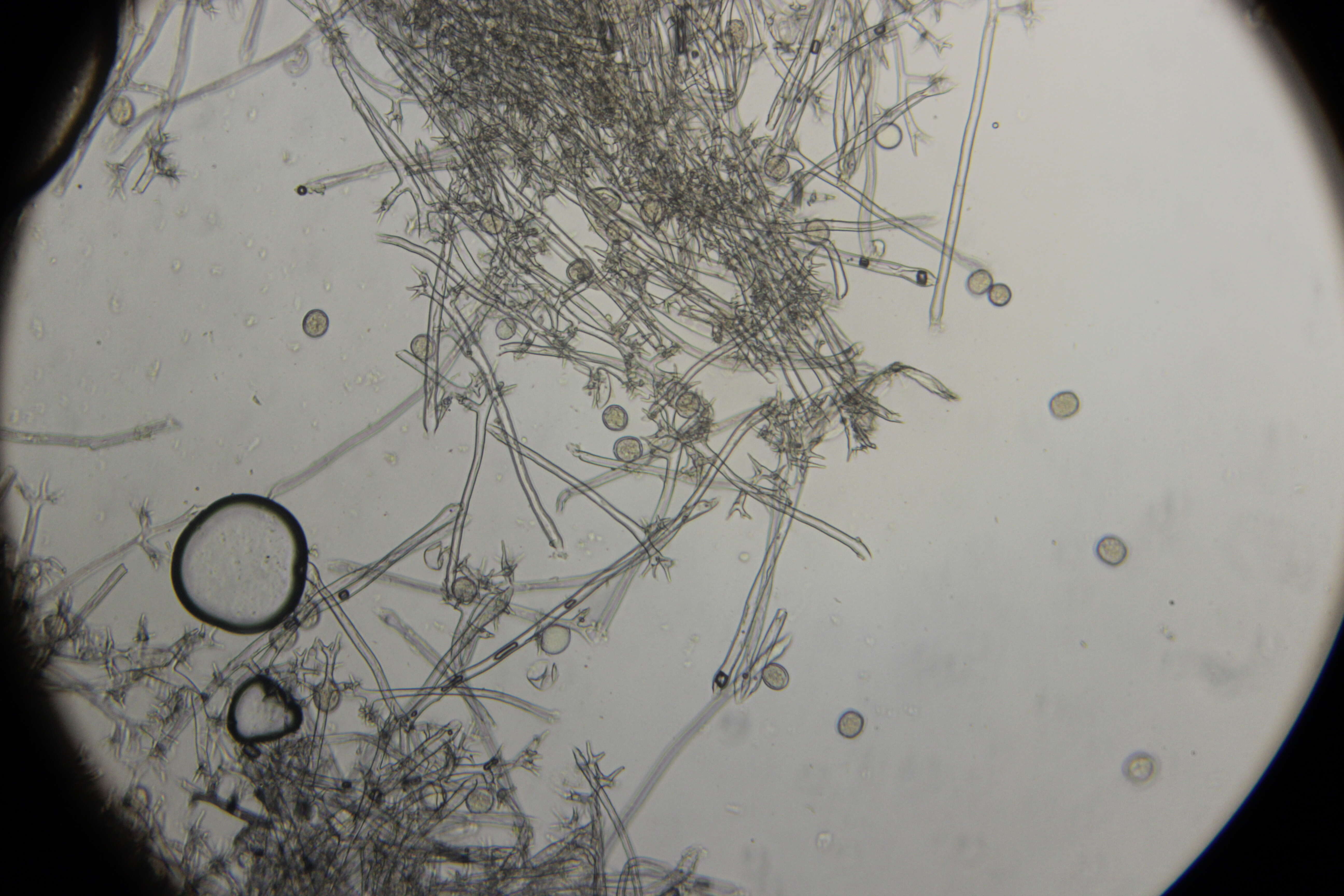
Description: English: Light microscopy of Saprolegnia showing the different stages of development for the oogonium of Saprolegnia. Showing a young, immature, and mature oogonium. The mature oogonium has eggs inside. Scale bar = 0.01mm. Date: 21 May 2014, 09:18:37. Source: Jon Houseman and Matthew Ford. Author: Jon Houseman. Other versions: Labeled. : This is a retouched picture, which means that it has been digitally altered from its original version. Modifications: Balance (Color, brightness, and contrast) and adjust background color.
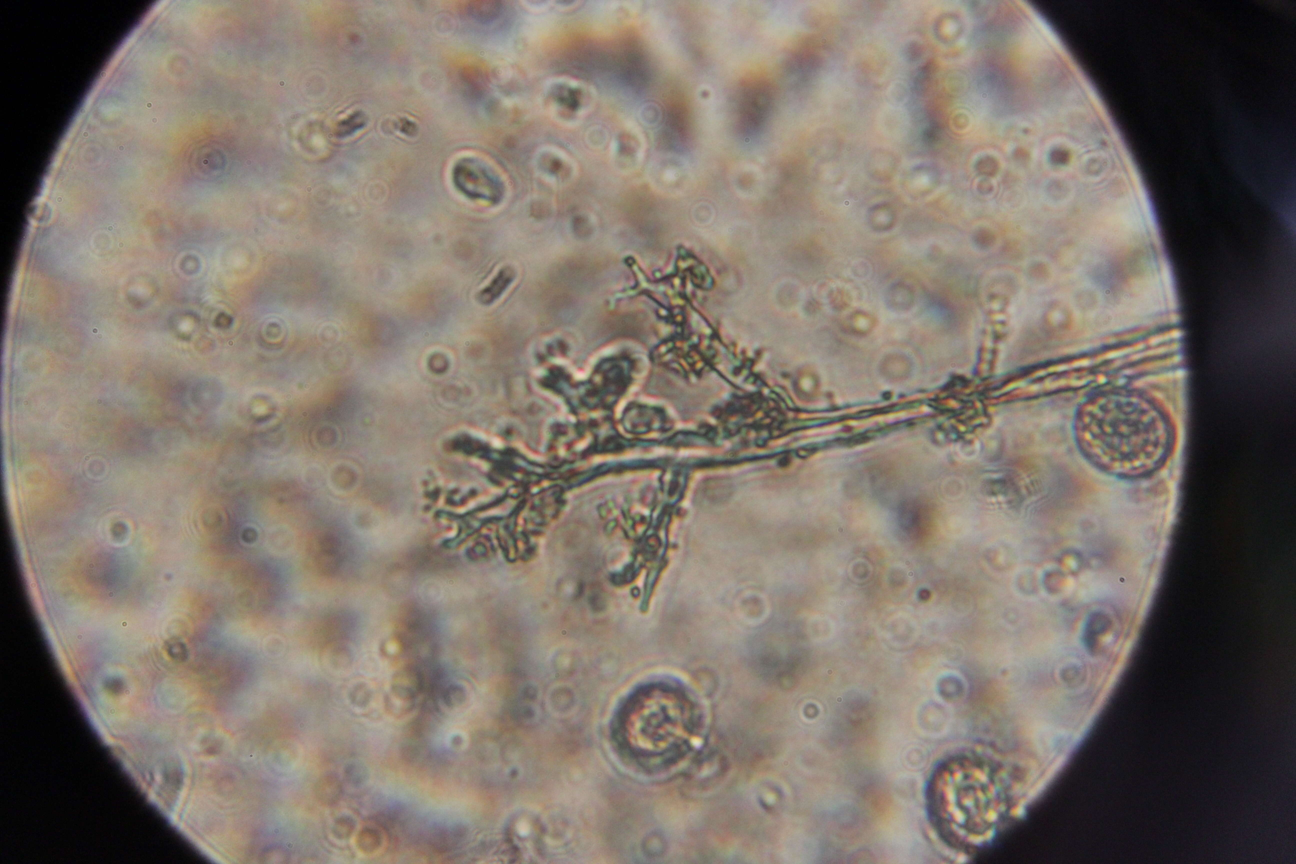
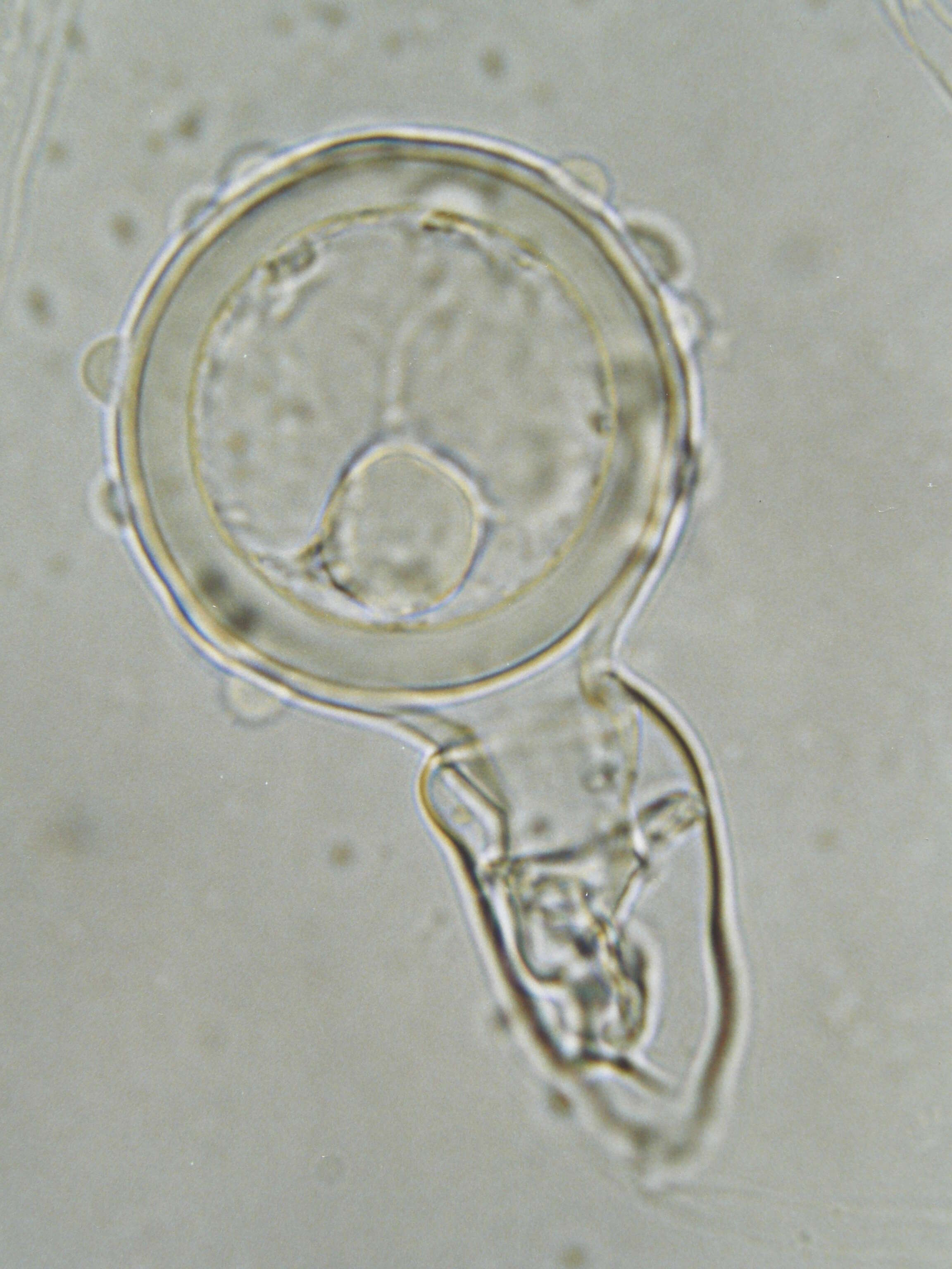
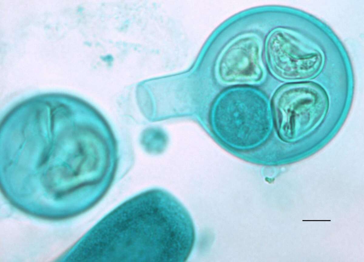
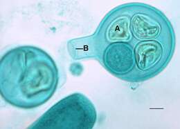
Description: English: Light microscopy of Saprolegnia with an oogonium and zygotes inside with a septum to enclose the entrance. A=Zygote, B=Septum. Scale bar = 0.01mm. Date: 26 May 2014, 09:17:05. Source: Jon Houseman and Matthew Ford. Author: Jon Houseman. Other versions: Original (unlabeled). : This is a retouched picture, which means that it has been digitally altered from its original version. Modifications: Balance (Color, brightness, and contrast) and adjust background color.
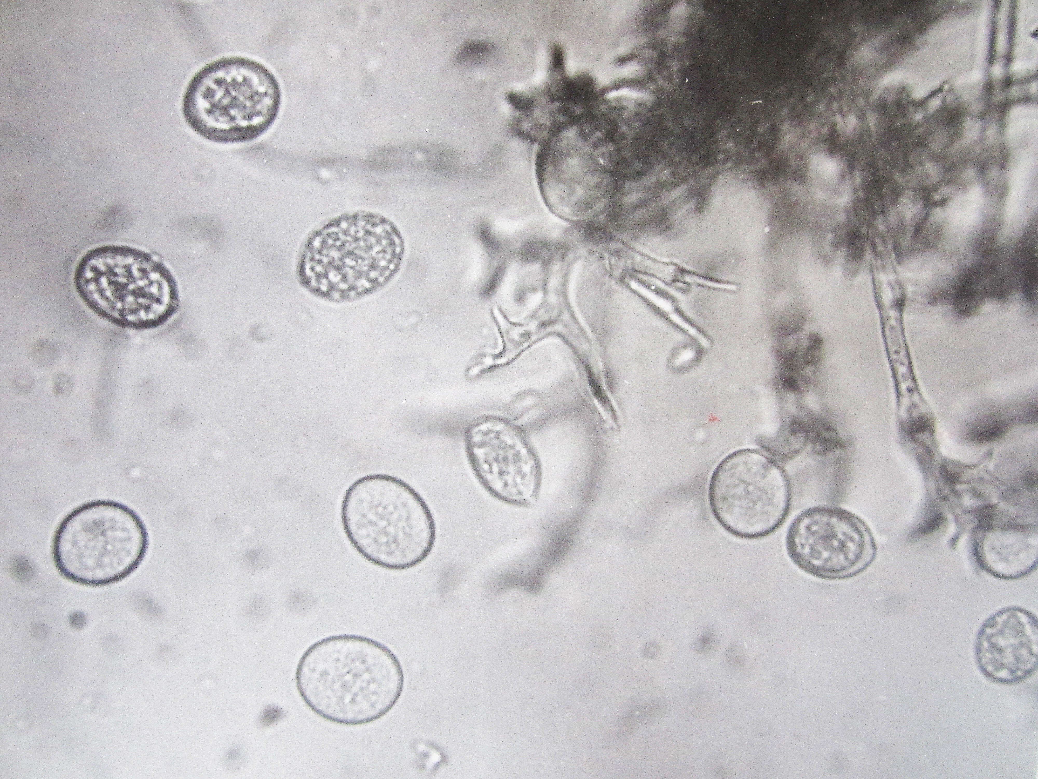
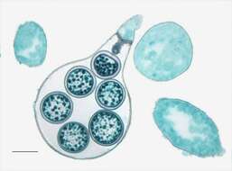
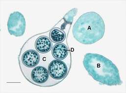
Description: English: Light microscopy of Saprolegnia showing the different stages of development for the oogonium of Saprolegnia. Showing a young, immature, and mature oogonium. The mature oogonium has eggs inside. A=Immature oogonium, B= Developing oogonium, C=Oogonium, D=Egg.Scale bar = 0.01mm. Date: 26 May 2014, 09:17:04. Source: Jon Houseman and Matthew Ford. Author: Jon Houseman. Other versions: Original (unlabeled). : This is a retouched picture, which means that it has been digitally altered from its original version. Modifications: Balance (Color, brightness, and contrast) and adjust background color.
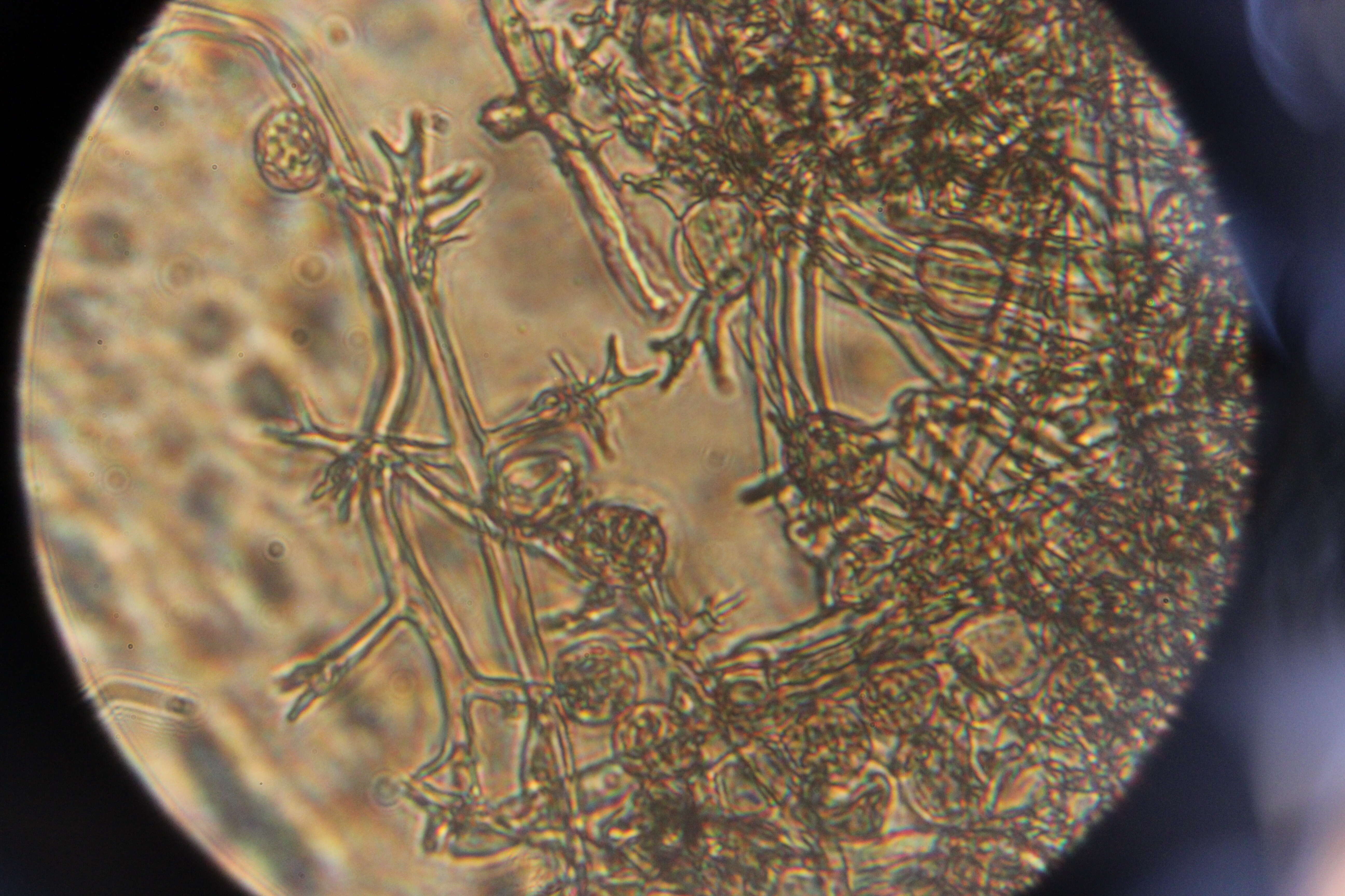
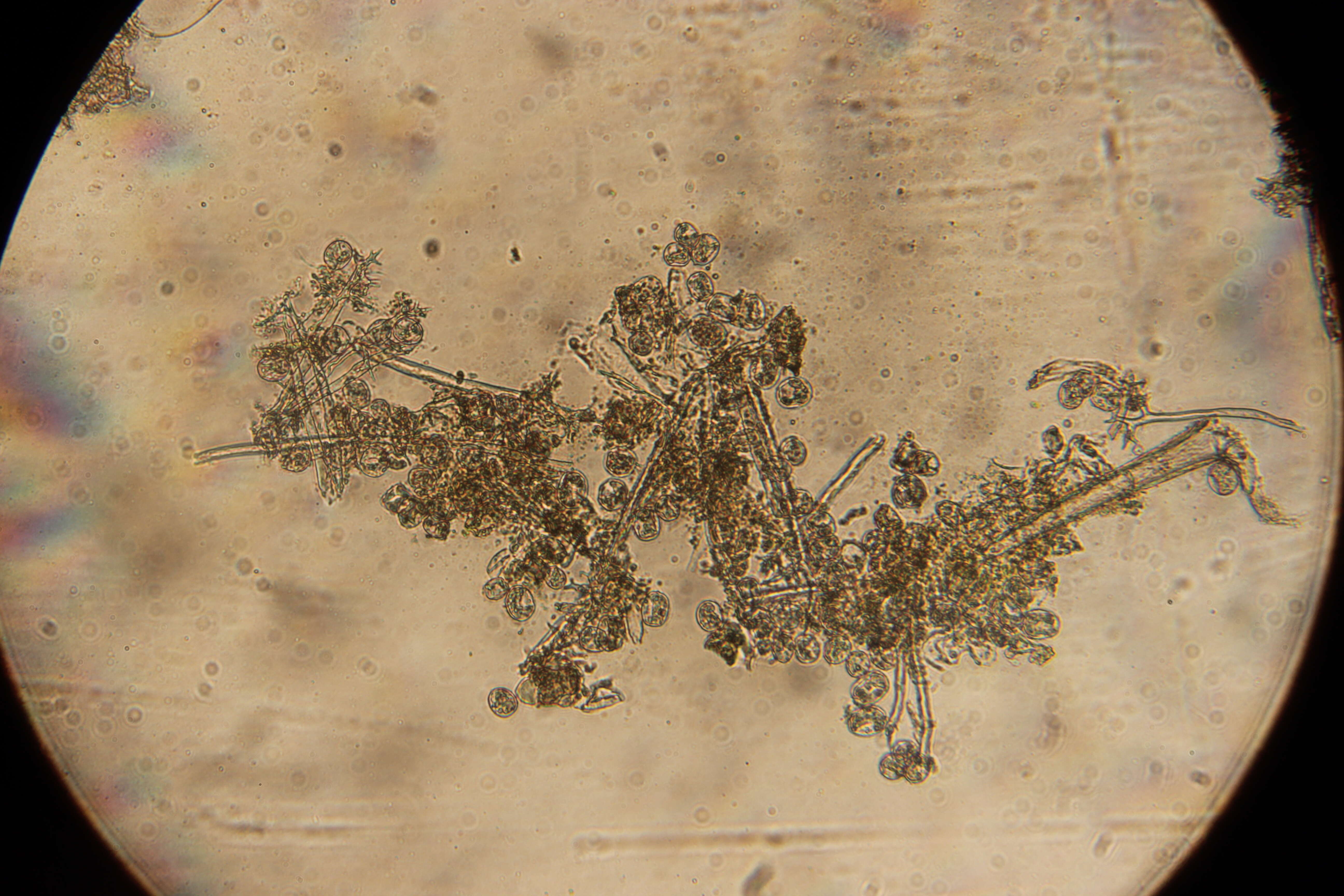
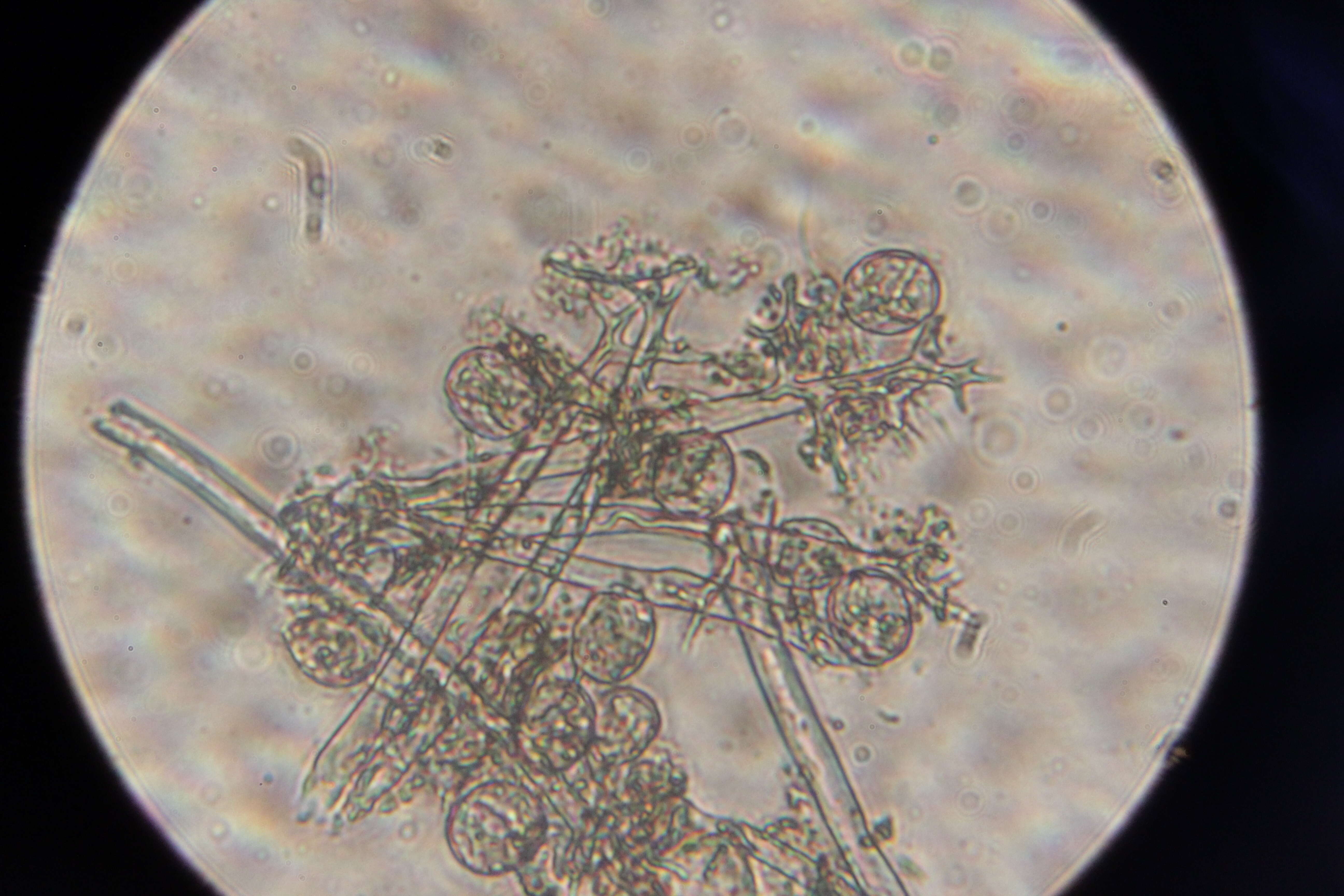
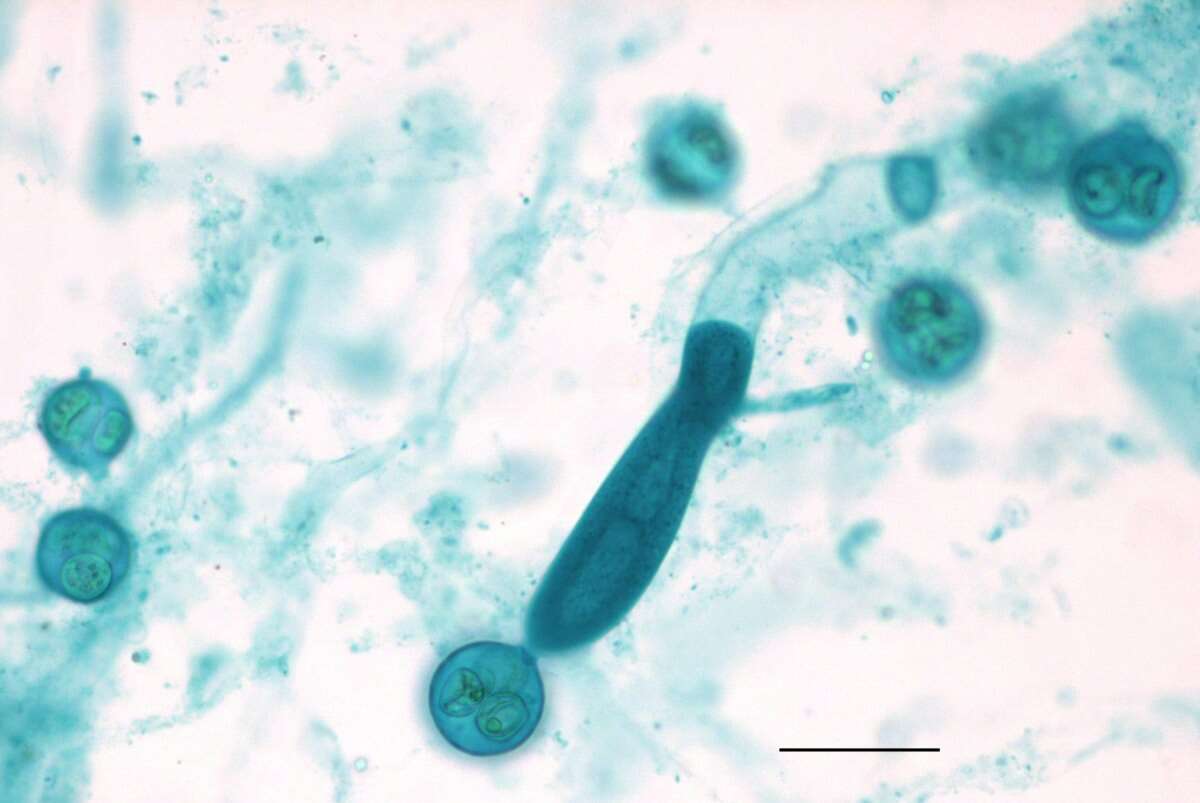
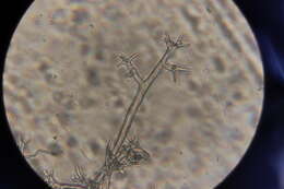
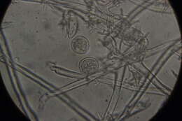
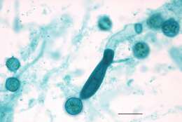
Description: English: Light microscopy of Saprolegnia showing a close up view of an oognium with zygotes inside that is in contact with a zoosporangium filled with zoospores (that are contained by a septum) and the vegetative hypha. Scale bar = 0.2mm. Date: 21 May 2014, 09:18:26. Source: Jon Houseman and Matthew Ford. Author: Jon Houseman. Other versions: Labeled. : This is a retouched picture, which means that it has been digitally altered from its original version. Modifications: Balance (Color, brightness, and contrast) and adjust background color.
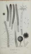
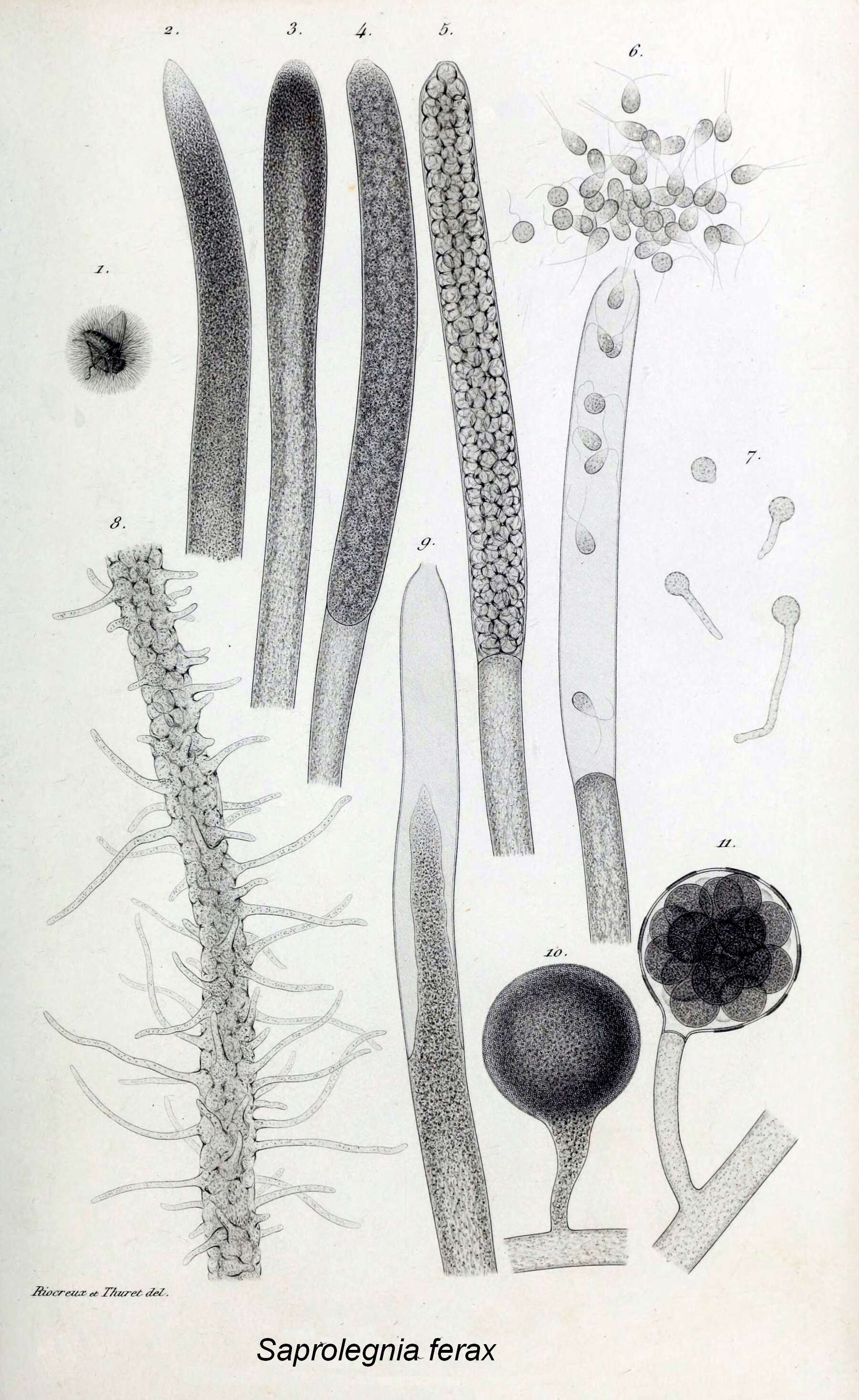
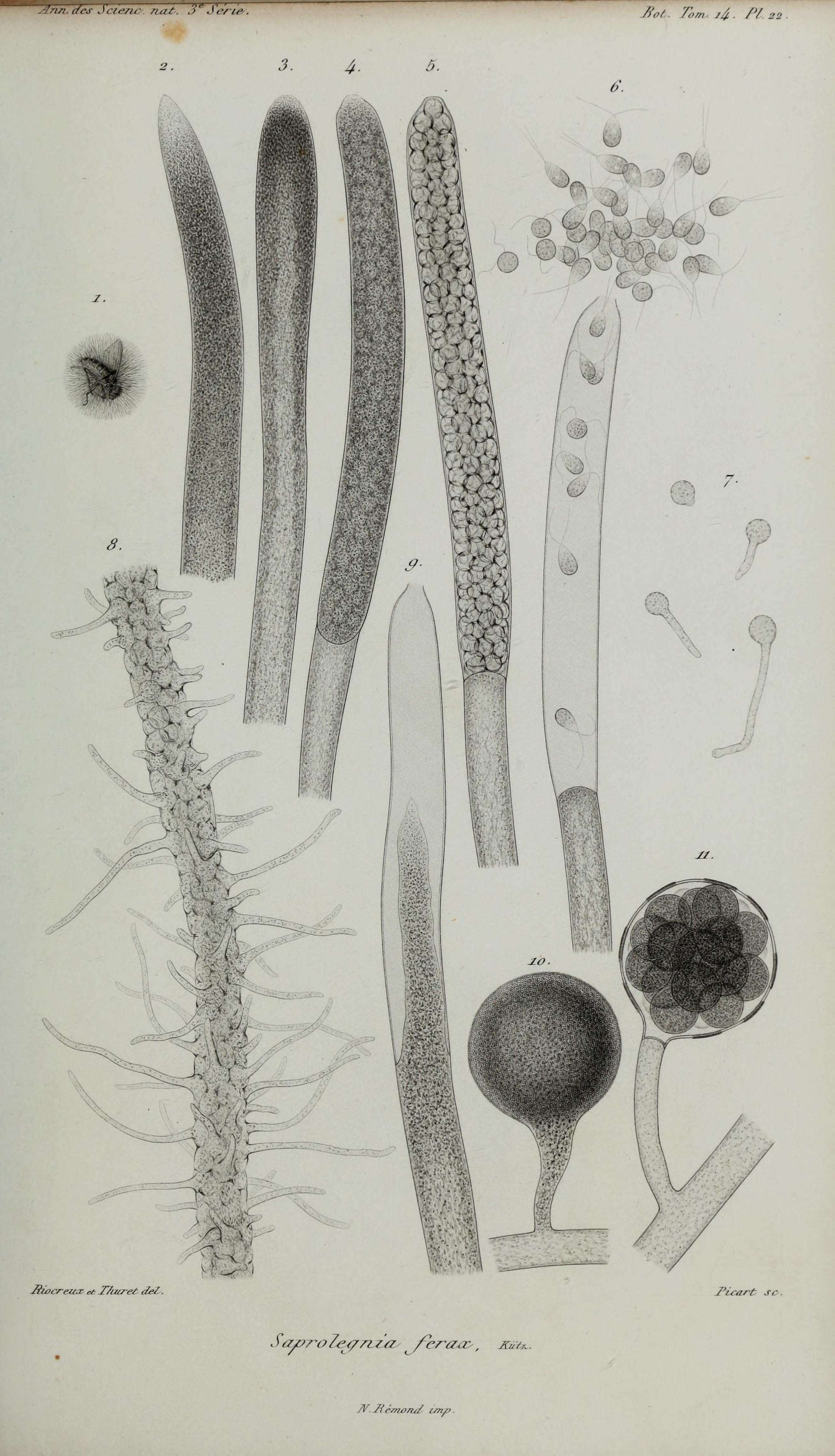
Description: English: Title: Annales des Sciences Naturelles Botaniques Identifier: annalesdesscienc3141850pa (find matches) Year: 1850 (1850s) Authors: Subjects: Publisher: Paris Contributing Library: Natural History Museum Library, London Digitizing Sponsor: BHL-SIL-FEDLINK View Book Page: Book Viewer About This Book: Catalog Entry View All Images: All Images From Book Click here to view book online to see this illustration in context in a browseable online version of this book. Text Appearing Before Image: c/^Jcuvu- n. Bot Tom 1&. /'/ Text Appearing After Image: Siocreux es TAï/reS de/ Picart S
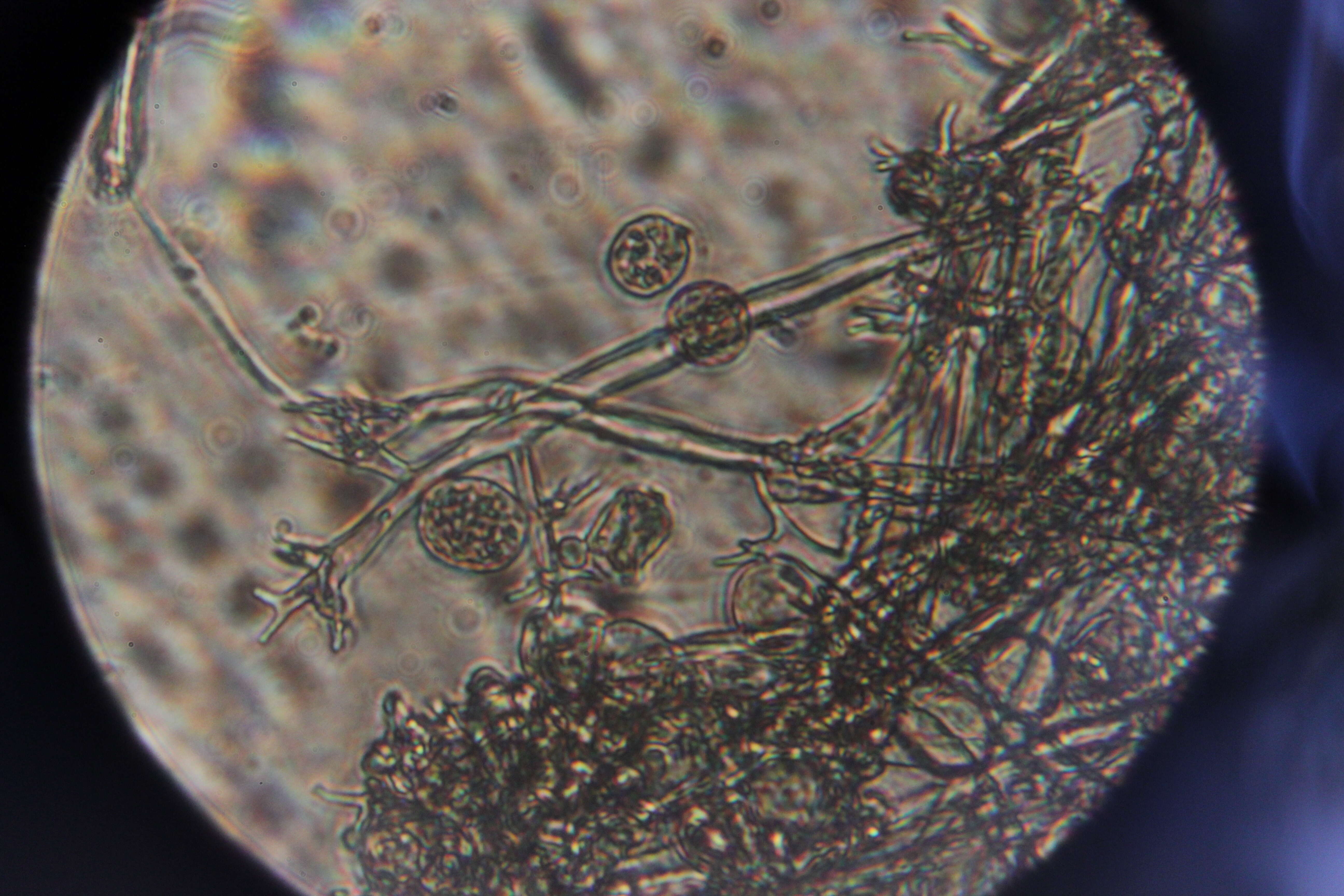
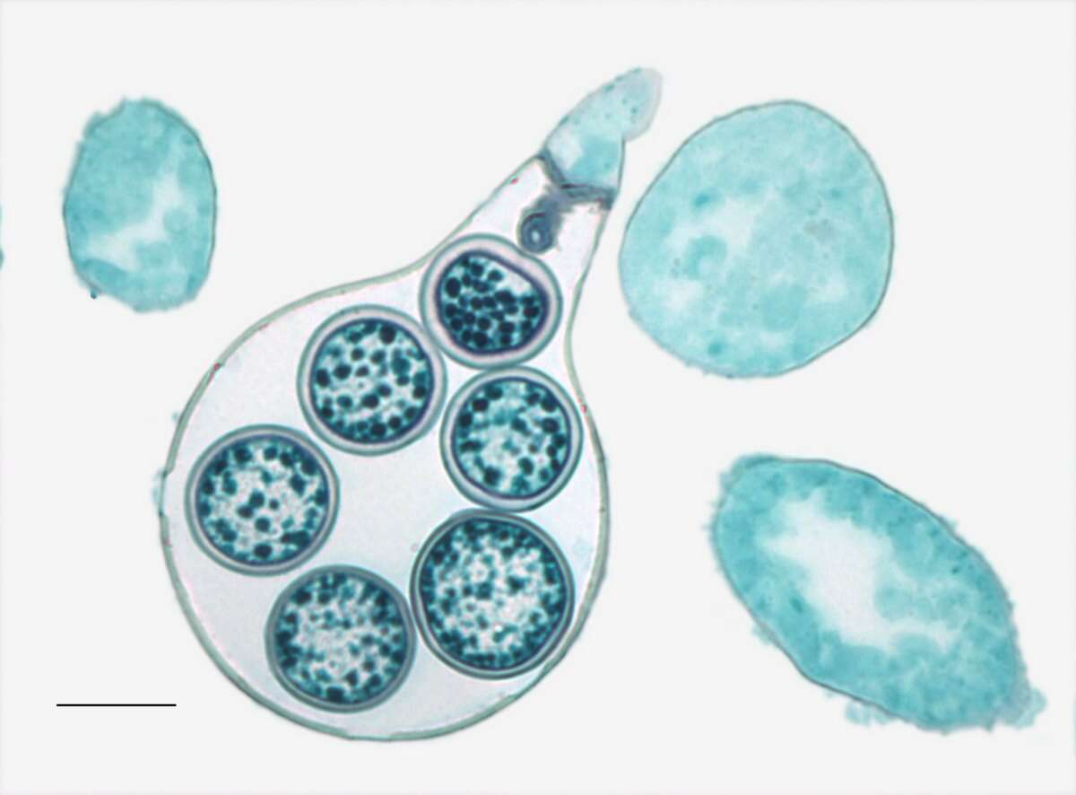
Description: English: Light microscopy of Saprolegnia with an oogonium and zygotes inside with a septum to enclose the entrance. Scale bar = 0.01mm. Date: 21 May 2014, 09:18:32. Source: Jon Houseman and Matthew Ford. Author: Jon Houseman. Other versions: Labeled. : This is a retouched picture, which means that it has been digitally altered from its original version. Modifications: Balance (Color, brightness, and contrast) and adjust background color.
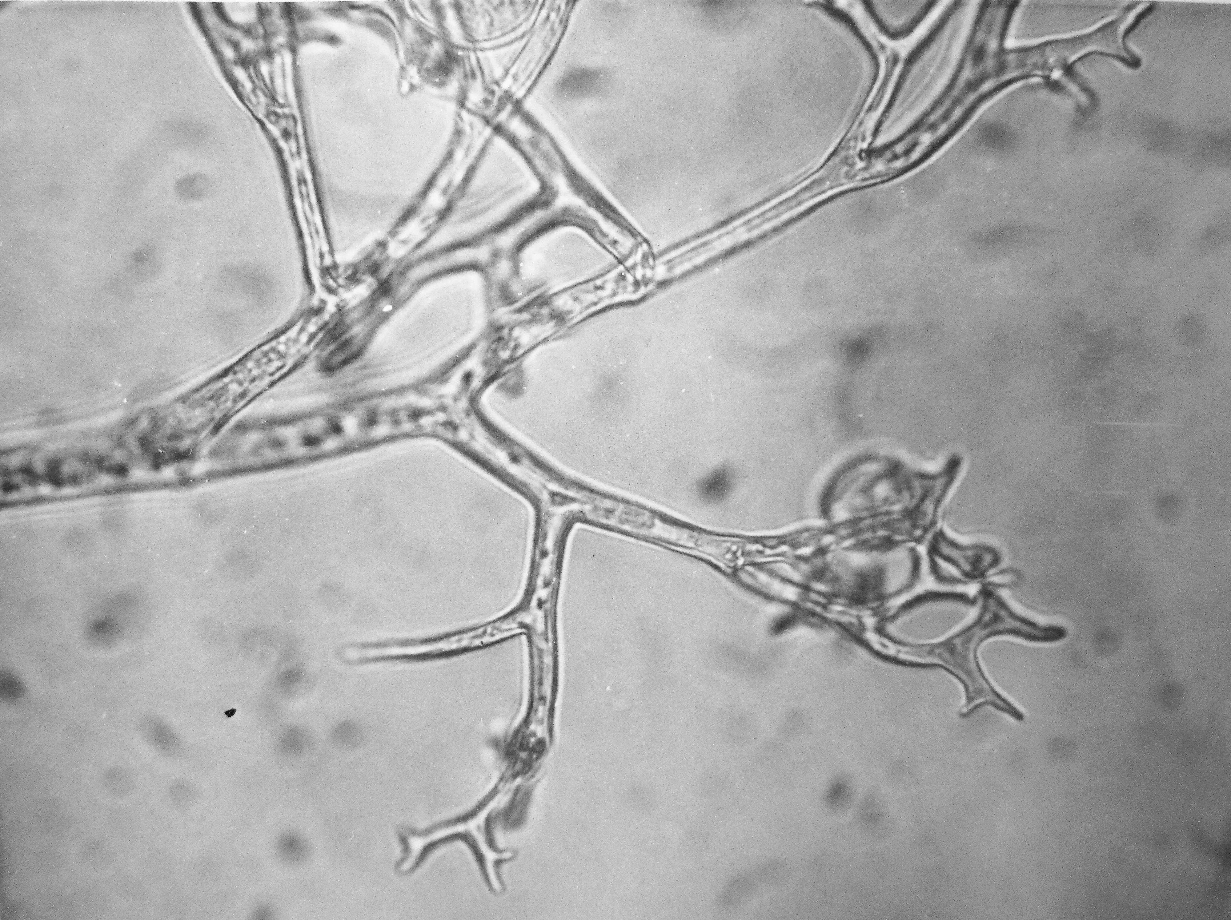
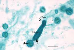
Description: English: Light microscopy of Saprolegnia showing a close up view of an oognium with zygotes inside that is in contact with a zoosporangium filled with zoospores (that are contained by a septum) and the vegetative hypha. A=Zygote, B=Zoosporangium, C=Vegitative hypha, D=Zoosporangium septum, E=Oogonium septum, F=Oogonium. Scale bar = 0.2mm. Date: 26 May 2014, 09:17:20. Source: Jon Houseman and Matthew Ford. Author: Jon Houseman. Other versions: Original (unlabeled). : This is a retouched picture, which means that it has been digitally altered from its original version. Modifications: Balance (Color, brightness, and contrast) and adjust background color.


