-
-
-
-
-
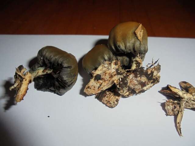
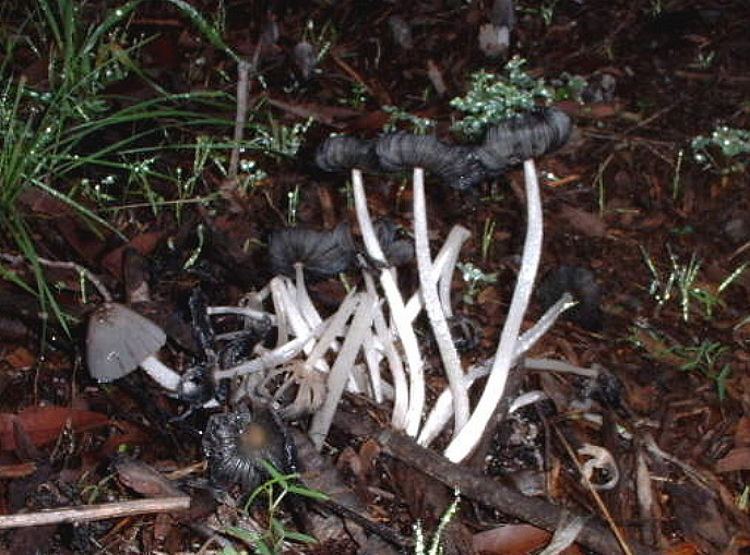
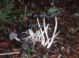
Mushroom Observer Image 739967: Psilocybe weraroa var. subsecotioides nom. prov.
-
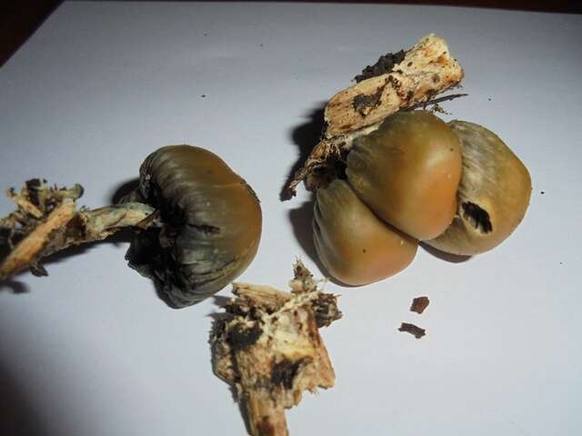
Mushroom Observer Image 739968: Psilocybe weraroa var. subsecotioides nom. prov.
-
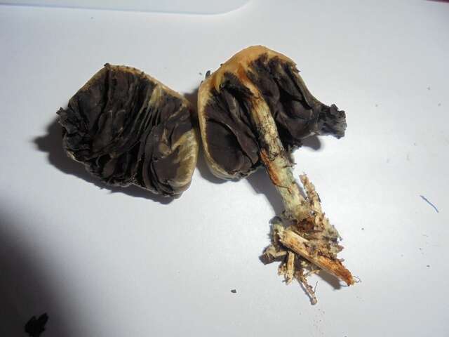
Mushroom Observer Image 739969: Psilocybe weraroa var. subsecotioides nom. prov.
-
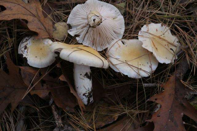
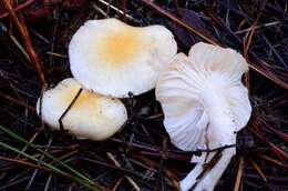
Mushroom Observer Image 391918: Hygrophorus flavodiscus Frost
-
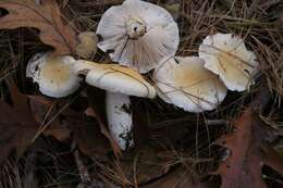
Mushroom Observer Image 571495: Hygrophorus flavodiscus Frost
-
-
Figures 1–3.1 Holotype of Bulbothrix continua 2 Detail of the shiny emaculate upper cortex 3 Holotype of Bulbothrix enormis. Scale bars = 1 cm (1,3), 1 mm (2).
-
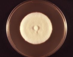
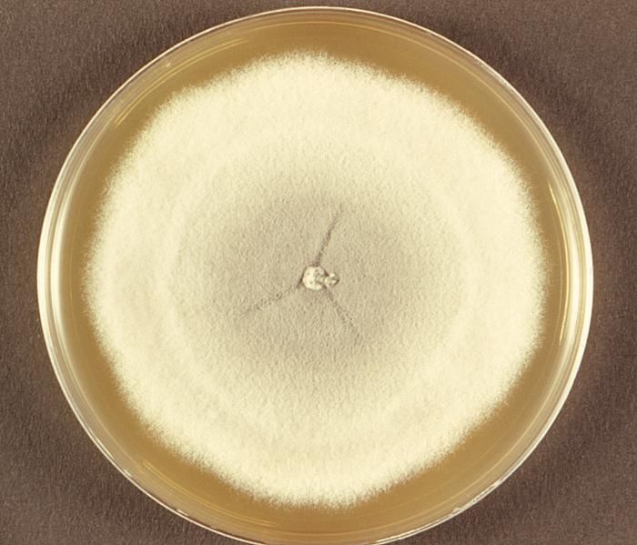
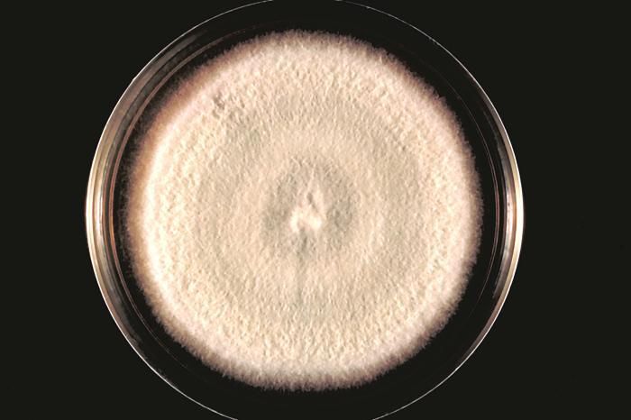
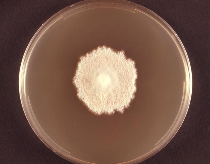
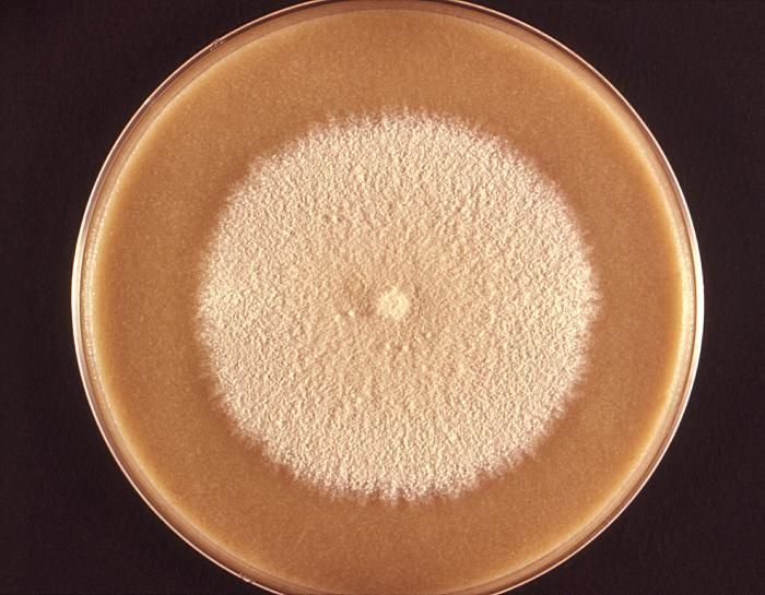
This photograph depicted a single, large colony of Acremonium falciforme fungal organisms, which had been grown on Sabouraud dextrose agar. A. falciforme colonies feature a variety of characteristics including a folded or, as it was in this case, a flat surface, and often have a raised center. Early colonies exhibit a soft, velvety texture, which becomes more cottony as the colony ages. Colonial coloration ranges from white, as seen here, to pale gray or pink.Created: 1974
-
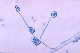
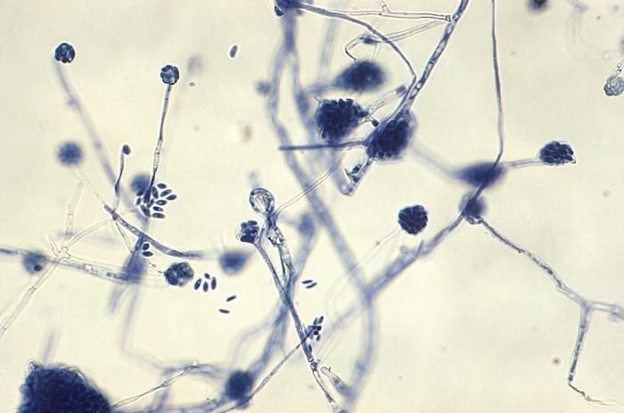
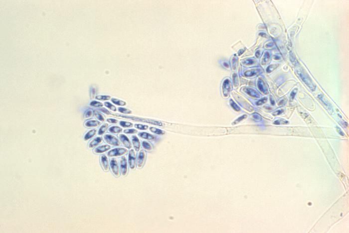
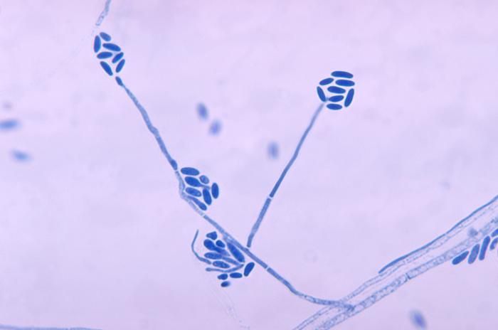
Magnified 1000X, this photomicrograph revealed some of the ultrastructural morphology exhibited by numbers of multiseptate, unbranched conidiophores of the fungal organism, Acremonium falciforme, each topped by a cluster of curved conidia.Created: 1974
-
Magnified 1000X, this photomicrograph revealed some of the ultrastructural morphology exhibited by two multiseptate, unbranched conidiophores of the fungal organism, Acremonium falciforme, each topped by a cluster of curved conidia.Created: 1974
-
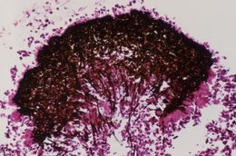
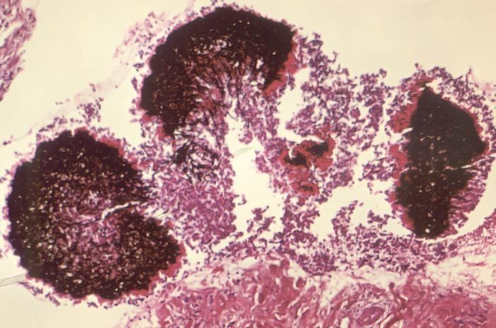
Magnified 250X, this photomicrograph revealed some of the histopathologic cytoarchitectural features exhibited by a single fungal granule of Acremonium falciforme, found in this Gomeri and H&E-stained tissue sample.Created: 1974
-
Magnified 100X, this photomicrograph revealed some of the histopathologic cytoarchitectural features exhibited by three granules of the fungus, Acremonium falciforme, found in this Gomeri and H&E-stained tissue sample.Created: 1974
-
This photomicrograph shows conidia and conidiophores of the fungus Acremonium falciforme.Created: 1970
-
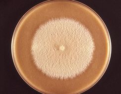
This image depicts a plate culture with a colony of the fungus Acremonium falciforme.Created: 1970
-
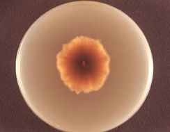
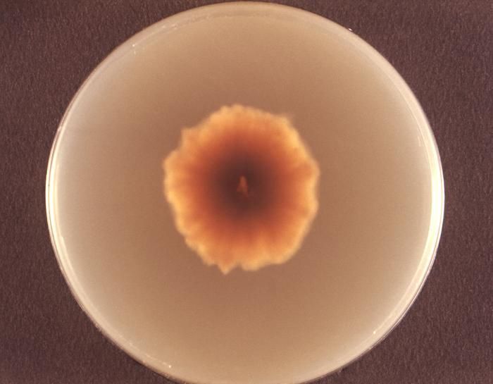
Viewed from the back, i.e., reverse, this image depicted a Petri dish containing Sabouraud's (SAB) dextrose agar, upon which a Microsporum persicolor fungal colony had been cultured. As seen in this reverse view, the colonial coloration can be yellow, or may even be a red-brown. From the front, as depicted in PHIL 10904 and 10906, the colonies can be white, or depending upon the Microsporum sp., may run the gamut, sporting a yellow, beige or cinnamon color, and display a flat, or glabrous, woolly or powdery texture.Created: 1973
-
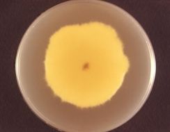
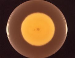
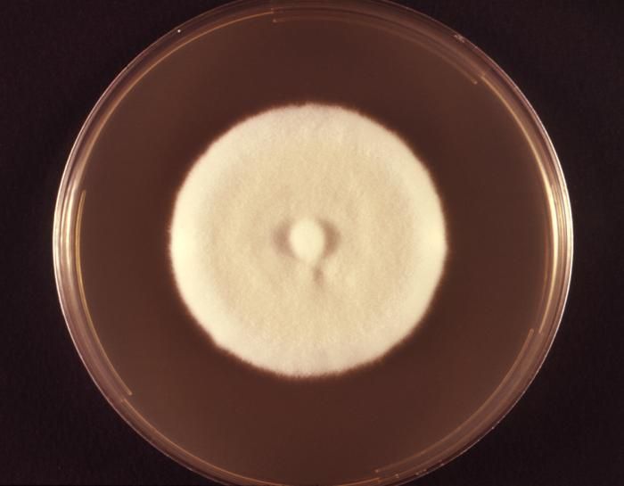
Photographed from the front, this image depicted a Petri dish containing cereal agar, upon which a Microsporum persicolor fungal colony had been cultured. As was the case here, the colonies can be white, or depending upon the Microsporum sp., may run the gamut, sporting a yellow, beige or cinnamon color, and display a flat, or glabrous, woolly or powdery texture. Taxonomically, M. persicolor is a member of the phylum Ascomycota. See PHIL 10905 for a reverse view of this colony, i.e., viewed from behind.Created: 1973
-
Viewed from the back, i.e., reverse, this image depicted a Petri dish containing Sabouraud's (SAB) dextrose agar, upon which a Microsporum persicolor fungal colony had been cultured. As seen in this reverse view, the colonial coloration can be yellow, or may even be a red-brown. From the front, as depicted in PHIL 10906, the colonies can be white, or depending upon the Microsporum sp., may run the gamut, sporting a yellow, beige or cinnamon color, and display a flat, or glabrous, woolly or powdery texture.Created: 1973
-
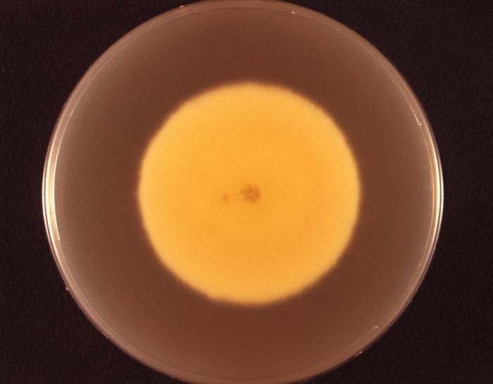
Photographed from the front, this image depicted a Petri dish containing cereal agar, upon which a Microsporum persicolor fungal colony had been cultured. As was the case here, the colonies can be white, or depending upon the Microsporum sp., may run the gamut, sporting a yellow, beige or cinnamon color, and display a flat, or glabrous, woolly or powdery texture. Taxonomically, M. persicolor is a member of the phylum Ascomycota. See PHIL 10903 for a reverse view of this colony, i.e., viewed from behind.Created: 1973
-
Viewed from the back, i.e., reverse, this image depicted a Petri dish containing Sabouraud's (SAB) dextrose agar, upon which a Microsporum persicolor fungal colony had been cultured. As seen in this reverse view, the colonial coloration can be yellow, or may even be a red-brown. From the front, as depicted in PHIL 10904, the colonies can be white, or depending upon the Microsporum sp., may run the gamut, sporting a yellow, beige or cinnamon color, and display a flat, or glabrous, woolly or powdery texture.Created: 1973
-
Photographed from the front, this image depicted a Petri dish containing cereal agar, upon which a Microsporum persicolor fungal colony had been cultured. As was the case here, the colonies can be white, or depending upon the Microsporum sp., may run the gamut, sporting a yellow, beige or cinnamon color, and display a flat, or glabrous, woolly or powdery texture. Taxonomically, M. persicolor is a member of the phylum Ascomycota.Created: 1973